. 2014 May 8;5(5):e1213.
doi: 10.1038/cddis.2014.172.

Fas-associated factor (Faf1) is a novel CD40 interactor that regulates CD40-induced NF-κB activation via a negative feedback loop

Affiliations

Fas-associated factor (Faf1) is a novel CD40 interactor that regulates CD40-induced NF-κB activation via a negative feedback loop

T Elmetwali et al. Cell Death Dis. .

Abstract

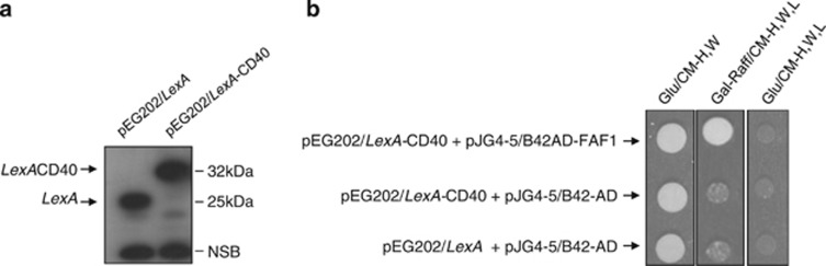
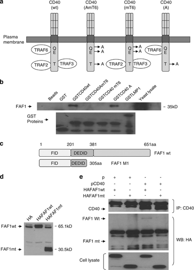
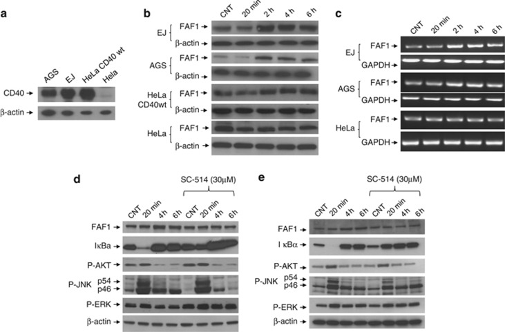
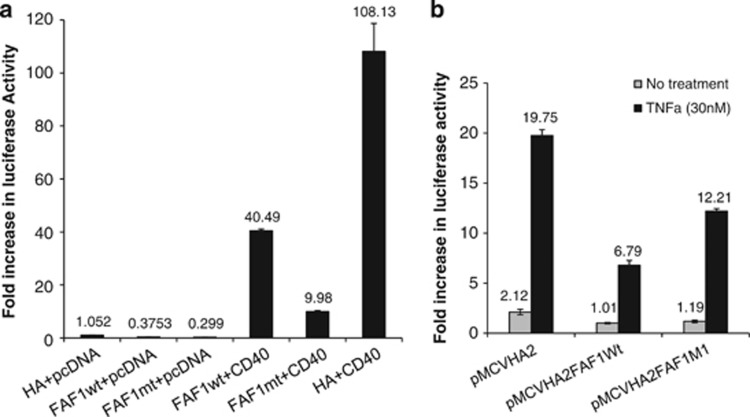
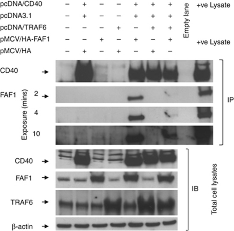
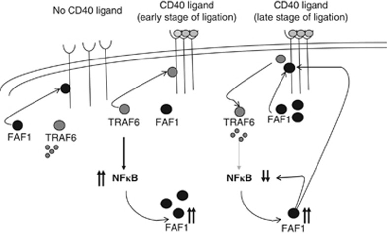
CD40-induced signalling through ligation with its natural ligand (CD40L/CD154) is dependent on recruitment of TRAF molecules to the cytoplasmic domain of the receptor. Here, we applied the yeast two-hybrid system to examine whether other proteins can interact with CD40. Fas-Associated Factor 1(FAF1) was isolated from a HeLa cDNA library using the CD40 cytoplasmic tail (216-278 aa) as a bait construct. FAF1 was able to interact with CD40 both in vitro and in vivo. The FAF1 N-terminal domain was sufficient to bind CD40 and required the TRAF6-binding domain within the cytoplasmic tail of CD40 for binding. CD40 ligation induced FAF1 expression in an NFκB-dependent manner. Knockdown of FAF1 prolonged CD40-induced NFκB, whereas overexpression of FAF1 suppressed CD40-induced NFκB activity and this required interaction of FAF1 with the CD40 receptor via its FID domain. Thus, we report a novel role for FAF1in regulating CD40-induced NFκB activation via a negative feedback loop. Loss of FAF1 function in certain human malignancies may contribute to oncogenesis through unchecked NFκB activation, and further understanding of this process may provide a biomarker of NFκB-targeted therapies for such malignancies.


Figures

Figure 1
Figure 1
CD40–FAF1 interaction in yeast two-hybrid system. (a) EGY48 yeast strain cells harbouring the pEG202/LexA empty plasmid or the pEG202/LexA-CD40 bait construct were grown under glucose inducible media. Protein lysates were prepared, and expression of the CD40 bait construct was then confirmed by western blot analysis using the anti-LexA antibody. (b) EGY48 yeast strain cells harbouring pEG202/LexA+pJG4-5/B42-AD, or pEG202/LexA-CD40+pJG4-5/B42-AD or pEG202/LexA-CD40+pJG4-5/B42-AD-FAF1 were plated on the selective media indicated above. EGY48 cells that harbour pEG202/LexA-CD40+pJG4-5/B42-AD-FAF1 were only able to grow on Gal-Raff/CM agar media lacking the amino acids histidine (H), tryptophan (W) and leucine (L) but not on the Glu/CM-H,W.L medium. EGY48 yeast cells transformed with empty plasmids of pEG202/LexA (202) and pJG4-5/B42-AD (4-5) were used as negative controls
Figure 2
Figure 2
CD40–FAF1 interaction in vitro. (a) Schematic diagram of the different point mutations within the GST-CD40 (wt) construct. A T254→A mutation (CD40A) abolishes TRAF2 and TRAF3 binding to CD40 but does not affect interaction with TRAF6, while Q234E235→AA (CD40mT6) double mutation disrupts interaction only with TRAF6. Finally, a CD40AmT6 mutant is unable to bind TRAF2, TRAF3 or TRAF6. (b) GST pull-down of FAF1 by CD40 protein was performed as previously described. Denaturated samples were analysed by western blotting using anti-HA and anti-GST specific antibodies. GST-LMP1 and GST proteins were used as negative controls. Protein lysate from EGY48 yeast cells harbouring the pJG4-5/B42-AD Hela cDNA FAF1 clone growing in Gal-Raff/CM-W liquid medium was used as a positive control. (c) Schematic representation of FAF1 domain structure, highlighting the Fas-interacting domain (FID) and the death-effector domain interacting domain (DEDID). (d) HEK293 cells were transected with 0.5 μg of empty pMCV/HA (HA) vector or pMCV/HA-FAF1wild-type (HA-FAF1wt) or pMCV/HA-FAF1mutant (FAF1mt; 1-305aa) (HAFAF1mt) for 36 h. HA-tagged FAF1 proteins were then examined by immunoblotting using an anti-HA specific antibody. (e) HEK293 cells were co-transfected with 2 μg of either empty pcDNA3.1 (pcD) and pMCV/HA-FAF1wt (HA-FAF1wt) or empty pcDNA3.1 (P) and pMCV/HA-FAF1mutant (HAFAF1mt) or pcDNA3.1/CD40 (pCD40) and pMCV/HA-FAF1wt, or pcDNA3.1/CD40 and pMCV/HA-FAF1mt for 36h. Protein lysates were prepared and co-immunoprecipitation was performed as previously described. Denaturated samples were resolved by SDS-PAGE and subjected to immunoblotting using anti-HA and anti-CD40 specific antibodies. Protein lysates were used as a positive control
Figure 3
Figure 3
Endogenous FAF1–CD40 interaction in EJ and AGS cells. Total protein lysates (1.5 mg/ml) of either EJ or AGS cells in co-immunoprecipitation (Co-IP) lysis buffer were prepared and precleared for 1 h at 4 oC with 30 μl Protein G sepharose beads (Amersham) (1:1 slurry, prewashed in co-immunoprecipitation lysis buffer), and 40 μg of each precleared total protein lysate was tested for FAF1 and CD40 expression to ensure equal input of total protein lysate in each tube. Lysates were then incubated with either 4 μg of purified mouse IgG1K isotype control (ebioscience cat, 14-4714-82) (Isotype ab) or mouse monoclonal anti-CD40 antibody (MABTECH) (CD40 ab) overnight at 4 oC. Immunoprecipitation was then performed as previously described. Denaturated samples were resolved by SDS-PAGE and subjected to immunoblotting using anti-FAF1and anti-CD40 specific antibodies
Figure 4
Figure 4
CD40 ligation induces FAF1 upregulation in an NFκB-dependent manner. (a) CD40-positive carcinomas (EJ, AGS, Hela cells stably expressing CD40) and CD40-negative carcinoma Hela cells were plated for 24 h in 60 mm tissue culture-treated dishes. Protein lysates were then prepared and analysed for CD40 expression and β-actin using specific antibodies against CD40 and β-actin. (b) Cells were plated for 24 h in 60 mm tissue culture-treated dishes, then treated with 1 μg/ml rsCD40L for 20 min, 2, 4 and 6 h or left untreated as a negative control. Cells were washed with PBS then lysed in situ for protein lysate preparation. FAF1 expression was examined by western blot analysis. (c) Total RNA was extracted from EJ, AGS and HeLa cells at the indicated time points, and 2 μg RNA was reverse transcribed for cDNA synthesis. RT-PCR using 1 μl cDNA was performed utilizing specific primers for FAF1 and GAPDH as a loading control. RT-PCR products were resolved by 1.2% agarose gel electrophoresis. (d) EJ cells and (e) AGS cells were pretreated with the NFκB inhibitor, SC-514 (30 μM) for 6 h. Culture media were then replaced with fresh media containing rsCD40L (1 μg/ml) and NFκB inhibitor SC-514 (30 μM) for 20 min, 4 h or 6 h or left untreated with rsCD40L as a negative control, then lysed in situ. Protein extracts were examined by western blotting for FAF1, IκBα, P-AKT, P-JNK, P-ERK expression. β-Actin was used as loading control
Figure 5
Figure 5
FAF1 inhibits CD40-induced NFκB and FAF1 knockdown results in prolonged NFκB activation. (a) HEK293 cells transiently transfected with 100 ng of the reporter plasmids were co-transfected with the empty pEGFPC1 (GFP, 500 ng) and empty pcDNA3.1 (P, 500 ng) or the empty pcDNA3.1 (500 ng) and pEGFPC1/FAF1 (GFP-FAF1, 50 ng or 500 ng) or empty pEGFPC1 (GFP, 500 ng) and the pcDNA3.1/CD40 (pCD40, 500 ng) or pcDNA3.1/CD40 (500 ng) and pEGFPC1/FAF1 (50 ng or 500 ng). Twenty-four hours later, NFκB activity was measured by luciferase reporter assay. Results are a mean of three triplicate samples±S.D. (b) Protein lysates were probed with anti-CD40 and anti-GFP antibodies to examine the expression of CD40 and GFP-FAF1 protein fusion, respectively. (c) EJ and (d) AGS cells were transfected with either control siRNA or FAF1 siRNA (50 pM) for 48 h then treated with rsCD40 (1 μg/ml) for 20 min, 2 h and 4 h or left untreated as a negative control and then lysed in situ. Protein extracts were then examined by western blotting for FAF1, IκBα, P- IκBα, using specific antibodies. β-Actin was used as a loading control
Figure 6
Figure 6
The FAF1 N-terminal domain is sufficient to inhibit CD40-induced NFκB activity. HEK293 cells transiently transfected with reporter plasmids (100 ng) were (a) co-transfected with the empty pMCV/HA (HA) and empty pcDNA3.1 (pcDNA) or the empty pcDNA3.1 and pMCV/HA-FAF1wt (FAF1wt) or empty pcDNA3.1 and pMCV/HA-FAF1mt, or pcDNA3.1/CD40 (CD40) and pMCV/HA-FAF1wt, or pcDNA3.1/CD40 and pMCV/HA-FAF1mt or pcDNA3.1/CD40 and pMCV/HA empty vector for 24 h, or (b) transfected with the 0.5 μg of empty pMCV/HA or pMCVHA/FAF1wt or pMCV/HAFAF1mt (1–305 aa) for 24 h then treated with TNFα (30 nM) for 6 h. NFκB activity was measured by luciferase reporter assay. Results are a mean of three triplicate samples±S.D.
Figure 7
Figure 7
TRAF6 overexpression abrogates CD40–FAF1 interaction. HEK293 cells were co-transfected with 2 μg of each plasmid as indicated or left untransfected as a negative control. Thirty hours later, cells were lysed in situ in co-immunoprecipitation buffer (1% NP-40/PBS, 1 mM MgCl2, 0.5 mM CaCl2, 20 mM Hepes pH 7.4, 1 mM Na3VO4, 50 mM NaF, 5 mg/ml leupeptin, 1 mg/ml aprotinin, and 1mg/ml pepstatin) for 1 h on ice. Protein lysates were prepared, and co-immunoprecipitation was performed as previously described. Denaturated samples were resolved by SDS-PAGE and subjected to immunoblotting using specific antibodies against CD40, Haemagglutinin (HA) tag and TRAF6. Total protein lysates were used as a positive control in the immunoprecipitation (IP) blots. β-Actin was used as a loading control
Figure 8
Figure 8
Proposed mechanism of FAF1 regulation of CD40 ligand-induced NFκB activity. In the absence of CD40 ligand, the conformation of the CD40 receptor favours FAF1 binding. In this conformation, TRAF6 is not bound to the receptor and is therefore degraded resulting in low NFκB activity. In the early stage of CD40 ligation, receptor trimerisation promotes recruitment of TRAF6. This results in NFκB induction, which, in turn, upregulates FAF1 expression. Higher FAF1 protein levels result in NFκB inhibition by direct inhibition of the assembly of the IKK complex and, indirectly, by competing with the CD40-TRAF6 binding through the TRAF6-binding domain

Similar articles

Cited by

References

    1. van Kooten C, Banchereau J. CD40-CD40 ligand. J Leukoc Biol. 2000;67:2–17. - PubMed
    1. Eliopoulos AG, Young LS. The role of the CD40 pathway in the pathogenesis and treatment of cancer. Curr Opin Pharmacol. 2004;4:360–367. - PubMed
    1. Planken EV, Dijkstra NH, Willemze R, Kluin-Nelemans JC. Proliferation of B cell malignancies in all stages of differentiation upon stimulation in the CD40 system. Leukemia. 1996;10:488–493. - PubMed
    1. Funakoshi S, Longo DL, Beckwith M, Conley DK, Tsarfaty G, Tsarfaty I, et al. Inhibition of human B-cell lymphoma growth by CD40 stimulation. Blood. 1994;83:2787–2794. - PubMed
    1. Davies CC, Mak TW, Young LS, Eliopoulos AG. TRAF6 is required for TRAF2 dependant CD40 signal transduction in nonhemopoietic cells. Mol Cell Biol. 2005;25:9806–9819. - PMC - PubMed

Publication types

MeSH terms