. 2014 May 15;40(5):706-19.
doi: 10.1016/j.immuni.2014.03.011. Epub 2014 May 1.

Interleukin-10 receptor signaling in innate immune cells regulates mucosal immune tolerance and anti-inflammatory macrophage function

Affiliations

Interleukin-10 receptor signaling in innate immune cells regulates mucosal immune tolerance and anti-inflammatory macrophage function

Dror S Shouval et al. Immunity. .

Abstract

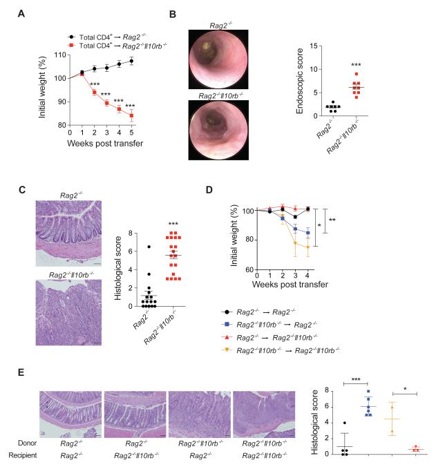
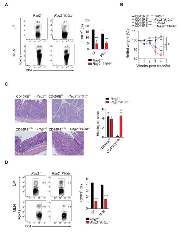
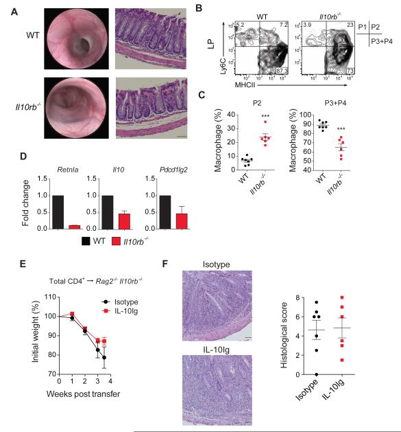
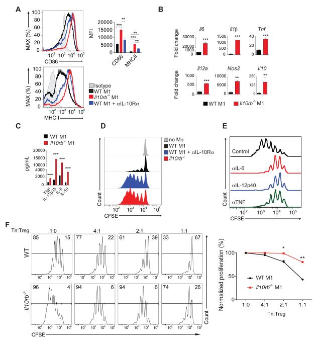
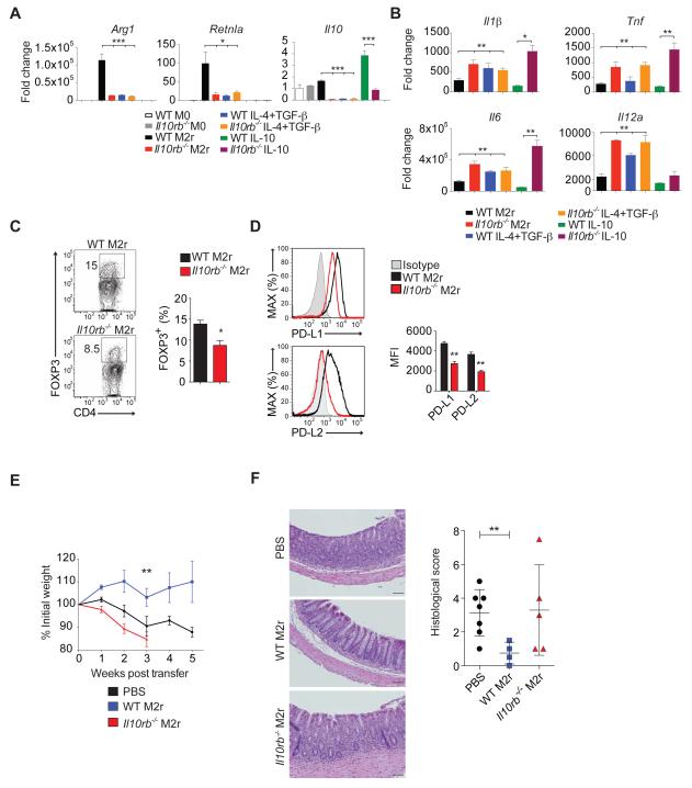
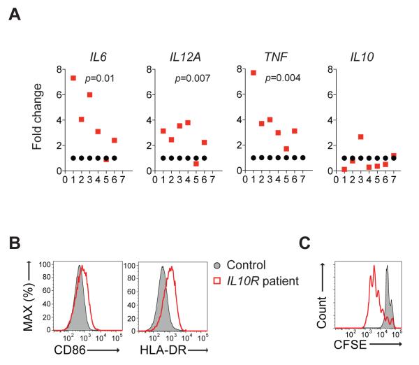
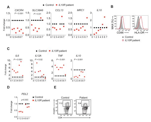
Intact interleukin-10 receptor (IL-10R) signaling on effector and T regulatory (Treg) cells are each independently required to maintain immune tolerance. Here we show that IL-10 sensing by innate immune cells, independent of its effects on T cells, was critical for regulating mucosal homeostasis. Following wild-type (WT) CD4(+) T cell transfer, Rag2(-/-)Il10rb(-/-) mice developed severe colitis in association with profound defects in generation and function of Treg cells. Moreover, loss of IL-10R signaling impaired the generation and function of anti-inflammatory intestinal and bone-marrow-derived macrophages and their ability to secrete IL-10. Importantly, transfer of WT but not Il10rb(-/-) anti-inflammatory macrophages ameliorated colitis induction by WT CD4(+) T cells in Rag2(-/-)Il10rb(-/-) mice. Similar alterations in the generation and function of anti-inflammatory macrophages were observed in IL-10R-deficient patients with very early onset inflammatory bowel disease. Collectively, our studies define innate immune IL-10R signaling as a key factor regulating mucosal immune homeostasis in mice and humans.


Figures

Figure 1
Figure 1. Transfer of WT CD4+ T cells into Rag2−/−Il10rb−/− mice induces severe colitis
Rag2−/− and Rag2−/−Il10rb−/− mice were injected i.p. with 1×10⁶ WT CD4+ T cells. (A) Mean % initial body weights ± SEM following transfer (n = 30 for each group). (B) Representative endoscopic images and scores ± SEM of Rag2−/− and Rag2−/−Il10rb−/− mice 5 weeks post-transfer. (C) Representative H&E section images (20X) and histological score ± SEM of Rag2−/− and Rag2−/−Il10rb−/− mice following transfer. (D) BM chimeras were generated by transferring Rag2−/− or Rag2−/−Il10rb−/− BM cells into lethally irradiated Rag2−/− or Rag2−/−Il10rb−/− recipients, and after 7 weeks WT CD4+ T cells were transferred into these mice. Mean weights ± SEM following T cells transfer displayed in (D) and images of representative H&E stained colonic sections (20X) and mean histological colitis scores ± SEM are displayed in (E). Scale bar = 200 μm. The data is representative of 2 or more independent experiments. Figures S1-S3 and S6 accompany.
Figure 2
Figure 2. Il10rb−/− innate immune cells impair WT Treg cells suppression and generation in vivo
(A) Frequency of Treg cells in LP and MLN of Rag2−/− and Rag2−/−Il10rb−/− mice that were transferred with unfractionated WT CD4+ T cell transfer. Representative flow cytometry plots of FOXP3+ cells among CD4+ T cells are followed by cumulative data in LP and MLN. (B) Mean % initial body weights ± SEM following transfer of WT T naïve (CD4+CD25CD45RBhi) cells alone or in combination with Treg cells (CD4+CD25+CD45RBlo) at a 1:1 ratio. (C) Representative H&E images (20X) of Rag2−/− and Rag2−/−Il10rb−/− recipient mice following transfer and mean histological colitis scores ± SEM. Scale bar = 200 μm. (D) Representative flow cytometry plots of the generation of inducible Treg cells in vivo assessed by FOXP3+ expression among CD4+ T cells in LP and MLN, 4 weeks after CD45RBhi transfer, followed by cumulative data. Results pooled from 2 independent experiments. Figure S4 and S6 accompany.
Figure 3
Figure 3. Reduction in anti-inflammatory intestinal macrophages of pre-colitic ll10rb−/− mice
(A) Endoscopic and histological (10X) images of WT and Il10rb−/− mice at 5 weeks of age. (B-C) Representative flow cytometry plots of macrophage subsets in LP of 5 week old WT and Il10rb−/− mice, followed by quantification of the pro- and anti-inflammatory populations. Pro-inflammatory population was defined as Ly6C+MHCII+ cells and anti-inflammatory as Ly6CMHCII+. (D) LP anti-inflammatory macrophages were sorted from WT (n = 20) and Il10rb−/− (n = 14) 5 week old mice and qRT-PCR was performed to quantify expression of various anti-inflammatory transcripts. Results representative of two independent experiments. (E) Rag2−/− Il10rb−/− were injected with 1 μg of IL-10Ig or isotype one day prior to WT CD4+ T cell transfer, and then twice weekly. Mean weights ± SEM shown in (E) and representative H&E sections images (20X) and histological scores ± SEM of both groups shown in (F). Scale bar = 200 μm. Results pooled from 2 independent experiments. Figures S5 and S6 accompany.
Figure 4
Figure 4. Il10rb−/− M1 BMDM exhibit a pronounced pro-inflammatory phenotype
(A) Mean Fluorescence Intensity (MFI) of MHCII and CD86 expression on M1 WT and Il10rb−/− BMDM or WT BMDM cultured with an IL-10Rα blocking antibody in M1 conditions. (B) Cytokine mRNA expression determined by qRT-PCR of BMDM cultured for 24 hours in M1 conditions; fold change is relative to unstimulated (M0) WT BMDM. (C) Cytokines concentrations determined by ELISA in supernatants of BMDM cultured for 48 hours under M1 conditions. (D) Representative flow cytometry plots of CFSE-labeled WT CD4+CD25 T naïve cells proliferation cultured without macrophages or in the presence of WT M1 BMDM, Il10rb−/− M1 BMDM or WT M1 BMDM cultured with an IL-10Rα blocking antibody. (E) Il10rb−/− M1 BMDM were cultured with sorted T naïve cells, in the presence of neutralizing antibodies to IL-6, IL-12p40 or TNF. (F) WT T naïve cells were cultured with WT or Il10rb−/− M1 BMDM in the presence of varying concentrations of WT Treg cells. Representative flow cytometry plot presented following cumulative data showing degree of proliferation normalized to conditions without Treg cells. All data are representative of two or more independent experiments. Figure S6 accompanies.
Figure 5
Figure 5. Loss of IL-10Rβ signaling impairs the generation and function of anti-inflammatory M2r BMDM
(A) qRT-PCR analysis of Arg1, Retnla (Fizz1) and Il10 transcripts produced by WT or Il10rb−/− BMDM cultured for 24 hours under different conditions. (B) Pro-inflammatory cytokines mRNA expression by WT and Il10rb−/− BMDM cultured in different conditions for 24 hours and then re-stimulated for 4 hours with LPS (100 ng/mL). (C) Representative flow cytometry plots and cumulative data of in vitro generation of FOXP3+ Treg cells among CD4+ T cells in the presence of WT or Il10rb−/− M2r macrophages. (D) Representative flow cyometry plots and cumulative MFI of PD-L1 and PD-L2 surface expression on WT and Il10rb−/− M2r BMDM. (E) 1×10⁶ WT or Il10rb−/− M2r BMDM or PBS were injected i.p. into Rag2−/−Il10rb−/− mice one day prior to WT CD4+ T cell transfer. Figure depicts mean % initial body weights ± SEM following transfer. (F) Representative H&E stained sections (20X) followed by histological scores ± SEM for treated groups. Scale bar = 200 μm. Results pooled from 2 or more independent experiments. Figure S6 accompanies.
Figure 6
Figure 6. Increased pro-inflammatory cytokine production and CD4+ T cell proliferation by human IL-10R-deficient pro-inflammatory macrophages
(A) qRT-PCR analysis of pro-inflammatory cytokines among seven patients with loss-of-function mutations in IL10R genes vs. healthy controls. Each red circle represents a unique patient, while each black rectangle represents an individual healthy control subject in the same experiment. Cytokine expression is normalized to corresponding healthy controls. (B) Flow cytometry plots demonstrating high CD86 and HLA-DR expression on M1 macrophages from an IL-10R-deficient patient, compared to healthy control. (C) Proliferation of CFSE-labeled CD4+CD25 T naïve cells obtained from an allogeneic healthy donor in the presence of IL-10R-deficient M1 macrophages from a patient compared to M1 macrophages obtained from a healthy control. Surface marker expression and proliferation data representative of 5 patients. Table S1 and Figure S6 accompany.
Figure 7
Figure 7. Impaired generation and function of anti-inflammatory macrophages in patients with defective IL-10R chains
(A) qRT-PCR analysis of M2 markers expressed in IL-4 stimulated monocyte-derived macrophages from IL-10R deficient patients vs. healthy subjects. (B) Surface expression of CD86 and HLA-DR by M2 macrophages generated from an IL-10R-deficient patient compared to healthy control. (C) qRT-PCR analysis of various cytokines expressed by M2 macrophages following re-stimulation with LPS. (D) PDL2 expression by M2 macrophages detected by qRT-PCR. (E) Flow cytometry plot illustrating in vitro Treg cells generation from CD4+ T cells in the presence of M2 macrophages from an IL-10R-deficieint patient compared to healthy control. Treg cell generation and flow cytometry data representative of 2 patients. Table S1 and Figure S6 accompany.

Comment in

Similar articles

Cited by

References

    1. Bain CC, Scott CL, Uronen-Hansson H, Gudjonsson S, Jansson O, Grip O, Guilliams M, Malissen B, Agace WW, Mowat AM. Resident and pro-inflammatory macrophages in the colon represent alternative context-dependent fates of the same Ly6Chi monocyte precursors. Mucosal immunology. 2013;6:498–510. - PMC - PubMed
    1. Begue B, Verdier J, Rieux-Laucat F, Goulet O, Morali A, Canioni D, Hugot JP, Daussy C, Verkarre V, Pigneur B, et al. Defective IL10 signaling defining a subgroup of patients with inflammatory bowel disease. The American journal of gastroenterology. 2011;106:1544–1555. - PubMed
    1. Bhattacharyya S, Sen P, Wallet M, Long B, Baldwin AS, Jr., Tisch R. Immunoregulation of dendritic cells by IL-10 is mediated through suppression of the PI3K/Akt pathway and of IkappaB kinase activity. Blood. 2004;104:1100–1109. - PubMed
    1. Chaudhry A, Samstein RM, Treuting P, Liang Y, Pils MC, Heinrich JM, Jack RS, Wunderlich FT, Bruning JC, Muller W, Rudensky AY. Interleukin-10 signaling in regulatory T cells is required for suppression of Th17 cell-mediated inflammation. Immunity. 2011;34:566–578. - PMC - PubMed
    1. Coccia M, Harrison OJ, Schiering C, Asquith MJ, Becher B, Powrie F, Maloy KJ. IL-1beta mediates chronic intestinal inflammation by promoting the accumulation of IL-17A secreting innate lymphoid cells and CD4(+) Th17 cells. The Journal of experimental medicine. 2012;209:1595–1609. - PMC - PubMed

Publication types

MeSH terms