. 2014 May 1;10(5):e1004058.
doi: 10.1371/journal.ppat.1004058. eCollection 2014 May.

Two novel human cytomegalovirus NK cell evasion functions target MICA for lysosomal degradation

Affiliations

Two novel human cytomegalovirus NK cell evasion functions target MICA for lysosomal degradation

Ceri A Fielding et al. PLoS Pathog. .

Abstract

NKG2D plays a major role in controlling immune responses through the regulation of natural killer (NK) cells, αβ and γδ T-cell function. This activating receptor recognizes eight distinct ligands (the MHC Class I polypeptide-related sequences (MIC) A andB, and UL16-binding proteins (ULBP)1-6) induced by cellular stress to promote recognition cells perturbed by malignant transformation or microbial infection. Studies into human cytomegalovirus (HCMV) have aided both the identification and characterization of NKG2D ligands (NKG2DLs). HCMV immediate early (IE) gene up regulates NKGDLs, and we now describe the differential activation of ULBP2 and MICA/B by IE1 and IE2 respectively. Despite activation by IE functions, HCMV effectively suppressed cell surface expression of NKGDLs through both the early and late phases of infection. The immune evasion functions UL16, UL142, and microRNA(miR)-UL112 are known to target NKG2DLs. While infection with a UL16 deletion mutant caused the expected increase in MICB and ULBP2 cell surface expression, deletion of UL142 did not have a similar impact on its target, MICA. We therefore performed a systematic screen of the viral genome to search of addition functions that targeted MICA. US18 and US20 were identified as novel NK cell evasion functions capable of acting independently to promote MICA degradation by lysosomal degradation. The most dramatic effect on MICA expression was achieved when US18 and US20 acted in concert. US18 and US20 are the first members of the US12 gene family to have been assigned a function. The US12 family has 10 members encoded sequentially through US12-US21; a genetic arrangement, which is suggestive of an 'accordion' expansion of an ancestral gene in response to a selective pressure. This expansion must have be an ancient event as the whole family is conserved across simian cytomegaloviruses from old world monkeys. The evolutionary benefit bestowed by the combinatorial effect of US18 and US20 on MICA may have contributed to sustaining the US12 gene family.


Conflict of interest statement

The authors have declared that no competing interests exist.

Figures
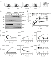
Figure 1
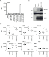
Figure 1. Regulation of NKG2DL during HCMV infection.
A. Fibroblasts (HF-CARs) were infected with RAds expressing IE1 or IE2 at an m.o.i. of 1. Cell surface expression of MICA, MICB, MICA/B, and ULBP2 were analyzed by flow cytometry at 72 h p.i. The results shown are representative of 2 independent experiments. B. Fibroblasts were infected with RAds expressing IE1 or IE2 at an m.o.i. of 1–8 or RAd control (CTRL) moi 1. Expression of MICA, MICB, MICA/B, and ULBP2 was analyzed by immunoblotting at 72 h p.i. The results shown are representative of 2 independent experiments. C. Fibroblasts were infected with RAds expressing IE1 or IE2 or RAds lacking an insert (RAd-CTRL). Infected cells were used as targets for IFN-α treated NK cells in a 51Cr release assay at an E∶T ratio of 1∶50. The results shown are representative of 2 independent experiments and were analyzed by 2-way ANOVA (** p<0.01, *** p = 0.001). D. Fibroblasts (HF-TERTs) were mock-infected or infected with HCMV strain Merlin or deletion mutants of this strain lacking UL16 (ΔUL16) or UL142 (ΔUL142). Cell surface expression of MICA, MICB, MICA/B, ULBP2, MHC-I, or murine immunoglobulin (mIgG) was analyzed between 6 and 120 h p.i. by flow cytometry. The results are shown as median fluorescence intensity (MFI) and are representative of 2 independent experiments.
Figure 2
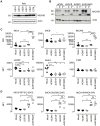
Figure 2. The HCMV gene function targeting MICA maps to the US18–22 gene locus.
A. Fibroblasts (HF-TERTs) were mock infected or infected with HCMV parent virus or a series of ‘block’ deletion mutants for 72 h. Cell surface expression of MICA was analyzed by immunostaining and flow cytometry. Results shown are the median fluorescence intensity (MFI) and are representative of two independent experiments. B. Fibroblasts (HF-TERTs) were mock infected or infected with HCMV parent virus or a deletion mutant lacking US18–22 (ΔUS18–22) for 72 h. Hydrophobic proteins were extracted using Triton X-114 and protein expression analyzed by immunoblotting. Data shown is representative of three independent experiments. C. Fibroblasts (HF-TERTs) were mock infected or infected with HCMV or a deletion mutant lacking US18–22 (ΔUS18–22) for 72 h. Cell surface expression was analyzed by immunostaining and flow cytometry. Results shown are the median fluorescent intensity (MFI) and from three independent experiments. The mean of the results was analysed by Student's t-test (*P<0.05, **P<0.01, ***P<0.001).
Figure 3
Figure 3. US18 and US20 down-regulate MICA levels.
A. Fibroblasts (HF-CARs) were infected with recombinant adenovirus expressing the individual US18–US22 genes for 72 h. Hydrophobic proteins were extracted using Triton X-114 and protein expression analyzed by immunoblotting. Data shown is representative of four similar independent experiments. B. Fibroblasts (HF-TERTs) were mock infected or infected with HCMV, ΔUS18, ΔUS20, ΔUS18&20 for 72 h. Hydrophobic proteins were extracted using Triton X-114, mock-treated (-) or treated with either Endoglycosidase H (E) or PNGase F (P) and protein expression analyzed by immunoblotting. Results are representative of two independent experiments. C. Fibroblasts (HF-TERTs) were mock infected or infected with HCMV, ΔUS18 or ΔUS18, ΔUS20 for 72 h. Cell surface expression was analyzed by immunostaining and flow cytometry. Results shown are the median fluorescence intensity (MFI) and are from four independent experiments. The mean of the results was analysed by Student's t-test (*P<0.05, **P<0.01, ***P<0.001). D. Dermal fibroblasts expressing different MICA alleles were mock infected or infected with HCMV, ΔUS18 or ΔUS18, ΔUS20 for 72 h. Cell surface expression was analyzed by immunostaining and flow cytometry. Results shown are the median fluorescence intensity (MFI) and are from three independent experiments. The mean of the results was analysed by Student's t-test (*P<0.05, **P<0.01, ns = not significant).
Figure 4
Figure 4. Comparison of US18 and US20 expressed during HCMV infection and by adenovirus expression vectors.
A. HF-TERTs were either mock-infected or infected with HCMV wt or HCMV with V5-tagged US18 and US20 genes for 1, 3 or 5 days at an m.o.i. of 10. Cells were harvested and hydrophobic proteins extracted with Triton-X114 before immunoblotting with an anti-V5, actin or UL141 antibodies. Results are representative of two independent experiments. B. HF-CARs were infected with adenovirus control or expressing US18 or US20. Cells were harvested and hydrophobic proteins extracted with Triton-X114 before immunoblotting with an anti-V5 and actin antibodies. Results are representative of five independent experiments. C. HF-TERTs were either mock-infected or infected with HCMV wt or HCMV with V5-tagged US18 and US20 genes for 3 days at an m.o.i. of 10. Cells were fixed, permeabilized and immunostained with anti-V5 antibodies and anti-mouse Alexa Fluor-594 secondary antibody, then counterstained with the nuclear stain DAPI and actin-binding phalloidin-Alexa Fluor 488. Results are representative of three independent experiments. D. HF-CARs were infected with adenovirus control or expressing US18 or US20. Cells were fixed, permeabilized and immunostained with anti-V5 antibodies and anti-mouse Alexa Fluor-594 secondary antibody, then counterstained with the nuclear stain DAPI and actin-binding phalloidin-Alexa Fluor 488. Results are representative of six independent experiments.
Figure 5
Figure 5. Effect of proteasomal and lysosomal inhibitors on MICA/B degradation by HCMV.
A. Fibroblasts (HF-TERTs) were mock-infected or infected with HCMV for 72 h. A proteasomal inhibitor (MG132) or lysososomal inhibitor (folimycin) was added 12 h prior to harvesting. Hydrophobic proteins were extracted using Triton X-114, and protein expression was analyzed by immunoblotting. The results are representative of 3 independent experiments. B. Fibroblasts (HF-TERTs) were either mock infected or infected with HCMV for 72 h. A proteasomal inhibitor (MG132) or lysososomal inhibitor (folimycin) was added 12 h prior to harvesting. Cell surface expression was analyzed by immunostaining and flow cytometry. The results shown are the median fluorescent intensity (MFI) and are representative of 3 independent experiments. C. Fibroblasts (HF-TERTs) were mock-infected or infected with HCMV for 72 h. The lysosomal inhibitors leupeptin or chloroquine were added 12 h prior to harvesting. Hydrophobic proteins were extracted using Triton X-114, and protein expression was analyzed by immunoblotting. The results are representative of 2 independent experiments. D. Fibroblasts (HF-TERTs) were either mock infected or infected with HCMV for 72 h. The lysosomal inhibitors leupeptin or chloroquine were added 12 h prior to harvesting. Cell surface expression was analyzed by immunostaining and flow cytometry. The results shown are the median fluorescent intensity (MFI) and are representative of 2 independent experiments.
Figure 6
Figure 6. Degradation of MICA-YFP by US18 and US20 in U373 cells.
U373 cells stably transfected with MICA-YFP were infected with control or US18-V5, US19-V5 or US20-V5 expressing adenovirus for 72 h in duplicate. Folimycin was added to one duplicate well 12 h prior to analysis. Cells were fixed and permeabilized and immunostained with a mouse antibody for the V5 epitope tag plus anti-mouse IgG-Alexa Fluor 594 secondary antibody and counterstained with the nuclear dye DAPI. Results are representative of three similar independent experiments.
Figure 7
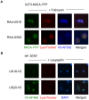
Figure 7. Targeting of MICA-YFP within lysosomes.
A. U373 cells stably transfected with MICA-YFP were infected with US18- or US20 expressing adenovirus for 72 h. Folimycin was added 12 h prior to analysis. Cells were incubated with Lysotracker Red DND-99 (1∶1000) for 30 mins, washed once in DMEM and twice in PBS, fixed and permeabilized, and immunostained with a mouse antibody for the V5 epitope tag plus anti-mouse IgG-Alexa Fluor 350 secondary antibody. Results are representative of two independent experiments. B. HF-TERTs were infected with US18-V5 tagged HCMV or US20 V5-tagged HCMV for 72 hr. Leupeptin was added 12 h prior to analysis. Cells were incubated with Lysotracker Red DND-99 (1∶1000) for 30 mins, washed once in DMEM and twice in PBS, fixed and permeabilized, and immunostained with a rabbit antibody for the V5 epitope tag plus anti-rabbit IgG-Alexa Fluor 488 secondary antibody and counterstained with the nuclear dye DAPI. Results are representative of two independent experiments.
Figure 8
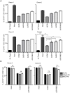
Figure 8. Loss of US18 and US20 increases NK degranulation in response to HCMV-infected targets.
A. Fibroblasts (HF-TERTs) were mock infected or infected with the Merlin strain of HCMV or mutants lacking US18 (ΔUS18), US20 (ΔUS20) or combination of US18 and US20 (ΔUS18, ΔUS20) for 72 h. Infected cells were incubated with donor PBMC for 5 h and NK degranulation assessed by % CD107+ cells within the CD3, CD56+ population by flow cytometry. Results are shown from 3 separate donors performed in triplicate and the mean of pooled results was analysed by Student's t-test (*P<0.05, **P<0.01, ***P<0.001). B. HF-TERTs were mock infected or infected with the Merlin strain of HCMV or the US18 and US20 deletion mutant for 72 h. Infected cells were incubated with isotype control or a MICA blocking antibody for 30 min prior to incubation with donor PBMC for 5 h. NK degranulation was assessed by % CD107+ cells within the CD3, CD56+ population by flow cytometry. Results are shown from 2 separate donors performed in triplicate and the results were analysed by Student's t-test (**P<0.01, ***P<0.001, ns = not significant).

Similar articles

Cited by

References

    1. Arvin AM, Fast P, Myers M, Plotkin S, Rabinovich R (2004) Vaccine development to prevent cytomegalovirus disease: report from the National Vaccine Advisory Committee. Clinical infectious diseases: an official publication of the Infectious Diseases Society of America 39: 233–239. - PubMed
    1. Baryawno N, Rahbar A, Wolmer-Solberg N, Taher C, Odeberg J, et al. (2011) Detection of human cytomegalovirus in medulloblastomas reveals a potential therapeutic target. The Journal of clinical investigation 121: 4043–4055. - PMC - PubMed
    1. Mitchell DA, Xie W, Schmittling R, Learn C, Friedman A, et al. (2008) Sensitive detection of human cytomegalovirus in tumors and peripheral blood of patients diagnosed with glioblastoma. Neuro-oncology 10: 10–18. - PMC - PubMed
    1. Hellstrand K, Martner A, Bergstrom T (2013) Valganciclovir in patients with glioblastoma. The New England journal of medicine 369: 2066. - PubMed
    1. Pierer M, Rothe K, Quandt D, Schulz A, Rossol M, et al. (2012) Association of anticytomegalovirus seropositivity with more severe joint destruction and more frequent joint surgery in rheumatoid arthritis. Arthritis and rheumatism 64: 1740–1749. - PubMed

Publication types

MeSH terms