. 2014 Jun;24(6):680-700.
doi: 10.1038/cr.2014.41. Epub 2014 May 2.

Pachytene piRNAs instruct massive mRNA elimination during late spermiogenesis

Affiliations

Pachytene piRNAs instruct massive mRNA elimination during late spermiogenesis

Lan-Tao Gou et al. Cell Res. 2014 Jun.

Erratum in

Abstract

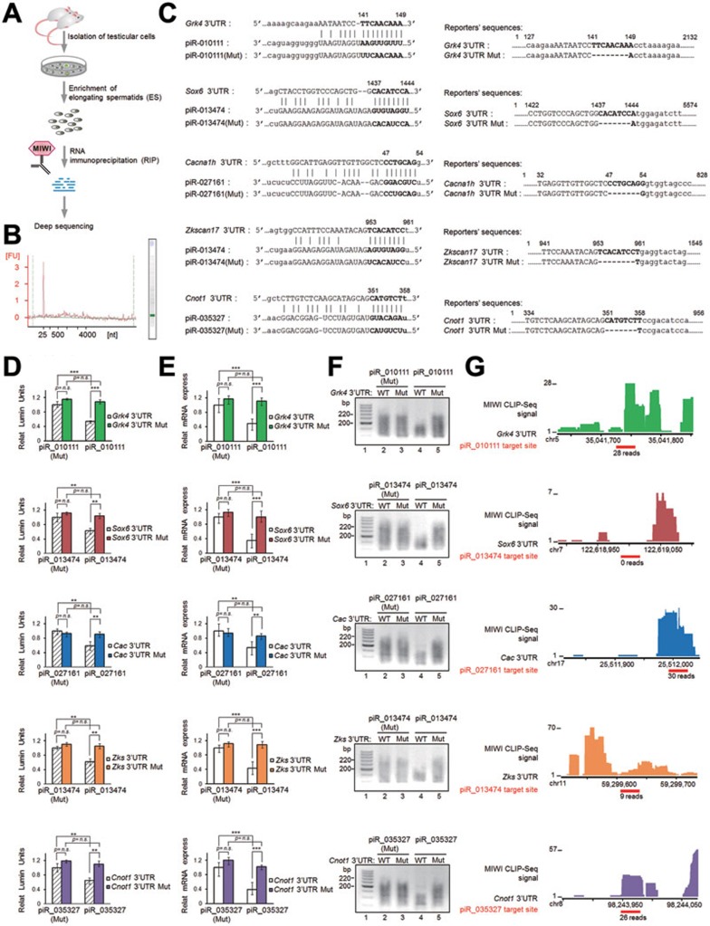
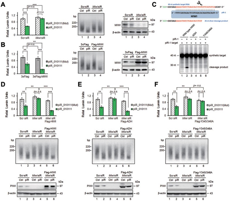
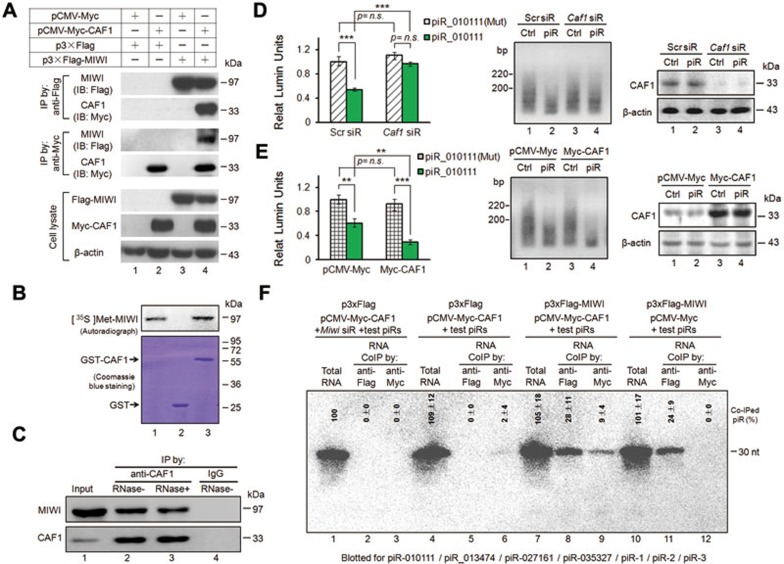
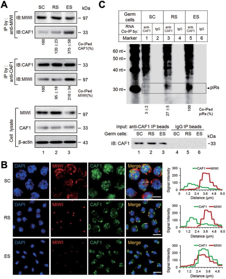
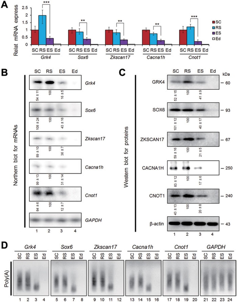
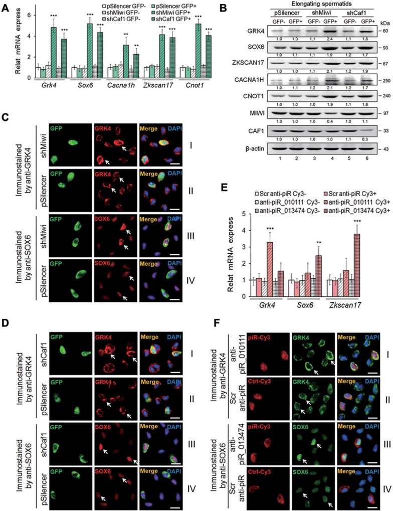
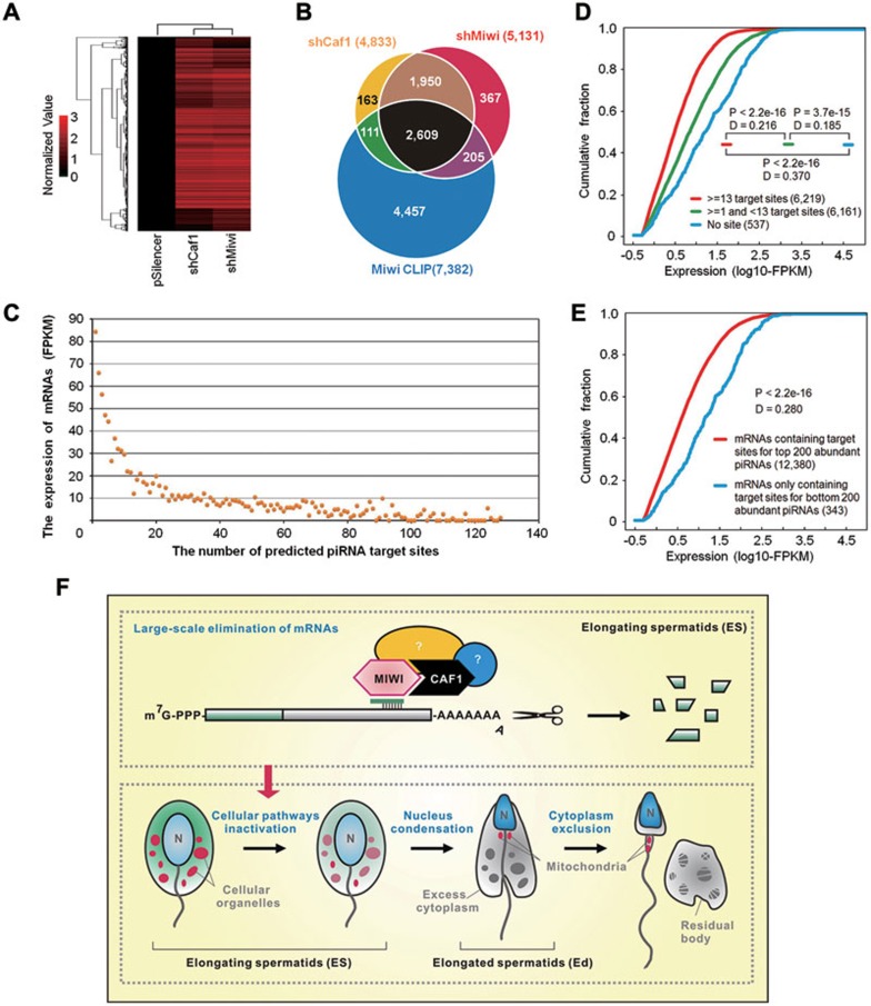
Spermatogenesis in mammals is characterized by two waves of piRNA expression: one corresponds to classic piRNAs responsible for silencing retrotransponsons and the second wave is predominantly derived from nontransposon intergenic regions in pachytene spermatocytes, but the function of these pachytene piRNAs is largely unknown. Here, we report the involvement of pachytene piRNAs in instructing massive mRNA elimination in mouse elongating spermatids (ES). We demonstrate that a piRNA-induced silencing complex (pi-RISC) containing murine PIWI (MIWI) and deadenylase CAF1 is selectively assembled in ES, which is responsible for inducing mRNA deadenylation and decay via a mechanism that resembles the action of miRNAs in somatic cells. Such a highly orchestrated program appears to take full advantage of the enormous repertoire of diversified targeting capacity of pachytene piRNAs derived from nontransposon intergenic regions. These findings suggest that pachytene piRNAs are responsible for inactivating vast cellular programs in preparation for sperm production from ES.


Figures

Figure 1
Figure 1
piRNAs act like miRNAs to induce mRNA deadenylation and decay. (A) Strategy to identify RNAs associated with MIWI in ES by RIP. (B) Urea-polyacrylamide gel electrophoresis analyses of MIWI RIPed-RNAs. Left, densities of RNAs at size range of measurement. Right, representative image of urea-polyacrylamide gel. (C) Predicted piRNA regulatory elements at the 3′UTRs of Grk4, Sox6, Cacna1h, Zkscan17, and Cnot1, and the sequences of synthetic piRNAs, negative control piRNA(Mut), and wild-type 3′UTR and mutated 3′UTR Mut-Renilla luciferase reporters. (D) Dual-luciferase reporter assays of the effect of piRNAs on reporter activities in GC-2spd(ts) cells, with cognate piRNA(Mut) serving as negative controls. (E) qRT-PCR analyses of the effect of piRNAs on reporter mRNA levels. (F) PAT assays of the effect of piRNAs on reporter Poly(A) tails. (G) Verification of piRNA target sites by MIWI CLIP. Top, the cluster of MIWI CLIP tags on the five validated piRNA target genes (one color per gene). The scales in genes were numbered following the dimension of their respective chromosomes. Bottom, the red thick lines indicated piRNA target sites. The indicated numbers at top left represent the highest reads of CLIP tags for cognate genes, and those under red thick lines show the reads of CLIP tags that perfectly matched to cognate target sites. The average values ± S.D. of three separate experiments were plotted. **P < 0.01, ***P < 0.001. Results shown are representative of three independent experiments.
Figure 2
Figure 2
MIWI is required for piRNA-induced target repression. (A and B) The effect of Miwi knockdown (A) or ectopic Flag-MIWI (B) on piR_010111-induced Grk4 reporter repression in GC-2spd(ts) cells. Left, dual-luciferase reporter assays. Middle, PAT assays. Right, western blot assays of MIWI proteins, with β-actin serving as a loading control. (C) In vitro slicer activity assays of wildtype HIWI (lanes 1 and 2), and its slicer activity-deficient ADH (lanes 3 and 4) and piRNA loading-deficient Y345/346A mutants (lanes 5 and 6). Left, Scheme showing the synthetic 40 nt RNA target for piR-1 as well as the expected cleavage products. Right, Slicer assay with HIWI complexes (purified with Flag antibody) and a 5′-[32P]-labeled RNA target bearing complementarity to piR-1. Lanes 1, 3, and 5, HIWI or its mutants purified from the cells transfected with Flag-HIWI or its mutant expression vectors; lanes 2, 4, and 6, HIWI/piR-1 complexes purified from GC-2spd(ts) cells cotransfected with respective HIWI constructs and piR-1. (D-F) Ectopic Flag-HIWI (D) or Flag-ADH HIWI mutant (E), but not Flag-Y345/346 HIWI mutant (F), rescued piR_010111-induced Grk4 reporter repression in Miwi siR-treated GC-2spd (ts) cells. Top, dual-luciferase reporter assays. Middle, PAT assays. Bottom, western blot assays of PIWI proteins, with β-actin serving as a loading control. The average values ± s.d. of three separate experiments were plotted. **P < 0.01, ***P < 0.001. Results shown are representative of three independent experiments.
Figure 3
Figure 3
CAF1 is a key partner of MIWI in piRNA-induced target repression. (A) Co-IP assays of the interaction between Myc-CAF1 and Flag-MIWI in GC-2spd (ts) cells. Anti-Flag IP (top) and anti-Myc IP pellets (middle) were respectively immunoblotted by anti-Myc and anti-Flag antibodies. Bottom, cell lysates immunoblotted by anti-Flag or anti-Myc antibodies, with β-actin serving as a loading control. (B) GST pull-down assays of the interaction between CAF1 and MIWI. The GST-fused CAF1 was generated in bacteria and purified. [35S]-Met-MIWI protein prepared by in vitro transcription/translation was incubated with GST (lane 2) or GST-CAF1 (lane 3), respectively. Input represents 10% of [35S]-Met-MIWI (lane 1). Samples were analyzed by autoradiography (top) or visualized by Coomassie blue staining (bottom). (C) Co-IP assays of the interaction between endogenous CAF1 and MIWI in adult mouse testes. Anti-CAF1 IP pellets from RNase A untreated (lane 2) or treated testis lysate (lane 3) immunoblotted by anti-MIWI (top) and anti-CAF1 (bottom) antibodies, respectively. Immunoblot of MIWI or CAF1 in testis lysate (lane 1) or IgG IP pellet (lane 4) served as positive and negative controls, respectively. (D and E) The effect of Caf1 knockdown (D) or ectopic Myc-CAF1 (E) on piR_010111-induced Grk4 reporter repression in GC-2spd (ts) cells. Left, dual-luciferase reporter assays. Middle, PAT assays. Right, western blot assays of CAF1 proteins, with β-actin serving as a loading control. (F) RIP combined with northern blot assays of pi-RISC complex assembly in GC-2spd (ts) cells. Cells were cotransfected with the indicated expression vectors and mouse test piRs plus Miwi siR (lanes 1-3) or test piR (lanes 4-12). Anti-Flag RIP (lanes 2, 5, 8, and 11) and anti-Myc RIP (lanes 3, 6, 9, and 12) were blotted with the probes of piR_010111, piR_013474, piR_027161, piR_035327, piR-1, piR-2, and piR-3. Total RNAs from respective transfected cells served as positive controls (lanes 1, 4, 7, and 10). The average values ± s.d. of three separate experiments were plotted. **P < 0.01, ***P < 0.001. Results shown are representative of three independent experiments.
Figure 4
Figure 4
pi-RISC is predominantly assembled in ES. (A) Co-IP assays of the interaction between CAF1 and MIWI in enriched-SC (lane 1), RS (lane 2), and ES (lane 3). Anti-MIWI IP (top) and anti-CAF1 IP pellets (middle) immunoblotted by anti-CAF1 and anti-MIWI antibodies, respectively. Bottom, respective cell lysates immunoblotted by anti-MIWI or anti-CAF1 antibodies, with β-actin serving as a loading control. (B) Double-immunostaining of MIWI (red) and CAF1 (green) in SC (top), RS (middle), and ES (bottom). Nuclei were counterstained with DAPI (blue). Colocalization of MIWI and CAF1 was shown as yellow. Scale bar, 10 μm. Z-stacks (right) was used to project the 6.0-μm side view (x axis) of MIWI (red) CAF1 (green) in the lined region. (C) RIP assays of CAF1-associated RNAs in SC (lane 1), RS (lane 3), and ES (lane5), with IgG-IPed RNA as negative controls (lanes 2, 4, and 6), and immunoblot of IP pellets with anti-CAF1 as loading references (bottom). Results shown are representative of three independent experiments.
Figure 5
Figure 5
Decay of piRNA targets occurs in ES. (A) qRT-PCR assays of the mRNA levels of the five piRNA targets (Grk4, Sox6, Zkscan17, Cacna1h, and Cnot1) in enriched SC (red), RS (cyan), ES (purple), and Ed (white), with GAPDH serving as an internal control. The average values ± s.d. of three separate experiments were plotted. **P < 0.01, ***P < 0.001. (B and C) Northern blot (B) or western blot analyses (C) of the expression of the five target genes in enriched SC (lane 1), RS (lane 2), ES (lane 3), and Ed (lane 4), with GAPDH (B) or β-actin (C) serving as loading controls. (D) PAT assays of Poly(A) tails of the five target mRNAs in enriched SC (lanes 1, 5, 9, 13, and 17), RS (lanes 2, 6, 10, 14, and 18), ES (lanes 3, 7, 11, 15, and 19), and Ed (lanes 4, 8, 12, 16, and 20), with GAPDH serving as a nontarget control (lanes 21-24). Results shown are representative of three independent experiments.
Figure 6
Figure 6
The components of pi-RISC are required for piRNA target repression. (A and B) The effect of Miwi or Caf1 knockdown on the expression of the five piRNA targets (Grk4, Sox6, Zkscan17, Cacna1h, and Cnot1) in ES. GFP+ cells were sorted from the ES isolated from shMiwi:GFP-, shCaf1:GFP-, or control pSilencer:GFP-transduced mice. (A) qRT-PCR assays of their mRNA levels, with GAPDH serving as an internal control. The respective treatments were indicated in parentheses. (B) Western blot assays of their protein levels in GFP+ cells from control pSilencer:GFP (lane 2), shMiwi:GFP (lane 4), or shCaf1:GFP (lane 6), with respective GFP cells (lanes 1, 3, and 5) as negative controls. β-actin served as an internal reference. (C and D) Immunostaining of GRK4 (panels I and II, red) and SOX6 proteins (panels III and IV, red) in ES transduced by shMiwi:GFP (C, panels I and III), shCaf1:GFP (D, panels I and III), or control pSilencer:GFP (C and D, panels II and IV). (E and F) The effect of inhibition of piRNA function on piRNA target expression in ES. (E) qRT-PCR assays of the mRNA levels of Grk4 (piR_010111 target), and Sox6 and Zkscan17 (piR_013474 targets) in Cy3+ ES, with GAPDH serving as an internal control. The respective treatments were indicated in parentheses. Cy3+ cells were sorted from ES isolated from mice electroporated by 2′-O-methyl anti-piR_010111, anti-piR_013474, or scrambled anti-piR, respectively. (F) Immunostaining of GRK4 (panels I and II, green) and SOX6 (panels III and IV, green) in ES electroporated by 2′-O-methyl anti-piR_010111 (panel I, red), anti-piR_013474 (panel III, red), or scramble anti-piR (panels II and IV, red), respectively. The average values ± s.d. of three separate experiments were plotted. **P < 0.01, ***P < 0.001. Scale bar, 10 μm. Results shown are representative of three independent experiments.
Figure 7
Figure 7
pi-RISC contributes to the mRNA eradication program during spermiogenesis. (A) Mouse transcriptome microarray profiling genes in ES altered by Miwi or Caf1 knockdown. The total RNAs for microarray assays were from sorted GFP+ ES isolated from mice transduced by pSilencer:GFP, shMiwi:GFP, or shCaf1:GFP, respectively. (B) Venn diagram showing the cross of genes upregulated by Miwi knockdown (red), Caf1 knockdown (yellow), and MIWI-associated mRNAs characterized by CLIP-seq (blue). (C) The levels of mRNAs in ES inversely correlated with the numbers of potential piRNA binding sites in their 3′UTRs. X axis represents the number of piRNA binding sites. Y axis shows average expression value (FPKM) of mRNAs with same piRNA target sites. (D and E) Comparison of the cumulative abundance (log10) among three groups of mRNAs with ≥ 13 sites (red line), 1-13 sites (green line) or no apparent site (blue line) (D), or between two groups of mRNAs matched to the top 200 (red line) or bottom 200 (blue line) abundant piRNAs (E), as indicated in parentheses. mRNA numbers in each group were indicated and P- and D-values determined by Kolmogorov-Smirnov test (R version 2.13.0) were shown in each panel. (F) A model for the function of MIWI in ES, in which MIWI is assembled into a functional pi-RISC complex with guider piRNAs and deadenylase CAF1, and mediates mRNA deadenylation and decay via a miRNA-like mechanism. Such pi-RISC-triggered large-scale elimination of mRNAs in ES may facilitate nucleus condensation and cytoplasm exclusion for the completion of spermatozoa formation in mammals. Results shown are representative of three independent experiments.

Comment in

Similar articles

Cited by

References

    1. Aravin A, Gaidatzis D, Pfeffer S, et al. A novel class of small RNAs bind to MILI protein in mouse testes. Nature. 2006;442:203–207. - PubMed
    1. Girard A, Sachidanandam R, Hannon GJ, Carmell MA. A germline-specific class of small RNAs binds mammalian Piwi proteins. Nature. 2006;442:199–202. - PubMed
    1. Grivna ST, Beyret E, Wang Z, Lin H. A novel class of small RNAs in mouse spermatogenic cells. Genes Dev. 2006;20:1709–1714. - PMC - PubMed
    1. Lau NC, Seto AG, Kim J, et al. Characterization of the piRNA complex from rat testes. Science. 2006;313:363–367. - PubMed
    1. Cox DN, Chao A, Baker J, Chang L, Qiao D, Lin H. A novel class of evolutionarily conserved genes defined by piwi are essential for stem cell self-renewal. Genes Dev. 1998;12:3715–3727. - PMC - PubMed

Publication types