. 2014 Apr 24;157(3):702-713.
doi: 10.1016/j.cell.2014.02.034.

A viral packaging motor varies its DNA rotation and step size to preserve subunit coordination as the capsid fills

Affiliations

A viral packaging motor varies its DNA rotation and step size to preserve subunit coordination as the capsid fills

Shixin Liu et al. Cell. .

Abstract

Multimeric, ring-shaped molecular motors rely on the coordinated action of their subunits to perform crucial biological functions. During these tasks, motors often change their operation in response to regulatory signals. Here, we investigate a viral packaging machine as it fills the capsid with DNA and encounters increasing internal pressure. We find that the motor rotates the DNA during packaging and that the rotation per base pair increases with filling. This change accompanies a reduction in the motor's step size. We propose that these adjustments preserve motor coordination by allowing one subunit to make periodic, specific, and regulatory contacts with the DNA. At high filling, we also observe the downregulation of the ATP-binding rate and the emergence of long-lived pauses, suggesting a throttling-down mechanism employed by the motor near the completion of packaging. This study illustrates how a biological motor adjusts its operation in response to changing conditions, while remaining highly coordinated.


Figures

Figure 1
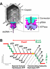
Figure 1. Overview of the φ29 Packaging Motor
(A) Cryo-EM reconstruction of the φ29 capsid (gray), connector (cyan), pRNA (magenta), and gp16 ATPase (blue). Reproduced from (Morais et al., 2008), with permission from Elsevier. (B) Mechanochemical model of the dwell-burst packaging cycle at low capsid filling.
Figure 2
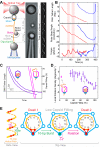
Figure 2. The φ29 Motor Rotates DNA during Packaging
(A) Left: Experimental geometry of the rotation assay. The packaging complex is tethered between two beads. Biotin-streptavidin linkages torsionally couple the rotor bead to the optically-trapped bead via dsDNA. A nick and a single-stranded DNA region ensure that the rotor bead is torsionally decoupled from the micropipette-bound bead. Middle: Micrograph of the experimental geometry. Right: Kymograph of the rotor bead position during packaging. (B) Sample traces displaying DNA tether length (top), rotor bead angle (middle), and torque stored in the DNA (bottom) during packaging. Pauses in translocation are concomitant with pauses in rotation (shaded light red). Slipping (shaded light blue) causes a reversal in the rotation direction. (C) Sample packaging traces with intact proheads (purple) and trepanated proheads (magenta). (D) Local DNA rotation density versus capsid filling. The data point obtained with trepanated proheads – corresponding to very low capsid filling conditions – is shown as a magenta square. Error bars represent SEM. (E) The geometric basis for DNA rotation at low capsid filling. Left: B-form DNA backbone diagram (only the 5′–3′ strand is shown). The φ29 motor forms specific contacts with pairs of phosphates (red) every 10 bases. Right: Top view of the motor (blue) and the DNA (orange). The same subunit contacts the DNA backbone phosphates in consecutive dwells. After a 10-bp burst, a 14° clockwise DNA rotation is needed to bring the DNA and the motor into perfect register. See also Figure S1 and Movie S1.
Figure 3
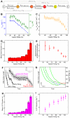
Figure 3. Motor Coordination Is Preserved at High Capsid Filling
(A) Experimental geometry of the high-resolution packaging assay. (B) Sample traces displaying individual packaging cycles at various levels of capsid filling. Raw 2500-Hz data are shown in gray and downsampled 100-Hz data in black. Stepwise fit to the data highlights dwells and bursts in red and green, respectively. (C) Cumulative probability distribution of the packaging cycle times. Each color corresponds to a certain filling level. (D) The nucleotide exchange scheme for each ATPase subunit. (E) Sample packaging traces at low external loads (7–10 pN) and various ATP concentrations. (F) Mean packaging velocity (pauses and slips removed) versus [ATP]. Each color represents a different filling level. The data are fit to the Hill equation, v = Vmax·[ATP]n/(KMn + [ATP]n). Inset: The Hill coefficient n from the fit. (G) Inverse of the mean packaging velocity versus the ratio of [ADP] to [ATP] at different filling levels. [ATP] is fixed at 250 μM. The data are fit to a competitive-inhibition model, 1v=1Vmax(1+KM[ATP]+KM[ADP]K1[ATP]). Inset: The dissociation constant for ADP, KI, from the fit. In panels F and G, error bars represent 95% confidence intervals (CI) estimated via bootstrapping. See also Figure S2.
Figure 4
Figure 4. High Capsid Filling Slows ATP Tight Binding and Induces Long-Lived Pauses
(A) The chemical cycle of each φ29 ATPase subunit. (B) Vmax (blue) and KM (green) versus capsid filling. (C) Vmax/KM versus capsid filling. (D) Mean dwell duration versus capsid filling at saturating [ATP]. (E) Apparent number of rate-limiting kinetic events during the dwell versus capsid filling. (F) Sample packaging traces at various filling levels highlighting regular dwells and long-lived pauses (LLPs). (G) Sample traces highlighting the locations of LLPs. For clarity only LLPs longer than 5 s are shown. LLPs from different packaging complexes are colored green, blue, black, and red. (H) Mean LLP duration versus capsid filling at saturating [ATP]. (I) Frequency of LLP occurrence versus capsid filling at saturating [ATP]. Error bars represent 95% CI. See also Figures S3 and S4.
Figure 5
Figure 5. Capsid Filling Modulates the Burst Duration
(A) Sample packaging traces collected at low capsid filling (15–30%) and various external loads. Raw 2500-Hz data are shown in gray and downsampled 100-Hz data in black. Stepwise fit to the data highlights dwells and bursts in red and green, respectively. (B) Mean burst duration versus external force at low capsid filling (15–30%). The data are fit to an Arrhenius-type equation, τburst(F) = τburst (0)·eFΔx/kT (dashed curve, Δx = 0.33±0.08 nm from the fit). Inset: Mean dwell duration versus external force. (C) Mean burst duration versus capsid filling. (D) The magnitude of the internal force as a function of capsid filling, obtained by applying the τburst-F curve (panel B) to the τburst-filling dependence (panel C). Error bars represent 95% CI.
Figure 6
Figure 6. Capsid Filling Modulates the Step Size of the Motor without Affecting Subunit Coordination
(A) Mean burst size versus capsid filling. (B) Sample packaging traces at high capsid filling and high external loads revealing fragmented bursts. Raw 2500-Hz data are shown in gray, downsampled 150-Hz data in black, and stepwise fits in blue. (C) Histogram of burst fragment sizes at high force and high filling. The distribution is well fit by two Gaussians that correspond to individual 2.3-bp steps and 4.6-bp burst fragments consisting of two consecutive 2.3-bp steps. (D) DNA rotation densities inferred from the observed burst sizes (gray) and the measured rotation density values (purple and magenta) as a function of capsid filling. (E) Illustration of how a smaller burst size results in a larger amount of DNA rotation (compare to Figure 2E). Error bars represent 95% CI. See also Figure S5.
Figure 7
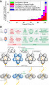
Figure 7. The φ29 Motor Adjusts Its Operation in Response to Increasing Capsid Filling
(A) Average time needed to package 100 bp of DNA at different filling levels. Each color denotes a distinct mechanism that contributes to the slowing down of the motor as the capsid fills. (B) Characteristics of packaging at low capsid filling and high capsid filling. The φ29 packaging cycle consists of a dwell phase (red) during which five ATPs are loaded sequentially, and a burst phase (green) during which four subunits translocate DNA. In each cycle, the motor rotates the DNA to form specific electrostatic contacts with the DNA backbone. These contacts anchor the motor onto the DNA during the subsequent dwell and determine the identity of the special subunit. Although here rotation is depicted to occur at the end of the burst, alternative scenarios where rotation occurs elsewhere during the cycle cannot be strictly ruled out. Several mechanisms modulate the motor operation at high filling while preserving the overall motor coordination: (1) The ATP-tight-binding rate is down-regulated, prolonging the dwell; (2) The motor enters the LLP state; (3) The duration of the force-sensitive burst phase is prolonged, most likely due to the rising internal force; (4) The step size of the motor decreases, which results in smaller bursts; (5) The amount of DNA rotation per cycle increases.

Similar articles

Cited by

References

    1. Aathavan K, Politzer AT, Kaplan A, Moffitt JR, Chemla YR, Grimes S, Jardine PJ, Anderson DL, Bustamante C. Substrate interactions and promiscuity in a viral DNA packaging motor. Nature. 2009;461:669–673. - PMC - PubMed
    1. Berg HC. The rotary motor of bacterial flagella. Annu Rev Biochem. 2003;72:19–54. - PubMed
    1. Bryant Z, Stone MD, Gore J, Smith SB, Cozzarelli NR, Bustamante C. Structural transitions and elasticity from torque measurements on DNA. Nature. 2003;424:338–341. - PubMed
    1. Bustamante C, Marko JF, Siggia ED, Smith S. Entropic elasticity of lambda-phage DNA. Science. 1994;265:1599–1600. - PubMed
    1. Cao S, Saha M, Zhao W, Jardine PJ, Zhang W, Grimes S, Morais MC. Insights into the structure and assembly of the bacteriophage φ29 dsDNA packaging motor. J Virol. published online ahead of print. - PMC - PubMed

Publication types

MeSH terms

LinkOut - more resources