

N-cadherin/catenin complex as a master regulator of intercalated disc function
- PMID: 24766605
- PMCID: PMC6054126
- DOI: 10.3109/15419061.2014.908853
N-cadherin/catenin complex as a master regulator of intercalated disc function
Erratum in
- Cell Commun Adhes. 2014 Dec;21(6):291
Abstract
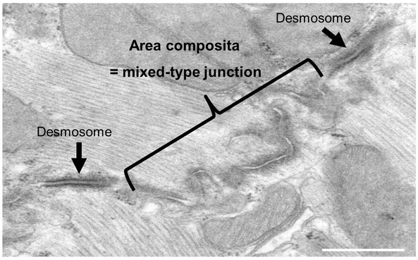
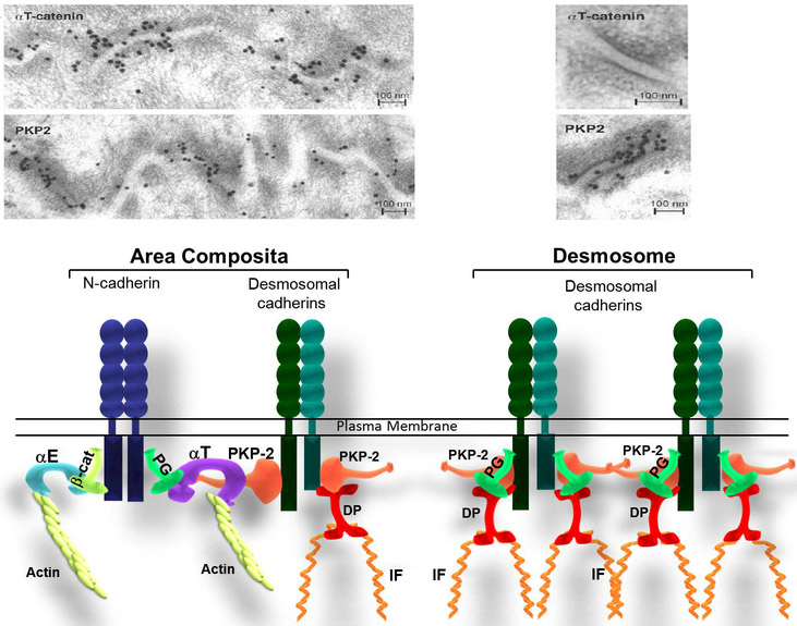
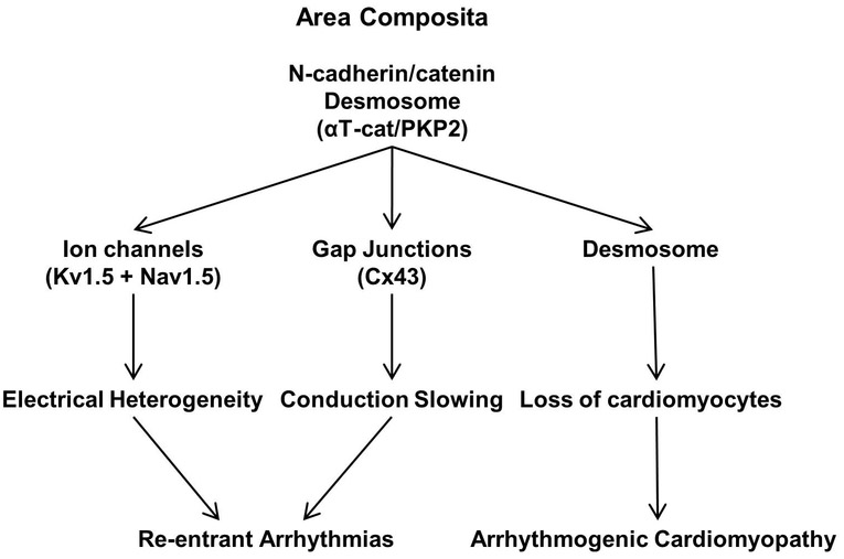
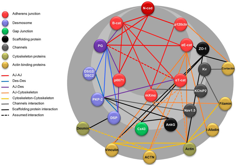
Intercellular adhesive junctions are essential for maintaining the physical integrity of tissues; this is particularly true for the heart that is under constant mechanical load. The correct functionality of the heart is dependent on the electrical and mechanical coordination of its constituent cardiomyocytes. The intercalated disc (ID) structure located at the termini of the rod-shaped adult cardiomyocyte contains various junctional proteins responsible for the integration of structural information and cell-cell communication. According to the classical description, the ID consists of three distinct junctional complexes: adherens junction (AJ), desmosome (Des), and gap junction (GJ) that work together to mediate mechanical and electrical coupling of cardiomyocytes. However, recent morphological and molecular studies indicate that AJ and Des components are capable of mixing together resulting in a "hybrid adhering junction" or "area composita." This review summarizes recent progress in understanding the in vivo function(s) of AJ components in cardiac homeostasis and disease.
Keywords: N-cadherin; adherens junction; arrhythmogenic cardiomyopathy; catenin; desmosome.
Conflict of interest statement
Conflict of interest
None
Figures
Similar articles
-
Loss of αT-catenin alters the hybrid adhering junctions in the heart and leads to dilated cardiomyopathy and ventricular arrhythmia following acute ischemia.J Cell Sci. 2012 Feb 15;125(Pt 4):1058-67. doi: 10.1242/jcs.098640. Epub 2012 Mar 15. J Cell Sci. 2012. PMID: 22421363 Free PMC article.
-
A new perspective on intercalated disc organization: implications for heart disease.Dermatol Res Pract. 2010;2010:207835. doi: 10.1155/2010/207835. Epub 2010 May 5. Dermatol Res Pract. 2010. PMID: 20585598 Free PMC article.
-
The N-cadherin interactome in primary cardiomyocytes as defined using quantitative proximity proteomics.J Cell Sci. 2019 Feb 11;132(3):jcs221606. doi: 10.1242/jcs.221606. J Cell Sci. 2019. PMID: 30630894 Free PMC article.
-
Evolution of the cadherin-catenin complex.Subcell Biochem. 2012;60:9-35. doi: 10.1007/978-94-007-4186-7_2. Subcell Biochem. 2012. PMID: 22674066 Review.
-
Refining the molecular organization of the cardiac intercalated disc.Cardiovasc Res. 2017 Mar 1;113(3):259-275. doi: 10.1093/cvr/cvw259. Cardiovasc Res. 2017. PMID: 28069669 Review.
Cited by
-
Pathologic Proteolytic Processing of N-Cadherin as a Marker of Human Fibrotic Disease.Cells. 2022 Jan 4;11(1):156. doi: 10.3390/cells11010156. Cells. 2022. PMID: 35011717 Free PMC article.
-
αT-catenin: A developmentally dispensable, disease-linked member of the α-catenin family.Tissue Barriers. 2018;6(2):e1463896. doi: 10.1080/21688370.2018.1463896. Epub 2018 May 10. Tissue Barriers. 2018. PMID: 29746206 Free PMC article. Review.
-
Sex-Specific Response to A1BG Loss Results in Female Dilated Cardiomyopathy.Res Sq [Preprint]. 2024 Jul 18:rs.3.rs-4631369. doi: 10.21203/rs.3.rs-4631369/v1. Res Sq. 2024. PMID: 39070637 Free PMC article. Preprint.
-
Hypertension Induces Pro-arrhythmic Cardiac Connexome Disorders: Protective Effects of Treatment.Biomolecules. 2023 Feb 9;13(2):330. doi: 10.3390/biom13020330. Biomolecules. 2023. PMID: 36830700 Free PMC article. Review.
-
In vivo elongation of thin filaments results in heart failure.PLoS One. 2020 Jan 3;15(1):e0226138. doi: 10.1371/journal.pone.0226138. eCollection 2020. PLoS One. 2020. PMID: 31899774 Free PMC article.
References
-
- ANGST BD, KHAN LU, SEVERS NJ, WHITELY K, ROTHERY S, THOMPSON RP, MAGEE AI & GOURDIE RG 1997. Dissociated spatial patterning of gap junctions and cell adhesion junctions during postnatal differentiation of ventricular myocardium. Circ Res, 80, 88–94. - PubMed
-
- ASIMAKI A, TANDRI H, HUANG H, HALUSHKA MK, GAUTAM S, BASSO C, THIENE G, TSATSOPOULOU A, PROTONOTARIOS N, MCKENNA WJ, CALKINS H & SAFFITZ JE 2009. A new diagnostic test for arrhythmogenic right ventricular cardiomyopathy. N Engl J Med, 360, 1075–84. - PubMed
-
- BASSO C, BAUCE B, CORRADO D & THIENE G 2012. Pathophysiology of arrhythmogenic cardiomyopathy. Nat Rev Cardiol, 9, 223–33. - PubMed
-
- BAUCE B, NAVA A, BEFFAGNA G, BASSO C, LORENZON A, SMANIOTTO G, DE BORTOLI M, RIGATO I, MAZZOTTI E, STERIOTIS A, MARRA MP, TOWBIN JA, THIENE G, DANIELI GA & RAMPAZZO A 2010. Multiple mutations in desmosomal proteins encoding genes in arrhythmogenic right ventricular cardiomyopathy/dysplasia. Heart Rhythm, 7, 22–9. - PubMed
-
- BAURAND A, ZELARAYAN L, BETNEY R, GEHRKE C, DUNGER S, NOACK C, BUSJAHN A, HUELSKEN J, TAKETO MM, BIRCHMEIER W, DIETZ R & BERGMANN MW 2007. Beta-catenin downregulation is required for adaptive cardiac remodeling. Circ Res, 100, 1353–62. - PubMed
Publication types
MeSH terms
Substances
Grants and funding
LinkOut - more resources
Full Text Sources
Other Literature Sources
Research Materials
Miscellaneous