. 2014 Apr 3;2(4):491-506.
doi: 10.1016/j.stemcr.2014.02.011. eCollection 2014 Apr 8.

Huntingtin regulates mammary stem cell division and differentiation

Affiliations

Huntingtin regulates mammary stem cell division and differentiation

Salah Elias et al. Stem Cell Reports. .

Abstract

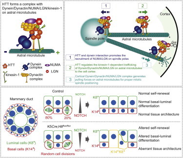
Little is known about the mechanisms of mitotic spindle orientation during mammary gland morphogenesis. Here, we report the presence of huntingtin, the protein mutated in Huntington's disease, in mouse mammary basal and luminal cells throughout mammogenesis. Keratin 5-driven depletion of huntingtin results in a decreased pool and specification of basal and luminal progenitors, and altered mammary morphogenesis. Analysis of mitosis in huntingtin-depleted basal progenitors reveals mitotic spindle misorientation. In mammary cell culture, huntingtin regulates spindle orientation in a dynein-dependent manner. Huntingtin is targeted to spindle poles through its interaction with dynein and promotes the accumulation of NUMA and LGN. Huntingtin is also essential for the cortical localization of dynein, dynactin, NUMA, and LGN by regulating their kinesin 1-dependent trafficking along astral microtubules. We thus suggest that huntingtin is a component of the pathway regulating the orientation of mammary stem cell division, with potential implications for their self-renewal and differentiation properties.


Figures

None
Graphical abstract
Figure 1
Figure 1
K5-Driven Loss of HTT Affects Basal and LC Populations (A) Mammary gland sections from virgin C57Bl6/J mice stained for HTT. (B) Quantitative real-time RT-PCR analysis of Htt gene and western blotting for HTT protein in basal and luminal mammary epithelial cells from 16-week-old virgin mice. Mw, molecular weight. (C) LacZ-stained mammary gland sections from 12-week-old virgin control and mutant K5Cre;Httflox/flox;R26 12-week-old virgin mice. (D) Quantitative real-time RT-PCR analysis of Htt gene expression in BCs and LCs from 16-week-old virgin mice. (E) Representative dot plots showing separation of luminal (CD31/CD45/CD24+/CD49F-low) and basal (CD31/CD45/CD24+/CD49F-high) epithelial cells from 16-week-old virgin mouse mammary glands by flow cytometry. (F) Number of BCs and LCs isolated per gland of 16-week-old virgin mice. (G) Percentages of CD49F-high cells in CD45/CD31/CD24+ cell populations. (H and I) Colonies formed by BCs (H) and LCs (I) isolated from mammary glands of 16-week-old virgin mice. Scale bars, 50 μm (A and C). Error bars, SEM.p < 0.05; ∗∗p < 0.01; ∗∗∗p < 0.001. See also Figure S1 and Table S1.
Figure 2
Figure 2
Loss of HTT Alters Basal to Luminal Specification (A and B) Quantitative real-time RT-PCR analysis of the indicated genes in BCs and LCs from 16-week-old mice. (C) Sections from 12-week-old mammary glands stained for K14 and K8. Arrows point to K8+K14+ epithelial cells. (D) Percentage of K8+, K14+, and K8+K14+ cells. (E) Sections from 12-week-old mammary glands stained for ERα. Right panel shows the percentages of ERα-positive cells. (F) Representative dot plots showing the frequency of SCA1+ and CD49B+ cells in the LC population in 16-week-old virgin mice. (G) Percentages of SCA1+CD49B, SCA1+CD49B+, and SCA1CD49B+ cells. (H) Ratio of SCA1+CD49B-to-SCA1CD49B+ cells. Scale bars, 10 μm. Error bars, SEM.p < 0.05; ∗∗p < 0.01; ∗∗∗p < 0.001. See also Table S1.
Figure 3
Figure 3
HTT Is Required for Epithelial Morphogenesis during Pregnancy and Lactation (A and B) Carmine-stained whole mounts of mammary glands and hematoxylin and eosin staining (H&E) at low (middle) and high (right) magnifications. The histograms show the quantification of the epithelial content. (C) Mammary gland sections stained for p-STAT5A and percentages of p-STAT5A-positive cells. (D) Mammary gland sections from 1-day lactating mice stained for WAP and quantitative real-time RT-PCR analysis of Csn2 and Wap gene expression. (E and F) Mammary gland sections from 18.5-day pregnant mice stained as indicated. Scale bars, 10 μm. Scale bars, 50 μm (A–D). Error bars, SEM. p < 0.05; ∗∗p < 0.01; ∗∗∗p < 0.001. See also Figure S2.
Figure 4
Figure 4
HTT Regulates Mitotic Spindle Orientation in Basal Cells in a Dynein-Dependent Manner (A) Mammary gland sections from 7.5-day pregnant mice. (B) Schemes illustrating measurement of the spindle angle α. (C) Mammary gland sections from 7.5-day pregnant mice. (D and E) Spindle angles of basal cells; values are expressed as a percentage of basal cells within each interval. Mean angle and number of measures (n) are shown. (F) Percentages of planar (0°–30°), oblique (30°–60°), and vertical (60°–90°) divisions. (G) Western blotting of mammary cell extracts. (H) HTT abundance at spindle poles (right). (I) Distribution and mean spindle angle in metaphase mammary cells. (J) Western blotting of cell extracts. The star indicates a contaminating band. (K) Distribution and mean spindle angles in metaphase mammary cells. (L) HTT (mCherry) labeling at spindle poles in metaphase mammary cells. Scale bars, 10 μm. Error bars, SEM. ns, not significant. ∗∗p < 0.01. ∗∗∗p < 0.001. See also Figure S3.
Figure 5
Figure 5
HTT Codistributes with Dynein/Dynactin/NUMA/LGN (A and B) Mammary cells stained as indicated. Scale bars, 10 μm. Telo, telophase; Ana, anaphase; Meta, metaphase; Prometa, prometaphase; Pro, prophase; Inter, interphase. (C) HTT/dynein/dynactin/NUMA/LGN complexes were immunoprecipitated from cells arrested in metaphase before lysis. Mouse IgG (mIgG) was used as a negative control. The immunoprecipitates (IP) were analyzed by western blotting. (D) Cartoon showing the HTT/dynein/dynactin/NUMA/LGN complex at the spindle pole and on astral microtubules in metaphase cells. See also Figure S4.
Figure 6
Figure 6
Loss of HTT Prevents Cortical Accumulation of Dynein-Dynactin-NUMA-LGN during Mitosis (A) Mammary cells stained as indicated. (B) Line-scan analysis (relative fluorescence intensity). (C) Western blotting of cell extracts. (D) Percentage of cells with cortical accumulation of dynein, P150Glued, NUMA, and LGN. (E) Mammary gland sections from 7.5-day pregnant mice. Gradients of color intensity were applied to NUMA and LGN stainings (insets). (F) DHC-GFP and GFP-LGN HeLa cells were video recorded. Maximum intensity and z projections are shown. (G) Percentage of HeLa cells with cortical accumulation of DHC and LGN. Scale bars, 10 μm. Error bars, SEM. ∗∗∗p < 0.001. See also Figure S5 and Movies S3, S4, S5, and S6.
Figure 7
Figure 7
Kinesin 1 Participates in HTT-Mediated Cortical Localization of the Dynein/Dynactin/NUMA/LGN Complex during Mitosis (A) Mammary cells stained as indicated. (B) HTT/dynein/kinesin 1 complexes were immunoprecipitated from cells arrested in metaphase before lysis. Mouse IgG (mIgG) was used as a negative control. The immunoprecipitates were analyzed by western blotting. (C) Mammary cells stained as indicated. (D) Kinesin 1 abundance at spindle poles. (E) Percentages of cells with kinesin 1 on astral microtubules. (F) Mammary cells stained as indicated. (G) Western blotting of cell extracts. (H) Quantification of the relative fluorescence intensities of P150Glued, dynein, and NUMA on astral microtubules. The intensities of the spindle (Ispindle) and of the total cell (Itotal) were determined with ImageJ software. Relative intensities on astral microtubules (Iastral, rel) were calculated, and control value was set to 100. (I) LGN abundance at the cell cortex. (J) Model for HTT-mediated regulation of mitotic spindle orientation. During mitosis, HTT is targeted to the spindle poles through its interaction with dynein and promotes the accumulation of NUMA and LGN (1). HTT regulates the kinesin 1-dependent trafficking of dynein, dynactin, NUMA, and LGN along astral microtubules to the cell cortex (2). Once at the cell cortex, the dynein/dynactin/NUMA/LGN complex generates pulling forces on astral microtubules for mitotic spindle positioning (3). Scale bars, 10 μm. Error bars, SEM. ∗∗p < 0.01; ∗∗∗p < 0.001. See also Figure S6.

Similar articles

Cited by

References

    1. Asselin-Labat M.L., Vaillant F., Sheridan J.M., Pal B., Wu D., Simpson E.R., Yasuda H., Smyth G.K., Martin T.J., Lindeman G.J., Visvader J.E. Control of mammary stem cell function by steroid hormone signalling. Nature. 2010;465:798–802. - PubMed
    1. Beleut M., Rajaram R.D., Caikovski M., Ayyanan A., Germano D., Choi Y., Schneider P., Brisken C. Two distinct mechanisms underlie progesterone-induced proliferation in the mammary gland. Proc. Natl. Acad. Sci. USA. 2010;107:2989–2994. - PMC - PubMed
    1. Bolte S., Cordelières F.P. A guided tour into subcellular colocalization analysis in light microscopy. J. Microsc. 2006;224:213–232. - PubMed
    1. Bouras T., Pal B., Vaillant F., Harburg G., Asselin-Labat M.L., Oakes S.R., Lindeman G.J., Visvader J.E. Notch signaling regulates mammary stem cell function and luminal cell-fate commitment. Cell Stem Cell. 2008;3:429–441. - PubMed
    1. Caviston J.P., Ross J.L., Antony S.M., Tokito M., Holzbaur E.L. Huntingtin facilitates dynein/dynactin-mediated vesicle transport. Proc. Natl. Acad. Sci. USA. 2007;104:10045–10050. - PMC - PubMed

Publication types

MeSH terms