

Phagocytosis of Staphylococcus aureus by human neutrophils prevents macrophage efferocytosis and induces programmed necrosis
- PMID: 24729616
- PMCID: PMC4011196
- DOI: 10.4049/jimmunol.1302692
Phagocytosis of Staphylococcus aureus by human neutrophils prevents macrophage efferocytosis and induces programmed necrosis
Abstract
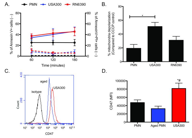
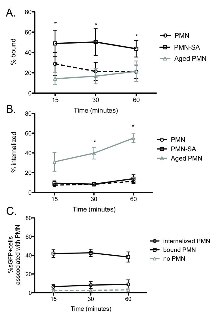
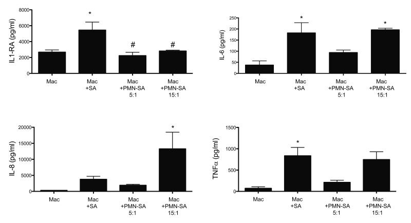
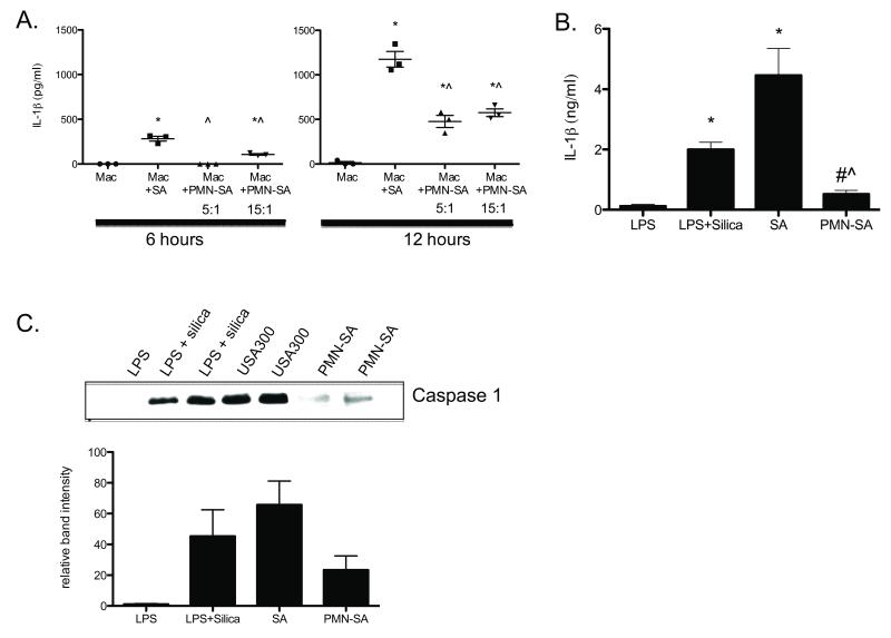
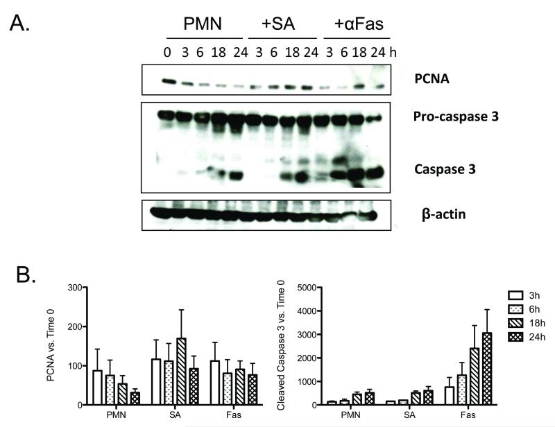
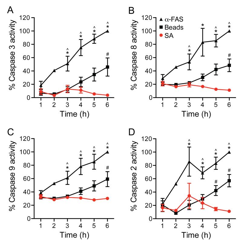
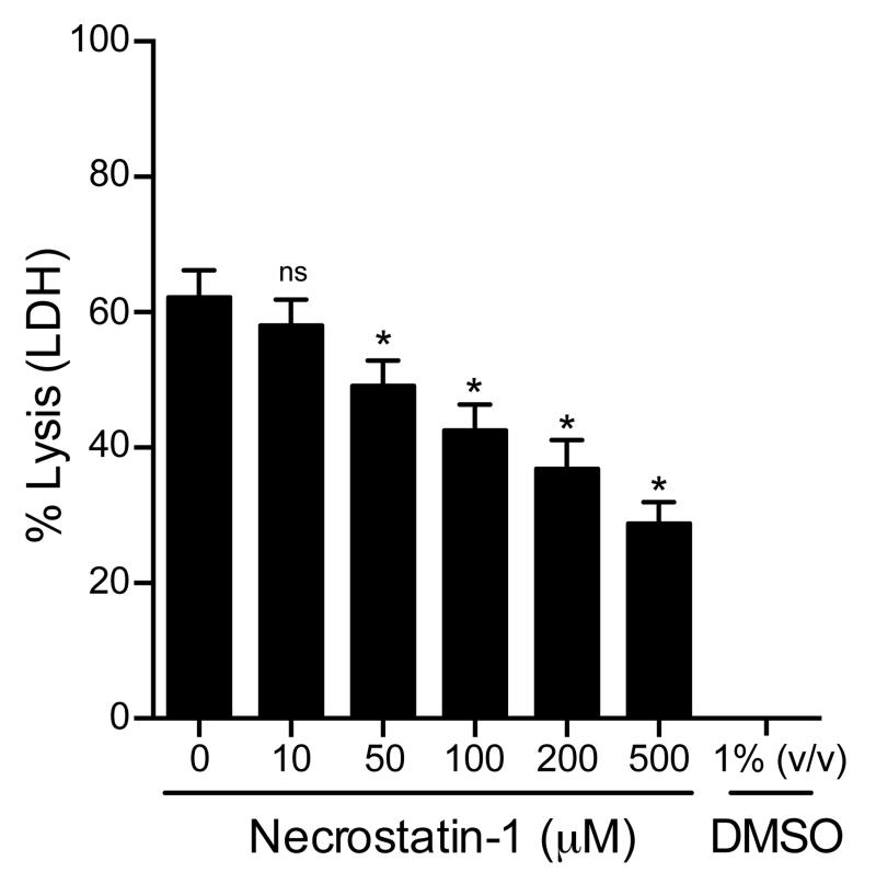
Community-associated methicillin-resistant Staphylococcus aureus (CA-MRSA) pose a significant threat to human health. Polymorphonuclear leukocytes (PMN) are the first responders during staphylococcal infection, but 15-50% of the initial ingested inoculum survives within the PMN phagosome and likely contributes directly or indirectly to disease pathogenesis. We hypothesize that surviving intracellular CA-MRSA undermine effective phagocyte-mediated defense by causing a decrease in macrophage uptake of PMN containing viable S. aureus and by promoting PMN lysis. In support of this hypothesis, PMN harboring viable CA-MRSA strain USA300 (PMN-SA) upregulated the "don't eat me" signal CD47, remained bound to the surface, and were inefficiently ingested by macrophages. In addition, coculture with PMN-SA altered the macrophage phenotype. Compared to macrophages fed USA300 alone, macrophages challenged with PMN-SA produced more IL-8 and less IL-1 receptor antagonist, TNF-α, activated caspase-1, and IL-1β. Although they exhibited some features of apoptosis within 3 h following ingestion of S. aureus, including phosphatidylserine exposure and mitochondrial membrane depolarization, PMN-SA had sustained levels of proliferating cell nuclear Ag expression, absence of caspase activation, and underwent lysis within 6 h following phagocytosis. PMN lysis was dependent on receptor-interacting protein 1, suggesting that PMN-SA underwent programmed necrosis or necroptosis. These data are the first demonstration, to our knowledge, that bacteria can promote sustained expression of proliferating cell nuclear Ag and that human PMN undergo necroptosis. Together, these findings demonstrate that S. aureus surviving within PMN undermine the innate immune response and may provide insight into the pathogenesis of S. aureus disease.
Figures
Similar articles
-
Frontline Science: Staphylococcus aureus promotes receptor-interacting protein kinase 3- and protease-dependent production of IL-1β in human neutrophils.J Leukoc Biol. 2019 Mar;105(3):437-447. doi: 10.1002/JLB.4HI0918-346R. Epub 2018 Dec 13. J Leukoc Biol. 2019. PMID: 30548986 Free PMC article.
-
Rapid neutrophil destruction following phagocytosis of Staphylococcus aureus.J Innate Immun. 2010;2(6):560-75. doi: 10.1159/000317134. Epub 2010 Jun 26. J Innate Immun. 2010. PMID: 20587998 Free PMC article.
-
Further Insight into the Mechanism of Human PMN Lysis following Phagocytosis of Staphylococcus aureus.Microbiol Spectr. 2021 Oct 31;9(2):e0088821. doi: 10.1128/Spectrum.00888-21. Epub 2021 Oct 27. Microbiol Spectr. 2021. PMID: 34704790 Free PMC article.
-
How methicillin-resistant Staphylococcus aureus evade neutrophil killing.Curr Opin Hematol. 2015 Jan;22(1):30-5. doi: 10.1097/MOH.0000000000000096. Curr Opin Hematol. 2015. PMID: 25394313 Free PMC article. Review.
-
Evasion of Neutrophil Killing by Staphylococcus aureus.Pathogens. 2016 Mar 17;5(1):32. doi: 10.3390/pathogens5010032. Pathogens. 2016. PMID: 26999220 Free PMC article. Review.
Cited by
-
Staphylococcus aureus Inhibits Neutrophil-derived IL-8 to Promote Cell Death.J Infect Dis. 2015 Sep 15;212(6):934-8. doi: 10.1093/infdis/jiv124. Epub 2015 Feb 26. J Infect Dis. 2015. PMID: 25722299 Free PMC article.
-
Toxin-induced necroptosis is a major mechanism of Staphylococcus aureus lung damage.PLoS Pathog. 2015 Apr 16;11(4):e1004820. doi: 10.1371/journal.ppat.1004820. eCollection 2015 Apr. PLoS Pathog. 2015. PMID: 25880560 Free PMC article.
-
IFN-γ targets macrophage-mediated immune responses toward Staphylococcus aureus.J Leukoc Biol. 2017 Mar;101(3):751-758. doi: 10.1189/jlb.4A1215-565RR. Epub 2016 Oct 5. J Leukoc Biol. 2017. PMID: 27707882 Free PMC article.
-
Frontline Science: Staphylococcus aureus promotes receptor-interacting protein kinase 3- and protease-dependent production of IL-1β in human neutrophils.J Leukoc Biol. 2019 Mar;105(3):437-447. doi: 10.1002/JLB.4HI0918-346R. Epub 2018 Dec 13. J Leukoc Biol. 2019. PMID: 30548986 Free PMC article.
-
In Vivo and In Vitro Assessments of the Antibacterial Potential of Chitosan-Silver Nanocomposite Against Methicillin-Resistant Staphylococcus aureus-Induced Infection in Rats.Biol Trace Elem Res. 2021 Jan;199(1):244-257. doi: 10.1007/s12011-020-02143-6. Epub 2020 Apr 18. Biol Trace Elem Res. 2021. PMID: 32306284
References
-
- Nauseef WM. How human neutrophils kill and degrade microbes: an integrated view. Immunol Rev. 2007;219:88–102. - PubMed
-
- Voyich JM, Braughton KR, Sturdevant DE, Whitney AR, Said-Salim B, Porcella SF, Long RD, Dorward DW, Gardner DJ, Kreiswirth BN, Musser JM, DeLeo FR. Insights into mechanisms used by Staphylococcus aureus to avoid destruction by human neutrophils. J Immunol. 2005;175:3907–3919. - PubMed
Publication types
MeSH terms
Substances
Grants and funding
LinkOut - more resources
Full Text Sources
Other Literature Sources
Medical
Research Materials