. 2014 Apr 17;40(4):542-53.
doi: 10.1016/j.immuni.2014.03.004. Epub 2014 Apr 3.

TIM-1 glycoprotein binds the adhesion receptor P-selectin and mediates T cell trafficking during inflammation and autoimmunity

Affiliations

TIM-1 glycoprotein binds the adhesion receptor P-selectin and mediates T cell trafficking during inflammation and autoimmunity

Stefano Angiari et al. Immunity. .

Abstract

Selectins play a central role in leukocyte trafficking by mediating tethering and rolling on vascular surfaces. Here we have reported that T cell immunoglobulin and mucin domain 1 (TIM-1) is a P-selectin ligand. We have shown that human and murine TIM-1 binds to P-selectin, and that TIM-1 mediates tethering and rolling of T helper 1 (Th1) and Th17, but not Th2 and regulatory T cells on P-selectin. Th1 and Th17 cells lacking the TIM-1 mucin domain showed reduced rolling in thrombin-activated mesenteric venules and inflamed brain microcirculation. Inhibition of TIM-1 had no effect on naive T cell homing, but it reduced T cell recruitment in a skin hypersensitivity model and blocked experimental autoimmune encephalomyelitis. Uniquely, the TIM-1 immunoglobulin variable domain was also required for P-selectin binding. Our data demonstrate that TIM-1 is a major P-selectin ligand with a specialized role in T cell trafficking during inflammatory responses and the induction of autoimmune disease.


Figures

Figure 1
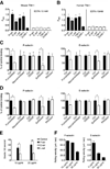
Figure 1. TIM-1 interacts with selectins in vitro requiring a specific glycosylation profile and mediates tethering and rolling on endothelial selectins under flow conditions
(A and B) Microtiter plates were coated with 5 µg/ml murine or human P-selectin, E-selectin or L-selectin, TIM-4 (positive control) (Meyers et al., 2005) or ICAM-1 (negative control) (Santiago et al., 2007B), and tested for their ability to bind recombinant mouse or human TIM-1 respectively. In some experiments, 10 mM EDTA was added to chelate divalent cations. Both murine (A) and human (B) TIM-1 bound to all three selectins, and binding was dependent on the presence of divalent cations (*P < 0.0001 compared to ICAM-1 binding). (C and D) Microtiter plate assays show the binding of recombinant mouse TIM-1 protein from CHO (shown in C) and 293T cells (shown in D) to P- and E-selectin after treatment with α1,(3,4)-fucosidase, tyrosine sulfatase, PNGase, OSGE and neuraminidase treatment (*P < 0.001). (E) Protein A-covered microspheres were coated with a murine TIM-1 Fc-chimera (Sizing et al., 2007) or a control mouse IgG Fc-chimera (control beads) and were infused into glass capillary tubes pre-coated with P-selectin, E-selectin or L-selectin, under physiological shear stress conditions (2 dyne/cm2) (*P < 0.002, **P < 0.0001). See also Movies S1 and S2. (F) The rolling velocity of TIM-1-covered beads declines as the concentration of P-selectin and E-selectin increases under physiological flow conditions. Data represent the means ± standard error of the mean (SEM) of three independent experiments. In (A), (B), (C) and (D), data represent means ± SEM of at least three independent experiments performed in triplicate for each condition.
Figure 2
Figure 2. The TIM-1 mucin domain is required for the capture and rolling of Th1 and Th17 cells on P-selectin under physiological flow conditions in vitro
(A–E) Treg cells or CD4+ T cells stimulated with concanavalin A (ConA) or polarized toward Th1, Th17 or Th2 phenotypes were infused into capillary tubes pre-coated with 5 µg/ml P-selectin or E-selectin under shear stress conditions (2 dyne/cm2) (*P < 0.002; **P < 0.0003; ***P < 0.0001). Bone marrow-derived PMNs, which do not express TIM-1, were used as negative controls. (B) Representative images of wild-type and Havcr1Δmucin Th1 cells rolling on P-selectin, showing a consistently lower number of rolling Havcr1Δmucin Th1 cells. See also Movies S3 and S4. (C and E) Rolling velocities of wild-type and Havcr1Δmucin ConA blasts, Th1, Th17, Th2 and Treg cells on P- and E-selectin. Data represent means ± SEM of four independent experiments. For rolling velocities, data represent means ± SEM of at least 100 cells per condition.
Figure 3
Figure 3. TIM-1 mediates tethering and rolling of Th1 and Th17 cells in vivo
Th1 and Th17 cells were generated from murine wild-type and Havcr1Δmucin CD4+ cells. Bone marrow-derived PMNs were used as negative controls. Cells were injected intravenously into the exposed mesenteric vessels of mice pre-treated with bovine thrombin to upregulate P-selectin on the vascular endothelium. (A) Havcr1Δmucin T cells have a significantly reduced ability to roll on P-selectin in vivo (*P < 0.002 compared to wild-type cells). See also Movie S5. (B) Representative images of Th1 cells rolling in mesenteric vessels. Cells are the white spots inside the venules (arrow tips). Scale bar: 100 µm. (C) The number of tethers (each new interaction with the vessel wall) are shown. (*P < 0.03 and **P < 0.004 compared to the corresponding wild-type cells). Data in (A) and (C) represent means ± SEM of 13-15 independent experiments for a total of 15-20 total venules/condition. (D and E) Havcr1Δmucin Th1 (D) and Th17 (E) cells display no defects in their rolling velocity in mesenteric venules, compared to wild-type cells. Rolling velocities represent the mean ± SEM of at least 100 cells per condition (left panel). The distribution of leukocyte rolling velocities is also shown (right panel).
Figure 4
Figure 4. The TIM-1 IgV domain is required for P-selectin-dependent rolling in vivo
(A–D) Wild-type Th1 cells were treated with control rat IgG or the blocking anti-TIM-1 antibodies RMT1-10 or 4A2.2, which recognize epitopes in the TIM-1 IgV domain (Xiao et al., 2007; Sizing et al., 2007). (A) Cells treated with RMT1-10 and 4A2.2 showed a strongly reduced ability to roll on P-selectin (*P < 0.005 and **P < 0.04 compared to cells treated with rat IgG). (B) RMT1-10 and 4A2.2 treatments also reduced the number of total tethers (*P < 0.008 and **P < 0.03 compared to control cells). Data in (A) and (B) represent means ± SEM of 3-4 independent experiments for a total of 6-8 total venules per condition. (C and D) Cells treated with both RMT1-10 (C) and 4A2.2 (D) showed no defects in their rolling velocity in mesenteric venules. Rolling velocities represent the mean ± SEM of at least 50 cells per condition. The distribution of leukocyte rolling velocities is also shown (right panels). (E) Microtiter plate assays showing the binding of full-length versus truncated TIM-1 proteins to P-selectin (*P < 0.001 compared to entire TIM-1 protein). ICAM-1 was used a negative control for TIM-1 binding. (F) Effect of RMT1-10 antibody on Havcr1Δmucin Th1 cell tethering and rolling in thrombin-treated mesenteric venules. Data represent means ± SEM of two experiments for a total of 5–6 venules per condition.
Figure 5
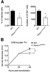
Figure 5. TIM-1 mediates T cell recruitment in the inflamed skin
Mice were sensitized with 1-fluoro-2,4-dinitrobenzene (DNFB) to induce cutaneous hypersensitivity (CHS) and were challenged again 5 days later on each side of the right ear pinnae with DNFB. The ear thickness was measured 0, 24 and 48 h after challenge. (A) 10 × 106 CFSE-labeled Th1 cells from wild-type and Havcr1Δmucin mice were transferred to CHS mice 24 h after the ear pinnae were painted, and Th1 accumulation was evaluated 24 h later by flow cytometry (*P < 0.02). (B) The injection of wild-type but not Havcr1Δmucin Th1 cells increased the inflammation response in the challenged ear (*P<0.01). In both A and B, results represent the mean ± SEM of 9-11 mice per condition.
Figure 6
Figure 6. TIM-1 controls T cell trafficking in the inflamed CNS and the induction of EAE
(A) Havcr1Δmucin Th1 and Th17 cells ability to roll and adhere firmly in LPS-inflamed brain pial vessels, in intravital microscopy experiments (**P < 0.008; *P < 0.01; ***P < 0.007). Data are mean ± SEM of three experiments for a total of 8-9 venules per condition. (B) Representative images of brain venules showing adhered Th1 cells as white spots inside the vessels (arrow tips). Scale bar = 100 µm. (C) 5 × 10⁶ MOG35-55-specific Th1 cells were labeled with CFSE and injected into C57BL/6J wild-type mice treated 5 h previously with 20 ng pertussis toxin (PTX). The accumulation of CSFE+ cells in the CNS was evaluated 60 h after cell transfer. Data represent means ± SEM of 12 mice per condition from two experiments (*P < 0.006). (D) 5 × 10⁶ MOG35-55-specific Th1 cells were transferred in C57BL/6J wild-type mice. Data represent the mean ± SEM of 10 mice per condition from two experiments (*P < 0.05). (E) EAE was actively induced by immunization with the MOG35-55 peptide. Data represent the mean ± SEM of 10 mice per condition from two independent experiments with similar results (*P < 0.05). (F) Hematoxylin & eosin and Spielmeyer stainings are shown at disease peak. (G) CD4+ T cells were isolated from the draining lymph nodes 7 days post-immunization with the MOG35-55 peptide and the proliferative response was determined. Data are shown as counts per minute (CPM) of [3H]-thymidine radioactivity and represent the mean ± SD of five mice per condition.
Figure 7
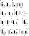
Figure 7. TIM-1 and PSGL-1 concur to control activated T cell rolling on P-selectin and T cell trafficking in inflamed tissues
(A) Th1 cells were infused into capillary tubes pre-coated with P-selectin or E-selectin (*P < 0.00001; **P<0.0003) Data represent the mean ± SEM of four experiments. (B) Rolling velocities in capillary tubes coated with P-selectin or E-selectin are shown (*P < 0.001 and **P < 0.008 compared to WT cells). Rolling velocities represent the mean ± SEM of at least 50 cells per condition. (C) Shown is the ability to tether and roll in thrombin-treated mesenteric venules. Data obtained by treating Havcr1Δmucin cells with antibody 4RA10 are also shown (*P < 0.001 and **P < 0.02 compared to WT cells). Data represent the mean ± SEM of 10 independent experiments for a total of 16-17 total venules per condition. (D) Rolling velocities are shown in mesenteric venules. Rolling velocities represent the mean ± SEM of at least 50 cells per condition. The distribution of leukocyte rolling velocities is also shown in vivo (right panel). (E) Selplg−/− Th1 cells treated with RMT1-10 showed a reduced ability to tether ad roll on P-selectin in vivo, compared to cells treated with the control rat IgG (*P < 0.05, left panel; *P < 0.002, right panel). Data represent the mean ± SEM of three experiments for a total of seven venules per condition. (F) Rolling and arrest interactions are shown in intravital microscopy experiments in LPS-treated cerebral vessels (*P < 0.001; **P < 0.04). (G and H) Accumulation of wild-type, Havcr1ΔmucinSelplg−/− and Selplg−/− Havcr1Δmucin Th1 cells in the inflamed ear pinnae of CHS mice (G) and in the CNS of pertussis toxin-treated mice (H) (*P < 0.01, **P < 0.001 and ***P < 0.03). Data represent the means ± SEM of five mice per condition from one representative experiment from a series of two with similar results. (I) Rolling and arrest of naïve CD4+ T cells in Peyer’s patches HEVs (*P < 0.03 compared to WT cell arrest). Data represent the mean ± SEM of four mice per condition for a total of nine venules per condition. (J) Accumulation of exogenous naïve CD4+ T cells in lymphoid organs of wild-type resting mice at 2h after transfer (*P < 0.003 and **P < 0.04 compared to WT cells). Data represent the mean ± SEM of six mice per condition from one representative experiment from a series of three with similar results.

Similar articles

Cited by

References

    1. Austrup F, Vestweber D, Borges E, Löhning M, Bräuer R, Herz U, Renz H, Hallmann R, Scheffold A, Radbruch A, Hamann A. P- and E-selectin mediate recruitment of T-helper-1 but not T-helper-2 cells into inflammed tissues. Nature. 1997;385:81–83. - PubMed
    1. Balasubramanian S, Kota SK, Kuchroo VK, Humphreys BD, Strom TB. TIM family proteins promote the lysosomal degradation of the nuclear receptor NUR77. Sci. Signal. 2012;5:ra90. - PMC - PubMed
    1. Butcher EC. Leukocyte-endothelial cell recognition: three (or more) steps to specificity and diversity. Cell. 1991;67:1033–1036. - PubMed
    1. D’Ambrosio D, Lecca P, Constantin G, Priami C, Laudanna C. Concurrency in leukocyte vascular recognition: developing the tools for a predictive computer model. Trends Immunol. 2004;8:411–416. - PubMed
    1. Deban L, Russo RC, Sironi M, Moalli F, Scanziani M, Zambelli V, Cuccovillo I, Bastone A, Gobbi M, Valentino S, et al. Regulation of leukocyte recruitment by the long pentraxin PTX3. Nat. Immunol. 2010;11:328–334. - PubMed

Publication types

MeSH terms