. 2014 Jun;88(12):6970-82.
doi: 10.1128/JVI.00384-14. Epub 2014 Apr 2.

Innate nuclear sensor IFI16 translocates into the cytoplasm during the early stage of in vitro human cytomegalovirus infection and is entrapped in the egressing virions during the late stage

Affiliations

Innate nuclear sensor IFI16 translocates into the cytoplasm during the early stage of in vitro human cytomegalovirus infection and is entrapped in the egressing virions during the late stage

Valentina Dell'Oste et al. J Virol. 2014 Jun.

Abstract

Intrinsic immune mechanisms mediated by constitutively expressed proteins termed "restriction factors" provide frontline antiviral defense. We recently demonstrated that the DNA sensor IFI16 restricts human cytomegalovirus (HCMV) replication by downregulating viral early and late but not immediate-early mRNAs and their protein expression. We show here that at an early time point during the in vitro infection of low-passage-number human embryonic lung fibroblasts, IFI16 binds to HCMV DNA. However, during a later phase following infection, IFI16 is mislocalized to the cytoplasmic virus assembly complex (AC), where it colocalizes with viral structural proteins. Indeed, upon its binding to pUL97, IFI16 undergoes phosphorylation and relocalizes to the cytoplasm of HCMV-infected cells. ESCRT (endosomal sorting complex required for transport) machinery regulates the translocation of IFI16 into the virus AC by sorting and trafficking IFI16 into multivesicular bodies (MVB), as demonstrated by the interaction of IFI16 with two MVB markers: Vps4 and TGN46. Finally, IFI16 becomes incorporated into the newly assembled virions as demonstrated by Western blotting of purified virions and electron microscopy. Together, these results suggest that HCMV has evolved mechanisms to mislocalize and hijack IFI16, trapping it within mature virions. However, the significance of this IFI16 trapping following nuclear mislocalization remains to be established.

Importance: Intracellular viral DNA sensors and restriction factors are critical components of host defense, which alarm and sensitize immune system against intruding pathogens. We have recently demonstrated that the DNA sensor IFI16 restricts human cytomegalovirus (HCMV) replication by downregulating viral early and late but not immediate-early mRNAs and their protein expression. However, viruses are known to evolve numerous strategies to cope and counteract such restriction factors and neutralize the first line of host defense mechanisms. Our findings describe that during early stages of infection, IFI16 successfully recognizes HCMV DNA. However, in late stages HCMV mislocalizes IFI16 into the cytoplasmic viral assembly complex and finally entraps the protein into mature virions. We clarify here the mechanisms HCMV relies to overcome intracellular viral restriction, which provides new insights about the relevance of DNA sensors during HCMV infection.


Figures

FIG 1
FIG 1
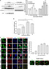
The HCMV genome is recognized by IFI16 at early time points following infection. (A) HELFs were mock infected (MOCK) or infected with HCMV (strain AD169, MOI of 2 PFU/cell), fixed in 1% paraformaldehyde at 12 hpi, and subjected to combined FISH with a BAC DNA probe containing the entire HCMV genome (red) and immunofluorescence analysis with anti-IFI16 antibodies (green). Cell nuclei are visualized in blue. Images were taken by confocal microscopy, and the far right hand picture shows 3D image reconstruction of stacks of confocal images. At least five fields were digitally reconstructed to generate the 3D images for each condition; representative images are shown. (B) HELFs were infected at an MOI of 5 and processed for ChIP assays 6 h later to test the association of endogenous IFI16 with HCMV and host DNA.
FIG 2
FIG 2
IFI16 accumulates in the cytoplasm of HCMV-infected cells at late time points postinfection. (A) HELFs were infected with HCMV at an MOI of 1 PFU/cell. Nuclear and cytoplasmic fractions were prepared at the indicated time points and subjected to Western blotting and subsequent densitometry for IFI16. The results were normalized to TBP and tubulin, respectively (*, P < 0.05; **, P < 0.01; ***, P < 0.001 [one-way analysis of variance, followed by Bonferroni's post test]). Nuclear fractions were stained for HCMV IEA as a positive control for viral infection (lower panel). (B) HELFs were infected with HCMV (MOI of 1 PFU/cell) or left untreated. Total RNA was isolated at the indicated time postinfection and assayed by quantitative real-time PCR to determine the relative levels of IFI16, normalized to the levels of cellular GADPH. The data shown are the average of three experiments ± the SD (*, P < 0.05; **, P < 0.01 [one-way analysis of variance, followed by Bonferroni's post test]). (C) Kinetics of IFI16 subcellular localization upon HCMV infection. HELFs were infected with HCMV at an MOI of 1 PFU/cell for the indicated time points and subjected to confocal microscopy analysis. IFI16 (green) and viral proteins (red) were visualized using primary antibodies, followed by secondary antibody staining, in the presence of 10% human serum. Nuclei are visualized in blue. The far right-hand picture of each panel shows a Z stack of confocal images, generating a 3D reconstruction, obtained as described in Fig. 1A (right panel). A graph shows the IFI16 delocalization levels. The bars indicate the percentages of cells positive for cytoplasmic IFI16 immunofluorescence over the course of infection. Images were acquired by using a microscope with a ×20 objective lens, and three random fields from two slides of each time point were counted using ImageJ software to calculate the ratio of cytoplasmic-IFI16 expressing cells to infected cells. The data represent means ± the SD (left panel). (D) IFI16 is mislocalized in HUVECs by the HCMV clinical isolate derivative VR1814. HUVECs were infected with HCMV (MOI of 1 PFU/cell), fixed in 1% paraformaldehyde at the indicated time points, and subjected to confocal immunofluorescence analysis as described in panel C. Images were acquired at ×63 magnification, and representative images are shown.
FIG 3
FIG 3
HCMV inhibition blocks the mislocalization of IFI16. HELFs were infected with wild-type or UV-inactivated HCMV (1 PFU/cell; 1.2 J/cm2 for two pulses) and treated with phosphonoformic acid (PFA; 100 μM) or ganciclovir (GCV; 100 μM) as indicated. Cells were fixed 72 h later in 1% paraformaldehyde and processed by immunofluorescence analysis for IFI16 (green) and gB (red) (left panel) or subjected to combined FISH with a BAC DNA probe containing the entire HCMV genome (red) and immunofluorescence analysis with anti-IFI16 antibodies (green); cell nuclei were visualized in blue (right panel). Images were taken by confocal microscopy (×63 magnification).
FIG 4
FIG 4
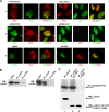
pUL97 mediates HCMV-induced IFI16 mislocalization. (A) pUL97 inhibition impairs IFI16 nuclear egress. HELFs were treated as described in detail below and in Results, fixed at the time points indicated below, and double stained with the appropriated antibodies. HELFs infected with a UL97 deletion mutant BAC (BAC ΔUL97) or with AD169 UL97+ as a control at an MOI of 1 PFU/ml were fixed and immunostained at 32 days or 96 hpi, respectively (row 1); HELFs were electroporated with a mixture of three different small interfering RNAs targeting UL97 (siRNA UL97) or with scrambled control siRNA (siRNA CTRL) and 24 h later infected with HCMV at an MOI of 1 PFU/cell for 72 h (row 2); HCMV-infected HELFs were treated with the pUL97 inhibitor Gö6976 (2 μM) or with an equal volume of vehicle control (DMSO) and immunostained after 72 h (row 3). (B) IFI16 interacts with pUL97. Total cell protein extracts from HELFs infected with HCMV at an MOI of 1 for 96 h were immunoprecipitated with polyclonal antibodies against IFI16 (left panel) or monoclonal antibodies against UL97 (right panel), and control antibody. Samples were then immunoblotted with antibodies for pUL97 and IFI16, respectively. Nonimmunoprecipitated whole-cell extract (Input) obtained from HCMV-infected cells was used to normalize the proteins subjected to immunoprecipitation. (C) Phosphorylation of IFI16 by pUL97 in vitro. HEK 293 cells were transfected with wild-type pUL97 (lane 1), catalytically active pUL97 (N-terminally truncated pUL97-181-707) (lane 2), or inactive C-terminally truncated pUL97 (pUL97-1-595, lane 3). At 48 h posttransfection, cells were lysed and subjected to immunoprecipitation (IP) with monoclonal antibodies for pUL97, followed by in vitro kinase reaction with recombinant IFI16 (rIFI16) as the substrate. Labeled phosphorylation products were separated by SDS-PAGE and visualized by exposing the blots to autoradiography film (upper panel). Lysate control samples taken prior to immunoprecipitation were used for Western blot analysis with the monoclonal antibodies for pUL97 to monitor the levels of expressed proteins (lower panel).
FIG 5
FIG 5
HCMV infection induces IFI16 sorting into multivesicular bodies (MVBs). (A) IFI16 colocalizes with components of MVBs and the ESCRT pathway in HCMV-infected cells. HELFs were infected with HCMV at an MOI of 1 PFU/cell; 96 h later cells were fixed, permeabilized, and costained with anti-IFI16, anti-TGN46, anti-Vps4A, and anti-HCMV gB antibodies. Nuclei were visualized in blue. Images were taken by confocal microscopy, and the far right hand panel shows a 3D image reconstruction of stacks of confocal images. At least five fields were digitally reconstructed for each condition, and a representative image is shown. (B) IFI16 interacts with Vps4A in HCMV-infected cells. Total cell protein extracts obtained by HELFs treated as described above were immunoprecipitated with antibodies against Vps4A (upper panel) or IFI16 (lower panel) and the appropriate control antibody (CTRL). Immunoprecipitated samples and whole-cell extracts (INPUT) were then immunoblotted using antibodies against Vps4A or IFI16. (C) Effect of blocking MVB biogenesis on IFI16 localization. HELFs cotransfected with a construct expressing Vps4A (Vps4WT) or the mutated form Vps4AE228Q were infected with HCMV (MOI of 1 PFU/cell) at 24 h posttransfection. Cells were fixed and photographed at 72 hpi. Vps4A was detected by anti-FLAG primary antibody (red) and IFI16 by the polyclonal anti-IFI16 antibody (green). Representative images were taken using ×63 magnification.
FIG 6
FIG 6
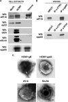
IFI16 is associated with purified HCMV particles. (A) HCMV particles (indicated as virions) were purified by sucrose gradient from supernatants of infected HELFs (192 hpi, MOI of 1) and analyzed by immunoblotting for the viral proteins IEA, UL44, and pp65, and the cellular proteins IFI16 or p53. Total cell extract from mock- or HCMV-infected cells were included as controls. (B) IFI16 interacts with pp65 in purified virions. HELFs were infected as described for panel A. Protein extracts were obtained from purified virions and immunoprecipitated with anti-pp65 (upper panel) or anti-IFI16 (lower panel) and the appropriate control antibodies (CTRL) and then immunoblotted with anti-IFI16 or anti-pp65 antibodies, respectively. Nonimmunoprecipitated whole-cell extracts (INPUT) were immunoblotted with anti-IFI16 or anti-pp65 antibodies and used to normalize the proteins subjected to immunoprecipitation. (C) Immunoelectron microscopy analysis of purified virions stained, in the presence of 10% human serum, for IFI16 and HCMV gB and pp65, or left unstained (SecAb). A 15-nm gold-conjugated secondary antibody was used to detect proteins. Scale bar, 100 nm.

Similar articles

Cited by

References

    1. Bieniasz PD. 2004. Intrinsic immunity: a front-line defense against viral attack. Nat. Immunol. 5:1109–1115. 10.1038/ni1125 - DOI - PubMed
    1. Roy CR, Mocarski ES. 2007. Pathogen subversion of cell-intrinsic innate immunity. Nat. Immunol. 8:1179–1187. 10.1038/ni1528 - DOI - PubMed
    1. Douglas JL, Gustin JK, Viswanathan K, Mansouri M, Moses AV, Fruh K. 2010. The great escape: viral strategies to counter BST-2/tetherin. PLoS Pathog. 6:e1000913. 10.1371/journal.ppat.1000913 - DOI - PMC - PubMed
    1. Takeuchi H, Matano T. 2008. Host factors involved in resistance to retroviral infection. Microbiol. Immunol. 52:318–325. 10.1111/j.1348-0421.2008.00040.x - DOI - PubMed
    1. Duggal NK, Emerman M. 2012. Evolutionary conflicts between viruses and restriction factors shape immunity. Nat. Rev. Immunol. 12:687–695. 10.1038/nri3295 - DOI - PMC - PubMed

Publication types

MeSH terms

Substances