. 2014 Jul;22(7):1320-1332.
doi: 10.1038/mt.2014.60. Epub 2014 Apr 3.

Maraba MG1 virus enhances natural killer cell function via conventional dendritic cells to reduce postoperative metastatic disease

Affiliations

Maraba MG1 virus enhances natural killer cell function via conventional dendritic cells to reduce postoperative metastatic disease

Jiqing Zhang et al. Mol Ther. 2014 Jul.

Abstract

This study characterizes the ability of novel oncolytic rhabdoviruses (Maraba MG1) to boost natural killer (NK) cell activity. Our results demonstrate that MG1 activates NK cells via direct infection and maturation of conventional dendritic cells. Using NK depletion and conventional dendritic cells ablation studies in vivo, we established that both are required for MG1 efficacy. We further explored the efficacy of attenuated MG1 (nonreplicating MG1-UV(2min) and single-cycle replicating MG1-Gless) and demonstrated that these viruses activate conventional dendritic cells, although to a lesser extent than live MG1. This translates to equivalent abilities to remove tumor metastases only at the highest viral doses of attenuated MG1. In tandem, we characterized the antitumor ability of NK cells following preoperative administration of live and attenuated MG1. Our results demonstrates that a similar level of NK activation and reduction in postoperative tumor metastases was achieved with equivalent high viral doses concluding that viral replication is important, but not necessary for NK activation. Biochemical characterization of a panel of UV-inactivated MG1 (2-120 minutes) revealed that intact viral particle and target cell recognition are essential for NK cell-mediated antitumor responses. These findings provide mechanistic insight and preclinical rationale for safe perioperative virotherapy to effectively reduce metastatic disease following cancer surgery.


Figures

Figure 1
Figure 1
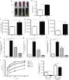
The in vivo efficacy of MG1 in the B16lacZ tumor model is not dependent on viral oncolysis. (a) B16lacZ cells were in vitro infected with indicated virus at different MOI. Forty-eight hours postinjection, cell viability was assessed by Alamar Blue. (b) B16lacZ cells were injected i.v. into B6 mice via tail vein at day 0. Virus treatment was given at day 1, 3, and 8. At day 14, lung metastases were stained and quantified. (c) Indicated virus treatment was administered at 1 day before B16lacZ tumor cell inoculation. Lung metastases were quantified at 3 days post–tumor injection. Data are representative of three similar experiments with n = 5–6/group (*P < 0.05; ***P < 0.001; ns, not significant).
Figure 2
Figure 2
Activation of NK cell function by MG1 plays a significant role in the reduction of B16lacZ lung metastases. (ae) B6 mice were treated with MG1 i.v. or PBS and sacrificed at 5 days after virus treatment. Spleens were isolated and assessed as follows: (a) photographs, (b) weight, (c) total number of lymphocytes, (d) total number of NK cells (TCRβ/CD122+), and (e) total number of DC (CD11c+). (fh) B6 mice were treated with MG1 (1 × 108 PFU) i.v or PBS and sacrificed at 1, 3, and 5 days after virus treatment. Spleens were harvested to assess NK cell (f) CD69 expression, (g) interferon-γ, (h) Granzyme B secretion, and (i) NK cytotoxicity (^P < 0.05; ^^P < 0.01; ^^^P < 0.001 comparing treatment to PBS controls; the data are displayed as the mean percent (±SD) of chromium release from triplicate wells for the indicated E:T ratios). (j) B6 mice were treated with MG1 i.v. or PBS at day −1. At day 0, all mice received 3 × 105 B16lacZ tumor cell inoculation via tail vein. NK cells were depleted using anti-NK1.1 i.v. at days −4, −1, and +1. Mice were sacrificed at 3 days post–cell injection and the number of lung tumor metastases was quantified. Data are representative of three similar experiments with n = 4–6/group (*P < 0.05; ***P < 0.001, ns, not significant).
Figure 3
Figure 3
Virus replication is important, but not necessary for NK cell activation and attenuation of B16lacZ lung metastases. PBS or indicated virus was injected i.v. into B6 mice. Twenty-four hours post–virus injection, spleens were harvested to assess (a) NK cell CD69 expression, (b) interferon-γ, (c) Granzyme B secretion via flow cytometry, and (d) ex vivo NK cytotoxicity (^P < 0.01; ^^P = 0.05 comparing treatment to PBS controls; the data are displayed as the mean percent (±SD) of chromium release from triplicate wells for the indicated E:T ratios). (e) B6 mice treated with PBS or indicated virus at day −1. At day 0, all mice received B16lacZ tumor cell inoculation. Mice were sacrificed at 3 days post–cell injection and the number of lung tumor metastases was quantified. Data are representative of three similar experiments with n = 4–6/group (*P < 0.05; **P < 0.01; ***P < 0.001; ns, not significant).
Figure 4
Figure 4
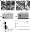
Viral particle structure, cellular recognition, viral proteins, and viral genomic RNA are retained in minimally UV-inactivated virus (MG1-UV2min). Electron Microscopy analysis of (a) purified virus and (b) 1 × 106 B16lacZ cells infected with virus (500 MOI). Scale bar: 1 cm = value indicated on EM figure; EC, extracellular space; IC, intracellular space. Western blot analysis of viral proteins from (c) 1 × 106 PFU purified virus preparations and (d) 2 × 105 B16lacZ infected with indicated virus (3 MOI). Cells were harvested at 18 hours postinjection; anti-rhabdovirus and GAPDH antibodies were used. Quantitative reverse transcription–PCR analysis of extracted genomic RNA from (e) purified virus preparations and (f) B16lacZ cells infected with virus at 6, 12, and 24 hours postinjection.
Figure 5
Figure 5
A nonreplicating, minimally UV-inactivated MG1 (MG1-UV2min) is capable of activating NK cells and attenuates lung metastases. PBS or indicated virus was injected i.v. into B6 mice. Twenty-four hours post–virus injection, spleens were harvested to assess (a) NK cell CD69 expression and (b) ex vivo NK cytotoxicity (^P = 0.05, ^^P = 0.01 comparing treatment to PBS controls; the data are displayed as the mean percent (±SD) of chromium release from triplicate wells for the indicated E:T ratios). (c) PBS or indicated virus was injected i.v. into B6 mice at day 0. At day 1, all mice received B16lacZ tumor cell inoculation. All mice were sacrificed at 3 days post–cell injection and the number of lung tumor metastases was quantified. (d) B6 mice were treated with MG1 i.v. or PBS at day −1. At day 0, all mice received 3 × 105 B16lacZ tumor cell inoculation via tail vein. NK cells were depleted using anti-NK1.1 i.v. or control IgG at days −4, −1 and +1. **P < 0.001; ***P = 0.05.
Figure 6
Figure 6
Maraba MG1 activates NK cell function through conventional dendritic cells. (a) In vitro NK cytotoxicity following NK cell-cDC coculture (^^^P < 0.001 comparing NK cells + DC + MG1 to NK cells + MG1). (b) In vitro infection and (c) activation by flow cytometry of cDC with indicated virus. (d) Quantification of lung tumor metastases at day 3 from B6 mice treated with live or attenuated MG1 i.v. at doses ranging from 1 × 105–7 PFU. Virus was administered at day −1. At day 0, all mice received 3 × 105 B16lacZ tumor cell inoculation via tail vein. (e) Electron microscopy analysis of 1 × 106 bone marrow–derived DCs infected with virus (500 MOI). Scale bar: 1 cm = value indicated on EM figure; EC, extracellular space; IC, intracellular space. (f) PBS or DT was injected intraperitoneally into CD11c-DTR Tg mice at day 0. Twenty-four hours following, MG1 and B16lacZ cells were injected i.v. Spleens were harvested at day 3 to assess ex vivo NK cytotoxicity (^^^P < 0.001 comparing MG1 treatment to PBS and MG1 + DT controls; the data are displayed as the mean percent (±SD) of chromium release from triplicate wells for the indicated E:T ratios). Data are representative of three similar experiments with n = 5–6/group (*P < 0.05).
Figure 7
Figure 7
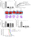
Recovery of metastatic disease by perioperative NK cells stimulation with UV-inactivated MG1. (a) Quantification of B16lacZ lung tumor metastases at 3 days in indicated treatment groups. (b) The ability of sorted NK cells to kill tumor targets from indicated treatment groups (^P < 0.05; ^^P < 0.005; ^^^P < 0.0001; the data are displayed as the mean percent (±SD) of chromium release from triplicate wells for the indicated E:T ratios). Assessment of 4T1 lung tumor metastases at 28 days from indicated treatment groups by (c) representative lung photographs and H&E staining of lung tissues, (d) lung weight, and (e) number of lung nodules. (f) Assessment of overall survival for B6 mice treated at day −1 with indicated virus, B16lacZ cells, and surgical stress at day 0. Data are representative of three similar experiments with n = 5–10/group (*P <0.05; **P < 0.01; ***P < 0.001; ns, not significant).
Figure 8
Figure 8
Maraba MG1 virus enhances natural killer cell function via infection and activation of conventional dendritic cells to reduce postoperative metastatic disease. Maraba MG1 administration results in cDC infection and activation, which in turn activates NK cell cytotoxicity. Preoperative administration of the live or attenuated MG1 results in NK cell activation via cDC, thereby preventing surgery-induced dysfunction and removal of tumor cell emboli and micrometastases in the postoperative period.

Similar articles

Cited by

References

    1. Vähä-Koskela MJ, Heikkilä JE, Hinkkanen AE. Oncolytic viruses in cancer therapy. Cancer Lett. 2007;254:178–216. - PMC - PubMed
    1. Russell SJ, Peng KW, Bell JC. Oncolytic virotherapy. Nat Biotechnol. 2012;30:658–670. - PMC - PubMed
    1. Stojdl DF, Lichty BD, tenOever BR, Paterson JM, Power AT, Knowles S, et al. VSV strains with defects in their ability to shutdown innate immunity are potent systemic anti-cancer agents. Cancer Cell. 2003;4:263–275. - PubMed
    1. Boisgerault N, Tangy F, Gregoire M. New perspectives in cancer virotherapy: bringing the immune system into play. Immunotherapy. 2010;2:185–199. - PubMed
    1. Parato KA, Lichty BD, Bell JC. Diplomatic immunity: turning a foe into an ally. Curr Opin Mol Ther. 2009;11:13–21. - PubMed

Publication types