

Novel monoclonal antibodies to normal and pathologically altered human TDP-43 proteins
- PMID: 24690345
- PMCID: PMC4023626
- DOI: 10.1186/2051-5960-2-33
Novel monoclonal antibodies to normal and pathologically altered human TDP-43 proteins
Abstract
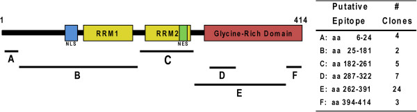
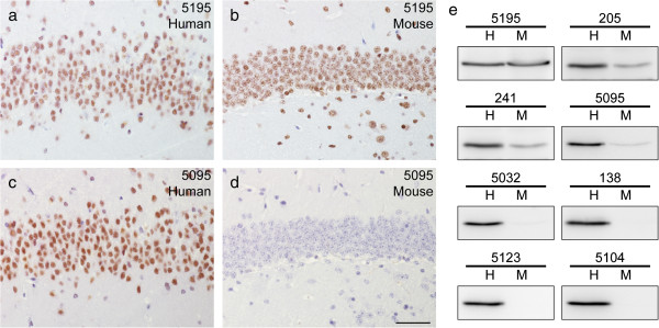
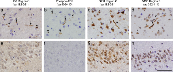
The RNA/DNA-binding protein, TDP-43, is the key component of ubiquitinated inclusions characteristic of amyotrophic lateral sclerosis (ALS) and the majority of frontotemporal lobar degeneration (FTLD-TDP) referred to collectively as TDP-43 proteinopathies. To further elucidate mechanisms of pathological TDP-43 processing and identify TDP-43 epitopes that could be useful as potential biomarkers of TDP-43 proteinopathies, we developed a panel of novel monoclonal antibodies (MAbs) directed at regions extending across the length of TDP-43. Here, we confirm previous observations that there is no or minimal accumulation of TDP-43 N-terminal domains in neocortical inclusions in human TDP-43 proteinopathy tissues and we identify a subset of these MAbs that are specific for human versus mouse TDP-43. Notably, one of these MAbs recognized an epitope that preferentially detected pathological TDP-43 inclusions with negligible reactivity for normal nuclear TDP-43 resembling anti-phospho-TDP-43 specific antibodies that only bind pathological TDP-43. Hence, we infer that this new MAb recognizes a phosphorylation independent but disease-specific pathologic conformation in abnormal TDP-43. These data suggest that the novel MAbs reported here will be useful for patient-oriented research as well as for studies of animal and cell-based models of TDP-43 proteinopathies including ALS and FTLD-TDP.
Figures

Similar articles
-
Epitope mapping of antibodies against TDP-43 and detection of protease-resistant fragments of pathological TDP-43 in amyotrophic lateral sclerosis and frontotemporal lobar degeneration.Biochem Biophys Res Commun. 2012 Jan 6;417(1):116-21. doi: 10.1016/j.bbrc.2011.11.066. Epub 2011 Nov 22. Biochem Biophys Res Commun. 2012. PMID: 22133678
-
Phosphorylation of S409/410 of TDP-43 is a consistent feature in all sporadic and familial forms of TDP-43 proteinopathies.Acta Neuropathol. 2009 Feb;117(2):137-49. doi: 10.1007/s00401-008-0477-9. Epub 2009 Jan 6. Acta Neuropathol. 2009. PMID: 19125255 Free PMC article.
-
Phosphorylated TDP-43 in frontotemporal lobar degeneration and amyotrophic lateral sclerosis.Ann Neurol. 2008 Jul;64(1):60-70. doi: 10.1002/ana.21425. Ann Neurol. 2008. PMID: 18546284 Free PMC article.
-
Molecular neuropathology of TDP-43 proteinopathies.Int J Mol Sci. 2009 Jan;10(1):232-246. doi: 10.3390/ijms10010232. Epub 2009 Jan 9. Int J Mol Sci. 2009. PMID: 19333444 Free PMC article. Review.
-
Amyotrophic lateral sclerosis and frontotemporal lobar degeneration: a spectrum of TDP-43 proteinopathies.Neuropathology. 2010 Apr;30(2):103-12. doi: 10.1111/j.1440-1789.2009.01091.x. Epub 2010 Jan 25. Neuropathology. 2010. PMID: 20102519 Free PMC article. Review.
Cited by
-
Targeting the TDP-43 low complexity domain blocks spreading of pathology in a mouse model of ALS/FTD.Acta Neuropathol Commun. 2024 Oct 3;12(1):156. doi: 10.1186/s40478-024-01867-z. Acta Neuropathol Commun. 2024. PMID: 39363348 Free PMC article.
-
Evaluation of blood-based, extracellular vesicles as biomarkers for aging-related TDP-43 pathology.Alzheimers Dement (Amst). 2022 Dec 15;14(1):e12365. doi: 10.1002/dad2.12365. eCollection 2022. Alzheimers Dement (Amst). 2022. PMID: 36540894 Free PMC article.
-
Progression of motor neuron disease is accelerated and the ability to recover is compromised with advanced age in rNLS8 mice.Acta Neuropathol Commun. 2016 Sep 29;4(1):105. doi: 10.1186/s40478-016-0377-5. Acta Neuropathol Commun. 2016. PMID: 27687289 Free PMC article.
-
TDP-43 Neuropathologic Associations in the Nun Study and the Honolulu-Asia Aging Study.J Alzheimers Dis. 2018;66(4):1549-1558. doi: 10.3233/JAD-180162. J Alzheimers Dis. 2018. PMID: 30452409 Free PMC article.
-
An insoluble frontotemporal lobar degeneration-associated TDP-43 C-terminal fragment causes neurodegeneration and hippocampus pathology in transgenic mice.Hum Mol Genet. 2015 Dec 20;24(25):7241-54. doi: 10.1093/hmg/ddv424. Epub 2015 Oct 16. Hum Mol Genet. 2015. PMID: 26476406 Free PMC article.
References
-
- Neumann M, Sampathu DM, Kwong LK, Truax AC, Micsenyi MC, Chou TT, Bruce J, Schuck T, Grossman M, Clark CM, McCluskey LF, Miller BL, Masliah E, Mackenzie IR, Feldman H, Feiden W, Kretzschmar HA, Trojanowski JQ, Lee VM. Ubiquitinated TDP-43 in frontotemporal lobar degeneration and amyotrophic lateral sclerosis. Science. 2006;2(5796):130–133. doi: 10.1126/science.1134108. doi:10.1126/science.1134108. - DOI - PubMed
-
- Geser F, Martinez-Lage M, Robinson J, Uryu K, Neumann M, Brandmeir NJ, Xie SX, Kwong LK, Elman L, McCluskey L, Clark CM, Malunda J, Miller BL, Zimmerman EA, Qian J, Van Deerlin V, Grossman M, Lee VM, Trojanowski JQ. Clinical and pathological continuum of multisystem TDP-43 proteinopathies. Arch Neurol. 2009;2(2):180–189. doi:10.1001/archneurol.2008.558. - PMC - PubMed
-
- Brettschneider J, Del Tredici K, Toledo JB, Robinson JL, Irwin DJ, Grossman M, Suh E, Van Deerlin VM, Wood EM, Baek Y, Kwong L, Lee EB, Elman L, McCluskey L, Fang L, Feldengut S, Ludolph AC, Lee VM, Braak H, Trojanowski JQ. Stages of pTDP-43 pathology in amyotrophic lateral sclerosis. Ann Neurol. 2013;2(1):20–38. doi: 10.1002/ana.23937. doi:10.1002/ana.23937. - DOI - PMC - PubMed
-
- Brettschneider J, Del Tredici K, Irwin DJ, Grossman M, Robinson JL, Toledo JB, Fang L, Van Deerlin VM, Ludolph AC, Lee VM, Braak H, Trojanowski JQ. Sequential distribution of pTDP-43 pathology in behavioral variant frontotemporal dementia (bvFTD) Acta Neuropathol. 2014;2(3):423–439. doi: 10.1007/s00401-013-1238-y. doi:10.1007/s00401-013-1238-y. - DOI - PMC - PubMed
Publication types
MeSH terms
Substances
Grants and funding
LinkOut - more resources
Full Text Sources
Other Literature Sources
Medical
Miscellaneous
