

Aldoketoreductase family 1B10 (AKR1B10) as a biomarker to distinguish hepatocellular carcinoma from benign liver lesions
- PMID: 24656094
- PMCID: PMC4030546
- DOI: 10.1016/j.humpath.2013.12.002
Aldoketoreductase family 1B10 (AKR1B10) as a biomarker to distinguish hepatocellular carcinoma from benign liver lesions
Abstract
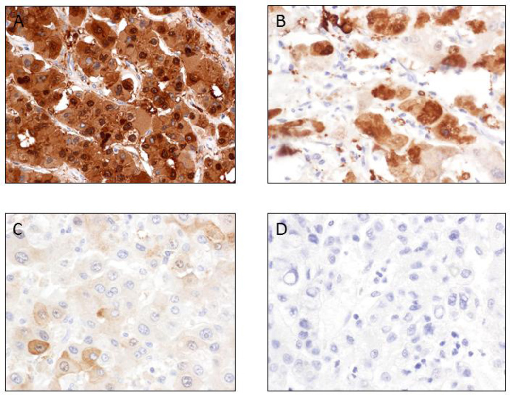
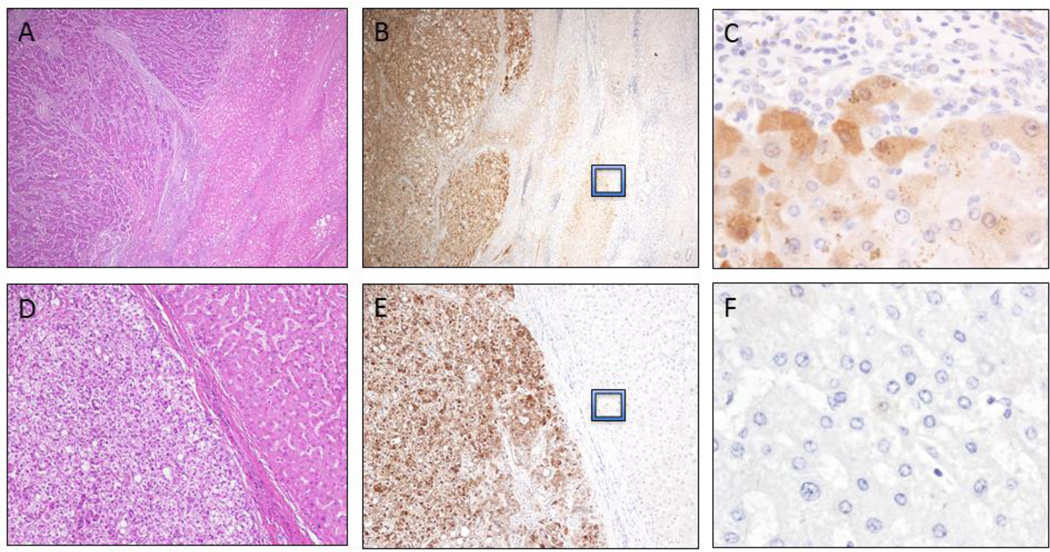
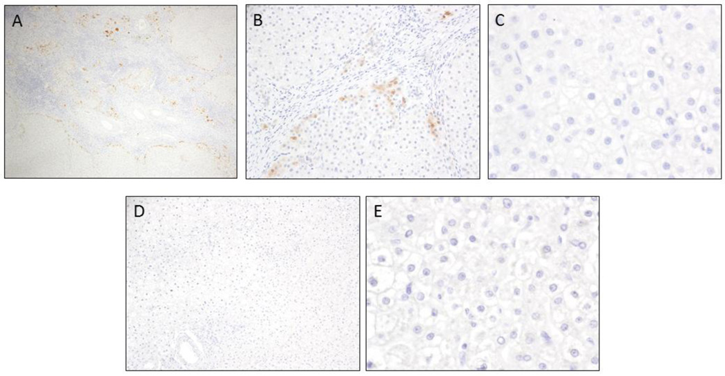
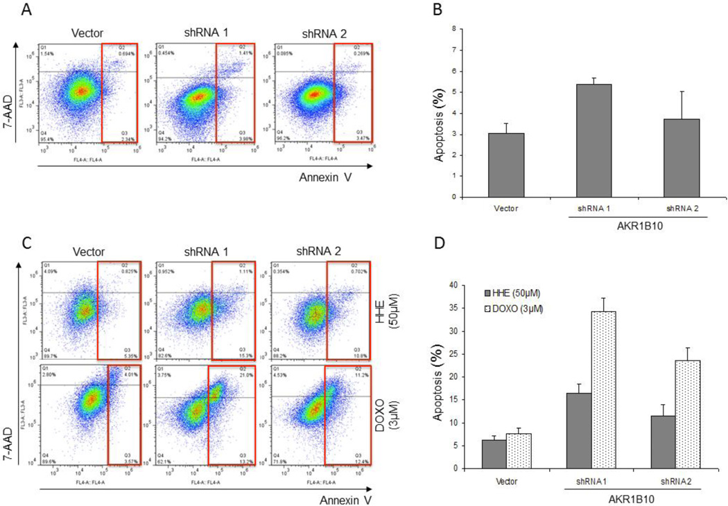
Hepatocellular carcinoma (HCC) is one of the most common highly aggressive malignant tumors worldwide. Aldoketoreductase 1B10 (AKR1B10) was first isolated from HCC and further identified to be over-expressed in many cancers from various organs. AKR1B10 contributes to detoxification of xenobiotics by lipid peroxidation and metabolizes physiological substrates such as farnesal, retinal, and carbonyls. Metabolizing these lipid substrates plays a crucial role in promoting carcinogenesis. In the present study, immunohistochemical analysis was performed to determine the prevalence/pattern of AKR1B10 expression in HCC and its usefulness to differentiate benign liver lesions from HCC. Oncogenic function of AKR1B10 was examined in hepatocellular carcinoma cells in vitro using Western blotting and shRNA knockdown approaches, with emphasis on cell apoptosis and response to chemotherapy. Immunohistochemistry analysis revealed AKR1B10 was overexpressed in 97% (86/89) of hepatocellular carcinomas, with minimal to no expression in adjacent hepatic tissue, while hepatic adenomas and focal nodular hyperplasia did not exhibit expression of AKR1B10. shRNA-mediated silencing of AKR1B10 expression in hepatocellular carcinoma cells resulted in (1) increased cell apoptosis, (2) decreased colony formation and size, and (3) enhanced cytoreductive response following exposure to doxorubicin chemotherapy. Our findings provide first time evidence that AKR1B10 is a unique biomarker involved in hepatocellular carcinogenesis via modulation of proliferation, cell apoptosis and chemoresistance and is a potential promising biomarker to differentiate HCCs from benign hepatic lesions.
Keywords: AKR1B10; Focal nodular hyperplasia; Hepatic adenoma; Hepatocellular carcinoma.
Copyright © 2014 Elsevier Inc. All rights reserved.
Conflict of interest statement
Figures

Similar articles
-
Overexpression and oncogenic function of aldo-keto reductase family 1B10 (AKR1B10) in pancreatic carcinoma.Mod Pathol. 2012 May;25(5):758-66. doi: 10.1038/modpathol.2011.191. Epub 2012 Jan 6. Mod Pathol. 2012. PMID: 22222635 Free PMC article.
-
Prognostic Significance of 14-3-3ε, Aldo-keto Reductase Family 1 B10 and Metallothionein-1 in Hepatocellular Carcinoma.Anticancer Res. 2018 Dec;38(12):6855-6863. doi: 10.21873/anticanres.13060. Anticancer Res. 2018. PMID: 30504401
-
High expression of aldo-keto reductase 1B10 is an independent predictor of favorable prognosis in patients with hepatocellular carcinoma.Gut Liver. 2014 Nov;8(6):648-54. doi: 10.5009/gnl13406. Epub 2014 Oct 7. Gut Liver. 2014. PMID: 25287169 Free PMC article.
-
Immunohistochemistry in the Diagnosis of Hepatocellular Carcinoma.Gastroenterol Clin North Am. 2017 Jun;46(2):311-325. doi: 10.1016/j.gtc.2017.01.006. Gastroenterol Clin North Am. 2017. PMID: 28506367 Review.
-
Diagnostic and Prognostic Potential of AKR1B10 in Human Hepatocellular Carcinoma.Cancers (Basel). 2019 Apr 5;11(4):486. doi: 10.3390/cancers11040486. Cancers (Basel). 2019. PMID: 30959792 Free PMC article. Review.
Cited by
-
HCCDB: A Database of Hepatocellular Carcinoma Expression Atlas.Genomics Proteomics Bioinformatics. 2018 Aug;16(4):269-275. doi: 10.1016/j.gpb.2018.07.003. Epub 2018 Sep 25. Genomics Proteomics Bioinformatics. 2018. PMID: 30266410 Free PMC article.
-
The role of hepatocyte nuclear factor 4-alpha in perfluorooctanoic acid- and perfluorooctanesulfonic acid-induced hepatocellular dysfunction.Toxicol Appl Pharmacol. 2016 Aug 1;304:18-29. doi: 10.1016/j.taap.2016.05.001. Epub 2016 May 3. Toxicol Appl Pharmacol. 2016. PMID: 27153767 Free PMC article.
-
Identification of a role for serum aldo-keto reductase family 1 member B10 in early detection of hepatocellular carcinoma.Oncol Lett. 2018 Dec;16(6):7123-7130. doi: 10.3892/ol.2018.9547. Epub 2018 Oct 3. Oncol Lett. 2018. PMID: 30546447 Free PMC article.
-
Compensatory upregulation of aldo-keto reductase 1B10 to protect hepatocytes against oxidative stress during hepatocarcinogenesis.Am J Cancer Res. 2019 Dec 1;9(12):2730-2748. eCollection 2019. Am J Cancer Res. 2019. PMID: 31911858 Free PMC article.
-
The hop-derived compounds xanthohumol, isoxanthohumol and 8-prenylnaringenin are tight-binding inhibitors of human aldo-keto reductases 1B1 and 1B10.J Enzyme Inhib Med Chem. 2018 Dec;33(1):607-614. doi: 10.1080/14756366.2018.1437728. J Enzyme Inhib Med Chem. 2018. PMID: 29532688 Free PMC article.
References
-
- Jemal A, Bray F, Center MM, Ferlay J, Ward E, Forman D. Global cancer statistics. CA Cancer J Clin. 2011;61(2):69–90. - PubMed
-
- Wong R, Corley DA. Racial and ethnic variations in hepatocellular carcinoma incidence within the United States. Am J Med. 2008;121(6):525–531. - PubMed
-
- Perz JF, Armstrong GL, Farrington LA, Hutin YJ, Bell BP. The contributions of hepatitis B virus and hepatitis C virus infections to cirrhosis and primary liver cancer worldwide. J Hepatol. 2006;45(4):529–538. - PubMed
-
- Ascha MS, Hanouneh IA, Lopez R, Tamimi TA, Feldstein AF, Zein NN. The incidence and risk factors of hepatocellular carcinoma in patients with nonalcoholic steatohepatitis. Hepatology. 2010;51(6):1972–1978. - PubMed
Publication types
MeSH terms
Substances
Grants and funding
LinkOut - more resources
Full Text Sources
Other Literature Sources
Medical
