. 2014 Jun;171(11):2827-41.
doi: 10.1111/bph.12623.

Signalling profiles of H3 relaxin, H2 relaxin and R3(BΔ23-27)R/I5 acting at the relaxin family peptide receptor 3 (RXFP3)

Affiliations

Signalling profiles of H3 relaxin, H2 relaxin and R3(BΔ23-27)R/I5 acting at the relaxin family peptide receptor 3 (RXFP3)

M Kocan et al. Br J Pharmacol. 2014 Jun.

Abstract

Background and purpose: Relaxin family peptide receptor 3 (RXFP3) is expressed in brain areas important for processing sensory information and feeding, suggesting that it may be a target for anti-anxiety and anti-obesity drugs. We examined the effects of H3 relaxin, the biased agonist H2 relaxin and the antagonist, R3(BΔ23-27)R/I5, on RXFP3 signalling to establish their suitability as tools to assess the physiological roles of RXFP3.

Experimental approach: The signalling profile of the RXFP3 ligands was determined using reporter gene assays, multiplexed signalling assays and direct examination of receptor-G protein and receptor-β-arrestin interactions using BRET.

Key results: H2 relaxin activated p38MAPK and ERK1/2 with lower efficacy than H3 relaxin, but had similar efficacy for JNK1/2 phosphorylation. H2 or H3 relaxin activation of p38MAPK, JNK1/2 or ERK1/2 involved Pertussis toxin-sensitive G-proteins. R3(BΔ23-27)R/I5 blocked H3 relaxin AP-1 reporter gene activation, but not H2 relaxin AP-1 activation or H3 relaxin NF-κB activation. R3(BΔ23-27)R/I5 activated the SRE reporter, but did not inhibit either H2 or H3 relaxin SRE activation. R3(BΔ23-27)R/I5 blocked H3 relaxin-stimulated p38MAPK and ERK1/2 phosphorylation, but was a weak partial agonist for p38MAPK and ERK1/2 signalling. p38MAPK activation by R3(BΔ23-27)R/I5 was G protein-independent. H3 relaxin-activated RXFP3 interacts with Gαi2 , Gαi3 , Gαo A and Gαo B whereas H2 relaxin or R3(BΔ23-27)R/I5 induce interactions only with Gαi2 or Gαo B . Only H3 relaxin promoted RXFP3/β-arrestin interactions that were blocked by R3(BΔ23-27)R/I5.

Conclusion and implications: Understanding signalling profile of drugs acting at RXFP3 is essential for development of therapies targeting this receptor.

Keywords: R3(BΔ23-27)R/I5; RXFP3 signalling; relaxin.


Figures

Figure 1
Figure 1
Structures of H3 relaxin (A); chimeric H3 relaxin B-chain/INSL5 A-chain (R3/I5) (B) and R3(BΔ23–27)R/I5 (C). The structures of H3 relaxin (2fhw) and R3/I5) (2k1v) were downloaded from the RSCB protein data bank and displayed using ICM-pro (Molsoft L.L.C., San Diego, CA, USA). The heterodimeric peptides contain an A-chain and B-chain linked by disulphide bonds (Bathgate et al., 2002; 2006a). Residues important for RXFP3 binding are in blue and for activation in green. Arg23 in R3(BΔ23–27)R/I5 is important for antagonist activity and shown in purple (Kuei et al., ; Hossain et al., 2009). (A) The NMR structure of H3 relaxin (Rosengren et al., 2006). (B) R3/I5 comprises the H3 relaxin B-chain and the human INSL5 A-chain (Sutton et al., 2004). NMR studies indicate that R3/I5 has a very similar structure to H3 relaxin (Haugaard-Jonsson et al., 2008). (C) The R3/I5 analogue, R3(BΔ23–27)R/I5 has the C-terminus of the B-chain truncated and with a terminal Arg residue (Kuei et al., ; Hossain et al., 2009).
Figure 2
Figure 2
Activation of the reporter genes AP-1 (A), NF-κB (B) and SRE (C) and inhibition of the forskolin-stimulated cAMP production (D) in CHO-RXFP3 or Flp-In CHO cells by H3 relaxin, H2 relaxin or R3(BΔ23–27)R/I5. CHO-RXFP3 or Flp-In CHO cells (negative control) were transiently co-transfected with either AP-1-SEAP, NF-κB-SEAP, or SRE-SEAP reporter genes and a constitutively active β-galactosidase reporter. Activation was determined by increased SEAP in the culture medium 24 h after stimulation. H3 relaxin activated AP-1, NF-κB and SRE (A, B, C), but only the AP-1 reporter was blocked by R3(BΔ23–27)R/I5 (A). H2 relaxin stimulated AP-1 and SRE and R3(BΔ23–27)R/I5 had no inhibitory effect (A, C). SRE activation was also detected following R3(BΔ23–27)R/I5 (C). FBS (10%) was used as a positive control in all experiments to demonstrate functional reporter genes. Data are expressed as percentage β-galactosidase activation and normalized to the FBS response. Data are mean ± SEM of six to eight independent experiments, conducted in triplicate. In the cAMP assay (D), forskolin-stimulated cAMP accumulation (30 μM) was inhibited in CHO-RXFP3 cells by H3 relaxin and to a lesser extent by H2 relaxin and R3(BΔ23–27)R/I5. R3(BΔ23–27)R/I5 blocked H3 relaxin, but not H2 relaxin-induced cAMP inhibition. Data are expressed as % response to forskolin. Vehicle represents signalling in the absence of forskolin and ligands. Data are mean ± SEM of three independent experiments, conducted in duplicate. Controls for CHO-RXFP3 cells were parallel treatments of the parent cell line Flp-In CHO. Data were analysed by one-way anova with Dunnett's post hoc test. *P ≤ 0.05 and ***P ≤ 0.001.
Figure 3
Figure 3
Time course of activation of p38MAPK (A, D), JNK1/2 (B, E) and ERK1/2 (C, F) by H3 relaxin, H2 relaxin or R3(BΔ23–27)R/I5 in CHO-RXFP3 (A, B, C) or Flp-In CHO (D, E, F) cells. Cells were treated with peptides for periods of up to 60 min, and p38MAPK (A, D), JNK1/2 (B, E) and ERK1/2 (C, F) activation was quantified using the appropriate phospho-‘kinase'-specific Surefire AlphaScreen kit. In CHO-RXFP3 cells (A, B, C), H2 relaxin (10−6 M) and H3 relaxin (10−6 M) activated all three kinases, but with different relative efficacies. H2 relaxin had lower efficacy than H3 relaxin for p38MAPK, but equivalent efficacy for JNK1/2 phosphorylation. ERK1/2 phosphorylation was activated by H3 relaxin > H2 relaxin. R3(BΔ23–27)R/I5 (10−6 M) stimulated p38MAPK, had no effect on JNK1/2 and weakly stimulated ERK1/2 with the same level of efficacy as H2 relaxin. Controls for CHO-RXFP3 cells were parallel treatments of the parent cell line Flp-In CHO (D, E, F). Data are mean ± SEM for three to six independent experiments.
Figure 4
Figure 4
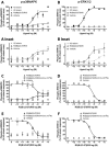
Concentration-dependent activation of p38MAPK (A, C, E) and ERK1/2 (B, D, F) by H3 relaxin, H2 relaxin or R3(BΔ23–27)R/I5 in CHO-RXFP3 cells. H3 relaxin, H2 relaxin or R3(BΔ23–27)R/I5 all activated p38MAPK and ERK1/2 (5 min treatment) with H3 relaxin having the highest efficacy. The effect of R3(BΔ23–27)R/I5 on H3 relaxin-stimulated p38MAPK or ERK1/2 activation was tested either by co-addition of R3(BΔ23–27)R/I5 and H3 relaxin (C, D) or by pre-incubation of cells with R3(BΔ23–27)R/I5 for 1 h prior to H3 relaxin treatment (E, F). R3(BΔ23–27)R/I5 inhibited H3 relaxin-stimulated p38MAPK and ERK1/2 activation in a concentration-dependent manner under both experimental conditions. Data are mean ± SEM for three to four independent experiments.
Figure 5
Figure 5
The role of PTX-sensitive G-proteins in p38MAPK (A, B, C), JNK1/2 (D, E, F) and ERK1/2 (G, H, I) activation by H3 relaxin, H2 relaxin or R3(BΔ23–27)R/I5 in CHO-RXFP3 cells. Cells were pretreated with PTX (100 ng·mL–1) for 18 h prior to stimulation with peptides for up to 30 min. Phosphorylation of p38MAPK (A, B, C), JNK1/2 (D, E, F) and ERK1/2 (G, H, I) was quantified using the appropriate phospho-‘kinase'-specific Surefire AlphaScreen kit. Activation of p38MAPK, JNK1/2 and ERK1/2 by H3 relaxin (A, D, G) or H2 relaxin (B, E, H) both at 10−6 M was blocked by PTX. R3(BΔ23–27)R/I5 (10−6 M) activated p38MAPK (C) and ERK1/2 (I) but not JNK1/2 (F); ERK1/2 but not p38MAPK activation was blocked by PTX pretreatment. Data are mean ± SEM for four to seven independent experiments. Data were analysed by Student's t-test. *P < 0.05; **P ≤ 0.05 & P ≥ 0.001; ***P < 0.001.
Figure 6
Figure 6
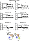
Detection of RXFP3 G protein interactions following treatment with H3 relaxin, H2 relaxin or R3(BΔ23–27)R/I5. Flp-In CHO cells were transiently co-transfected with RXFP3 Rluc8, Gγ2-Venus, Gβ1 and one of Gα subunits (Gαi2, Gαi3, GαoA, GαoB, Gαs, Gαq). Interactions between RXFP3 and G proteins were detected prior to and after treatment with H3 relaxin (10−6 M), H2 relaxin (10−6 M) or R3(BΔ23–27)R/I5 (10−6 M) using real-time BRET. H3 relaxin induced interactions between RXFP3 and Gαi2, Gαi3, GαoA or GαoB (A–D). H2 relaxin or R3(BΔ23–27)R/I5 induced interactions only between RXFP3 and Gαi2 or GαoB proteins and with a smaller signal compared with H3 relaxin (A, D). The ligand-induced BRET ratios were calculated by subtracting the ratio for the vehicle-treated sample from the BRET ratio for each ligand-treated sample as described. Data shown are mean ± SEM of four independent experiments. Illustration of the BRET interaction between receptor and G-protein subunits (G).
Figure 7
Figure 7
Detection of RXFP3 – β-arrestin interactions following treatment with H3 relaxin, H2 relaxin or R3(BΔ23–27)R/I5 (A, B) and role of β-arrestin in H3 relaxin, H2 relaxin and R3(BΔ23–27)R/I5 – stimulated ERK1/2 signalling (C). Flp-In CHO cells were transiently co-transfected with RXFP3-Rluc8 and β-arrestin 1-Venus or β-arrestin 2-Venus. Ligand-induced interactions between RXFP3 and β-arrestin 1 or β-arrestin 2 were detected using real-time BRET. H3 relaxin (10−6 M) induced RXFP3/β-arrestin 1 and RXFP3/β-arrestin 2 interactions whereas H2 relaxin (10−6 M) or R3(BΔ23–27)R/I5 (10−6 M) had no effect. To examine the role of β-arrestin in ERK1/2 signalling, CHO-RXFP3 cells were transfected with β-arrestin 1 WT (β-arr 1 WT) or dominant negative mutant V53D (β-arr 1 V53D) and tested 48 h later. ERK1/2 activation by H3 relaxin or R3(BΔ23–27)R/I5 (C) was sensitized by dominant negative β-arrestin 1 V53D whereas responses to H2 relaxin were slightly inhibited. Data shown are mean ± SEM of three independent experiments conducted in duplicate. Illustration of BRET between receptor and β-arrestins (D).
Figure 8
Figure 8
Effect of R3(BΔ23–27)R/I5 and PTX on H3 relaxin-induced RXFP3 – β-arrestin interactions. Flp-In CHO cells were transiently co-transfected with RXFP3-Rluc8 and β-arrestin 1-Venus or β-arrestin 2-Venus. Cells were pretreated with R3(BΔ23–27)R/I5 for 15 min. H3 relaxin(10−6 M)-induced RXFP3 – β-arrestin 1 (A) and RXFP3 – β-arrestin 2 (C) BRET were both completely blocked by R3(BΔ23–27)R/I5. H3 relaxin-induced interactions between RXFP3 and β-arrestin 1 (B) or β-arrestin 2 (D) were partially blocked by PTX suggesting some G protein-independent coupling. Data shown are mean ± SEM of three experiments (β-arrestin) or four to six independent experiments (PTX).
Figure 9
Figure 9
Signalling pathways activated by H3 relaxin, H2 relaxin and R3(BΔ23–27)R/I5 in CHO cells expressing RXFP3. H3 relaxin interacts with RXFP3 (A) to cause coupling of the receptor with GαoB, Gαi2, GαoA and Gαi3. RXFP3/β-arrestin interactions utilize both Gi/o-dependent and G protein-independent mechanisms. H3 relaxin induces strong ERK and weaker JNK and p38MAPK activation – all through Gi/o. Phosphorylation of MAPKs leads to reporter gene transcription. H3 relaxin also inhibits forskolin-stimulated cAMP accumulation and activates the NF-κB reporter. H2 relaxin (B) promotes coupling of RXFP3 to GαoB and Gαi2 but not GαoA, Gαi3 or β-arrestins. It promotes much less ERK phosphorylation than H3 relaxin, but similar JNK and p38MAPK activation – all through Gi/o proteins. H2 relaxin inhibits forskolin-stimulated cAMP accumulation and activates AP-1 and SRE but not NF-κB. R3(BΔ23–27)R/I5 (C) causes coupling of RXFP3 to GαoB and Gαi2 but not GαoA, Gαi3 or β-arrestins. It also causes ERK but not JNK phosphorylation and activates p38MAPK via Gi/o protein-independent pathways. It also inhibits forskolin-stimulated cAMP accumulation and activates the SRE reporter. R3(BΔ23–27)R/I5 (D) inhibits (red crosses) H3 relaxin-stimulated cAMP inhibition, p38MAPK and ERK phosphorylation and AP-1 reporter transcription. R3(BΔ23–27)R/I5 completely inhibits RXFP3 coupling to β-arrestins induced by H3 relaxin.

Similar articles

Cited by

References

    1. Ahn S, Shenoy SK, Wei H, Lefkowitz RJ. Differential kinetic and spatial patterns of beta-arrestin and G protein-mediated ERK activation by the angiotensin II receptor. J Biol Chem. 2004;279:35518–35525. - PubMed
    1. Alexander SPH, Benson HE, Faccenda E, Pawson AJ, Sharman JL. Spedding M, et al. The Concise Guide to PHARMACOLOGY 2013/14: G Protein-Coupled Receptors. Br J Pharmacol. 2013;170:1459–1581. - PMC - PubMed
    1. Ayoub MA, See HB, Seeber RM, Armstrong SP, Pfleger KD. Profiling epidermal growth factor receptor and heregulin receptor 3 heteromerization using receptor tyrosine kinase heteromer investigation technology. PLoS ONE. 2013;8:e64672. - PMC - PubMed
    1. Azzi M, Charest PG, Angers S, Rousseau G, Kohout T, Bouvier M, et al. Beta-arrestin-mediated activation of MAPK by inverse agonists reveals distinct active conformations for G protein-coupled receptors. Proc Natl Acad Sci U S A. 2003;100:11406–11411. - PMC - PubMed
    1. Baker JG, Hall IP, Hill SJ. Agonist and inverse agonist actions of beta-blockers at the human beta 2-adrenoceptor provide evidence for agonist-directed signaling. Mol Pharmacol. 2003;64:1357–1369. - PubMed

Publication types