. 2014 Mar 13;156(6):1193-1206.
doi: 10.1016/j.cell.2014.02.008.

Unified polymerization mechanism for the assembly of ASC-dependent inflammasomes

Affiliations

Unified polymerization mechanism for the assembly of ASC-dependent inflammasomes

Alvin Lu et al. Cell. .

Abstract

Inflammasomes elicit host defense inside cells by activating caspase-1 for cytokine maturation and cell death. AIM2 and NLRP3 are representative sensor proteins in two major families of inflammasomes. The adaptor protein ASC bridges the sensor proteins and caspase-1 to form ternary inflammasome complexes, achieved through pyrin domain (PYD) interactions between sensors and ASC and through caspase activation and recruitment domain (CARD) interactions between ASC and caspase-1. We found that PYD and CARD both form filaments. Activated AIM2 and NLRP3 nucleate PYD filaments of ASC, which, in turn, cluster the CARD of ASC. ASC thus nucleates CARD filaments of caspase-1, leading to proximity-induced activation. Endogenous NLRP3 inflammasome is also filamentous. The cryoelectron microscopy structure of ASC(PYD) filament at near-atomic resolution provides a template for homo- and hetero-PYD/PYD associations, as confirmed by structure-guided mutagenesis. We propose that ASC-dependent inflammasomes in both families share a unified assembly mechanism that involves two successive steps of nucleation-induced polymerization. PAPERFLICK:


Figures

Figure 1
Figure 1. AIM2 Promotes Formation of ASCPYD Filaments
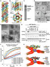
A. Domain composition and interaction hierarchy of NLRP3 and AIM2 inflammasomes. B. An electron micrograph of the AIM2PYD/ASCPYD binary complex. C. Gel filtration fractions of biotinylated AIM2PYD/ASCPYD complex as visualized by Coomassie Blue-stained SDS-PAGE (left) and streptavidin-alkaline phosphatase Western blot (right). D. Labeling of biotinylated AIM2PYD/ASCPYD binary complex by streptavidin-gold conjugate (6nm). E. Fluorescence polarization (FP) assay of AIM2PYD-nucleated ASCPYD filament formation. mP: unit for FP. Data are represented as mean±SD (N=3). F. Effect of dsDNA on AIM2FL-nucleated ASCPYD filament formation. 2 µM of AIM2FL monomer from gel filtration was incubated with or without equimolar300-bp dsDNA (assuming a 10-bp footprint of AIM2 for molar calculation) for 30 minutes before diluting to a working concentration 0.1 µM (ASCPYD:AIM2FL=10:1) for the FP assay. Data are represented as mean±SD (N=3). See also Figure S1.
Figure 2
Figure 2. NLPR3FL and NLRP3PYD-NBD but not NLRP3PYD Promote ASCPYD Filament Formation
A, B, C.Nucleation of ASCPYD filaments by titrating increasing amounts of NLRP3PYD (A), NLRP3PYD-NBD (B), NLRP3FL (C) as monitored by fluorescence polarization. Data are represented as mean±SD (N=3). D. A less aggregated gel filtration fraction of NLRP3 was subjected to ASCPYD polymerization assay with or without 5mM ATP. Data are represented as mean±SD (N=3). E. Streptavidin-gold (6nm) labeling of biotinylated NLRP3PYD-NBD/ASCPYD binary complex. See also Figure S2.
Figure 3
Figure 3. Cryo-EM Structure of the ASCPYD Filament at Near Atomic Resolution
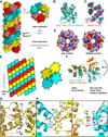
A. A cryo-EM image of ASCPYD filaments. B. Average power spectra of ASCPYD filaments in two twist bins (left and right halves) showing constant axial rise per subunit (blue arrow) and variable long-range twist features (red arrows). C. Filament segments can be divided into separate twist bins according to azimuth angle, or rotation per subunit. D. Cryo-EM reconstruction of the ASCPYD filament, superimposed with the final atomic model shown in three colors each for one start of the three-start helical assembly. E. A zoom up view of helix α6 shown in stick model and superimposed with the EM density. See also Figure S3 and Movie S1.
Figure 4
Figure 4. Detailed Cryo-EM Model of the ASCPYD Filament
A. The ASCPYD filament is a three-start helical assembly with C3 symmetry as shown in a surface representation. The three-start helical strands are denoted by red, cyan, and yellow, respectively, with alternating darker and lighter shades to show subunit boundaries. B. Comparison of the initial ASCPYD subunit model (gray, PDB: 1UCP) and the subunit structure after refinement against the cryo-EM density (cyan). C.Structures of ASC2PYD (magenta) and NLRP3PYD (orange) are similar to the ASCPYD subunit structure in the filament (cyan). D.Electrostatic surface representations of approximate cross sections of the filament. E. A schematic diagram of the ASCPYD filament and the three types of asymmetric interactions, defined in accordance with the previously observed DD/DD interactions. F. Comparison of the Type III interactions in the ASCPYD filament (cyan) and in the MyD88/IRAK4/IRAK2 DD complex (orange). G, H, I. Detailed interactions in Type I, II, and III interfaces, respectively. Side chains of interfacial residues are shown as stick models and labeled. See also Figure S4.
Figure 5
Figure 5. Structure-based Mutations Disrupts ASCPYD Filament Formation, AIM2PYD/ASCPYD Interaction and NLRP3PYD/ASCPYD Interaction in Vitro and in Cells
A. Size-exclusion chromatography of WT and mutant ASCPYD showing both filamentous (void) and monomeric fractions from a Superdex 200 column. Hyphen denotes ASCPYD and asterisk denotes a contaminant. B. Morphology of transfected WT and mutant eGFP-tagged ASCPYD constructs visualized by confocal laser scanning microscopy. The arrowhead depicts filaments. n: nucleus; scale bars = 10µm. C.Morphology of transfected eGFP-tagged ASCPYD visualized by confocal laser scanning microscopy. Top: ASCPYD with charge reversal mutation on a residue outside the filament interface. Bottom: ASCPYD with triple charge reversal mutation that rescued the defectiveness of the single mutants. Arrow heads depict filaments. D.A schematic model of AIM2PYD/ASCPYD or NLRP3PYD/ASCPYD filaments composed of a top AIM2PYD or NLRP3PYD layer extended by ASCPYD filament body. E,F. Mutations of conserved interfacial residues on AIM2PYD (E) and NLRP3PYD-NBD (F) reduced or abolished their ability to nucleate ASCPYD filaments. See also Figure S5.
Figure 6
Figure 6. Reconstitution of the Full Ternary AIM2 Inflammasome
A. The ASCPYD filament structure in a ribbon representation. The protruding C-termini for connecting to ASCCARD are labeled for the subunits at the right. B. ASCFL NMR structure (PDB: 2KN6) is superimposed on the ASCPYD model to show the outward located ASCCARD. C. Pull-down of the core AIM2 inflammasome in vitro as visualized on Coomassie-Blue stained SDS-PAGE. D, E.Electron micrographs of His-GFP-caspase-1CARD/ASCFL/AIM2PYD ternary complex labeled with anti-ASC gold (D) and Ni-NTA gold (E). F. Promotion ofHis-MBP-caspase-1CARD-Sumo (3 µM) polymerization by ASCFL or ASCCARD at sub stoichiometric ratios of 1:20 and 1:10 upon removal of His-MBP by TEV. ASCPYD did not enhance caspase-1CARD polymerization. G. Mutations in ASCPYD reduced its binding to ASCFL. ASCFL-Myc-His was co-transfected with WT and mutant ASCPYD-eGFP. Immuno precipitation and Western blotting was carried out using anti-His and anti-eGFP antibodies respectively. H. Model of inflammasome assembly. Up stream sensing proteins such as AIM2 and NLRP3 oligomerize upon activation to form a platform of PYDs that induces ASC filament assembly through PYD/PYD interactions. Multiple ASCCARD molecules cluster to promotecaspase-1 filament formation through CARD/CARD interactions. Proximity induced dimerization of the caspase domain activates the enzyme followed by auto-cleavage. See also Figure S6.
Figure 7
Figure 7. Morphology, Stoichiometry and ProIL-1β Processing in Inflammasomes
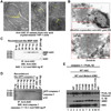
A. Morphology of anti-ASC immuno precipitated NLRP3 inflammasomes from uric acid crystal activated THP-1 cells analyzed by negative stain EM. Arrows denote filaments. B. Immuno gold EM on ultra thin cryo sections from ASCFL-eGFP transfected COS-1 cells. The ASC-containing compact structure is densely decorated by gold particles (10 nm). N, nucleus; NM, nuclear membrane. C, D. Quantification of immuno precipitated ASC-containing complex (IP) from uric acid crystal activated THP-1 cells using quantitative anti-ASC (C)and anti-caspase-1 p12 (D) Western blotting. Known amounts of recombinant His-MBP-ASC and His-GFP-caspase-1 were Western blotted to generate standard curves. The full-length caspase-1 and the cleaved p12 bands were both included in the quantification. E. AIM2 inflammasome reconstitution in HEK293T cells to define the functional consequence of structure-based mutations in AIM2. Cells were co-transfected with plasmids encoding proIL-1β and caspase-1 (lane 1), plus ASC alone (lane 2), or WT AIM2 alone (lane 11), or ASC together with WT or indicated AIM2 mutants (lanes 3 to 10). Maturation of proIL-1βinto biologically active IL-1β was detected by Western blotting using anti-IL-1β antibody (top panel).The expression levels of HA-ASC and Flag-AIM2 were detected by Western blotting using anti-HA and anti-Flag antibodies (lower panels). See also Figure S7.

Comment in

Similar articles

Cited by

References

    1. Bae JY, Park HH. Crystal structure of NALP3 protein pyrin domain (PYD) and its implications in inflammasome assembly. J Biol Chem. 2011;286:39528–39536. - PMC - PubMed
    1. Bai XC, Fernandez IS, McMullan G, Scheres SH. Ribosome structures to near-atomic resolution from thirty thousand cryo-EM particles. Elife. 2013;2:e00461. - PMC - PubMed
    1. Bammes BE, Rochat RH, Jakana J, Chen DH, Chiu W. Direct electron detection yields cryo-EM reconstructions at resolutions beyond 3/4 Nyquist frequency. J Struct Biol. 2012;177:589–601. - PMC - PubMed
    1. Chae JJ, Cho YH, Lee GS, Cheng J, Liu PP, Feigenbaum L, Katz SI, Kastner DL. Gain-of-function Pyrin mutations induce NLRP3 protein-independent interleukin-1beta activation and severe autoinflammation in mice. Immunity. 2011;34:755–768. - PMC - PubMed
    1. de Alba E. Structure and interdomain dynamics of apoptosis-associated speck-like protein containing a CARD (ASC) J Biol Chem. 2009;284:32932–32941. - PMC - PubMed

Publication types