Review
. 2014 Apr;15(4):250-63.
doi: 10.1038/nrn3708. Epub 2014 Mar 12.

Electrical synapses and their functional interactions with chemical synapses

Affiliations
Review

Electrical synapses and their functional interactions with chemical synapses

Alberto E Pereda. Nat Rev Neurosci. 2014 Apr.

Abstract

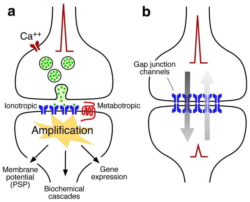
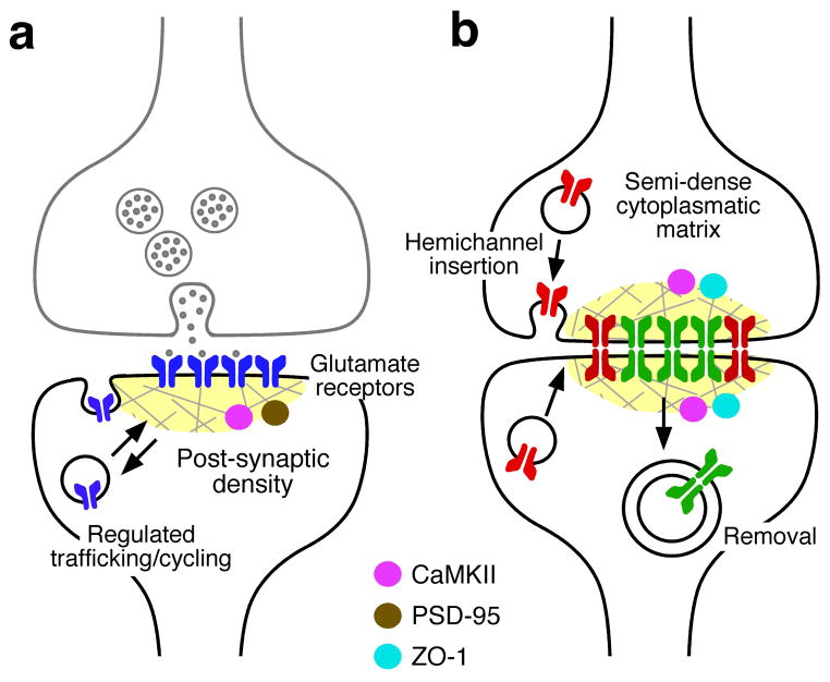
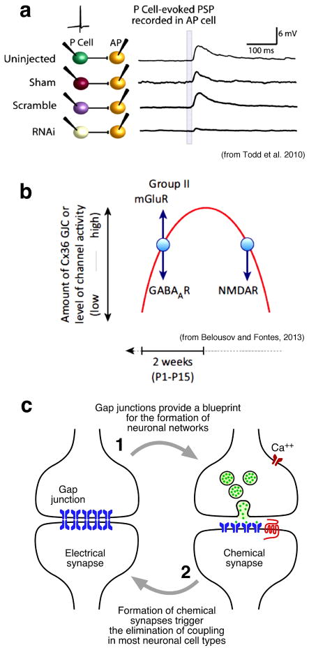
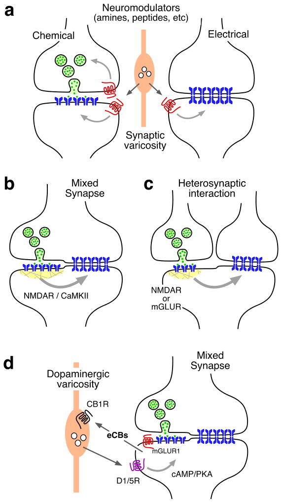
Brain function relies on the ability of neurons to communicate with each other. Interneuronal communication primarily takes place at synapses, where information from one neuron is rapidly conveyed to a second neuron. There are two main modalities of synaptic transmission: chemical and electrical. Far from functioning independently and serving unrelated functions, mounting evidence indicates that these two modalities of synaptic transmission closely interact, both during development and in the adult brain. Rather than conceiving synaptic transmission as either chemical or electrical, this article emphasizes the notion that synaptic transmission is both chemical and electrical, and that interactions between these two forms of interneuronal communication might be required for normal brain development and function.


Figures

Figure 1
Figure 1. The two main modalities of synaptic transmission
a, Chemical transmission requires sophisticated presynaptic molecular machinery that regulates neurotransmitter release in a probabilistic manner upon depolarization of the presynaptic terminal, in this case by the arrival of an action potential. A similarly complex postsynaptic molecular machinery is also required. This includes the presence of inotropic and metabotropic receptors that are capable of detecting and translating the presynaptic message (neurotransmitter) into various postsynaptic events, ranging from changes in resting potential to gene expression. b, Electrical transmission is mediated by clusters of intercellular channels called gap junctions that communicate the interior of two adjacent cells, directly allowing the bi-directional passage of electrical currents carried by ions (arrows), as well intracellular messengers and small metabolites (not illustrated here). Electrical synapses are bi-directional in nature: when a “presynaptic” action potential propagates to the “postsynaptic” cell, the membrane resting potential of the “postsynaptic” cell simultaneously propagates to the “presynaptic” cell (arrows).
Figure 2
Figure 2. Trafficking of channels at chemical and electrical synapses
a, Glutamate receptors are trafficked in and out of synapses. Postsynaptic densities provide a scaffold that helps to regulation this trafficking. PSD-95 and CaMKII are both abundant components of postsynaptic densities. Regulated trafficking of AMPA receptors (blue) is thought to underlie the modification of synaptic strength at glutamatergic synapses. b, Gap junction channels at electrical synapses turnover. New connexons are trafficked to the membrane in vesicles as unpaired hemichannels, where they are inserted at the periphery of the gap junction plaque and dock with hemichannels in the apposed membrane. They are internalized as small clusters of entire channels (green) into either of the coupled cells from regions near the center of the plaque. Proteins in the “semi dense cytoplasmatic matrix” act as scaffold. ZO-1 is a structural component whereas CaMKII seems to be a non-obligatory component of the macromolecular complex with functions that might be similar to those at postsynaptic densities of chemical synapses.
Figure 3
Figure 3. Electrical and chemical synapses interact during development
a, Blockade of electrical synapse formation in leech embryos perturbs the formation of chemical synapses. Chemical synaptic potentials in an AP cell in response to a single spike in a P cell (marked by the gray bar on the AP recordings) under control conditions (uninjected, sham, scrambled) and after injection of double-stranded RNA (RNAi) that interferes with the translation of INX-1 in embryonic P cells, at a developmental time (~50% embryonic development) at which innexin-based gap junctions are forming but chemical synapses have not yet formed. [Data from Todd et al., 2010 (figure will be redrawn by the journal’s graphic art department).] b, Effect of signaling through GABAA receptor (GABAAR), metabotropic glutamate receptor mGluR) and NMDA receptor (NMDAR) on gap junction communication during development. Red line represents the increase (upward phase) and decrease (downward phase) in the amount of neuronal gap junction communication (GJC) and expression of connexin 36 (Cx36) during development. Blue arrows show the direction of the change in gap junction communication after activation of the receptor. P1 and P15 indicate postnatal days 1 and 15. [Taken from Belousov and Fontes et al., 2013 (figure will be redrawn by the journal’s graphic art department)] c, Modalities of interactions between electrical and chemical synapses during development.
Figure 4
Figure 4. Modalities of interactions between electrical and chemical synapses in the adult nervous system
a, Neurotransmitter modulators released by nearby synaptic terminals (orange) regulate the synaptic strength of chemical and electrical synapses via activation of G-protein coupled metabotropic receptors. Regulation at chemical synapses could occur pre- or postsynaptically. b, Electrical and chemical synapses co-exist at mixed synapses. Glutamatergic synapses regulate the strength of electrical synapses via a postsynaptic mechanism that includes the activation of NMDA receptors (NMDAR) and CaMKII. c, Regulation of electrical synapses by glutamatergic transmission could also be heterosynaptic. Nearby glutamatergic synapses can regulate electrical transmission via NMDAR or mGLUR activation. d, Another mechanism of interaction at goldfish mixed synapses results when synaptic activity leads to mGluR activation, which in turn triggers endocannabinoid (eCB) release from the postsynaptic cell, and activates cannabinoid type-1 receptors (CB1Rs) on nearby dopaminergic fibers. CB1R activation leads to dopamine release that, by activating postsynaptic dopamine D1/5 receptors (D1/5R) and increasing PKA activity, is responsible for simultaneous enhancement of electrical and glutamatergic synaptic transmission.
Figure 5
Figure 5. Interactions between electrical synapses and inhibitory chemical synapses
a, Inhibitory GABAergic synapses are often located in the vicinity of gap-junctions between dendro-dendritic processes (spines) of two neurons. b, By locally increasing membrane conductance, inhibitory synaptic chloride conductances (gCl) produced by activation of GABARs briefly shunt excitatory currents to temporarily reduce effective electrical coupling between two coupled neurons.
Figure 6
Figure 6. Interactions between chemical and electrical synapses and pathological processes
a, Chemical and electrical synapses interact after injury. Release of glutamate from injured neurons causes neuronal damage via NMDAR activation and Ca++ overload at the site of the lesion. Simultaneous enhancement of coupling via mGluR-dependent increased expression of Cx36 extends neuronal damage by facilitating the passage of ‘death signals’ to neurons adjacent to the lesion or “penumbra” area (from Belousov and Fontes et al., 2013). b, Summary of potential interactions of chemical and electrical synapses during pathological processes. The lack, or dysfunction, of gap junction channels at early developmental stages might lead to defective formation of critical neural circuits formed by chemical synapses, underlying developmentally-related neurological conditions. During adulthood, release of glutamate from compromised neurons after injury or stroke enhances coupling which leads to neuronal death in uncompromised areas adjacent to the lesion. Dysregulation of electrical synapses strength by neurotransmitters might contribute to cognitive disorders.

Similar articles

Cited by

References

    1. The Synapse by Bernardo Sabatini Morgan Sheng | 9781936113026 | Hardcover | Barnes & Noble.

    1. Bennett MVL, Zukin RS. Electrical coupling and neuronal synchronization in the Mammalian brain. Neuron. 2004;41:495–511. - PubMed
    1. Zoli M, et al. The emergence of the volume transmission concept. Brain Res Brain Res Rev. 1998;26:136–47. - PubMed
    1. Faber DS, Korn H. Electrical field effects: their relevance in central neural networks. Physiol Rev. 1989;69:821–63. - PubMed
    1. Connors BW, Long MA. Electrical synapses in the mammalian brain. Annu Rev Neurosci. 2004;27:393–418. - PubMed

Publication types

LinkOut - more resources