. 2014 Apr;15(4):393-401.
doi: 10.1038/ni.2846. Epub 2014 Mar 9.

Negative regulation of Hif1a expression and TH17 differentiation by the hypoxia-regulated microRNA miR-210

Affiliations

Negative regulation of Hif1a expression and TH17 differentiation by the hypoxia-regulated microRNA miR-210

Haopeng Wang et al. Nat Immunol. 2014 Apr.

Abstract

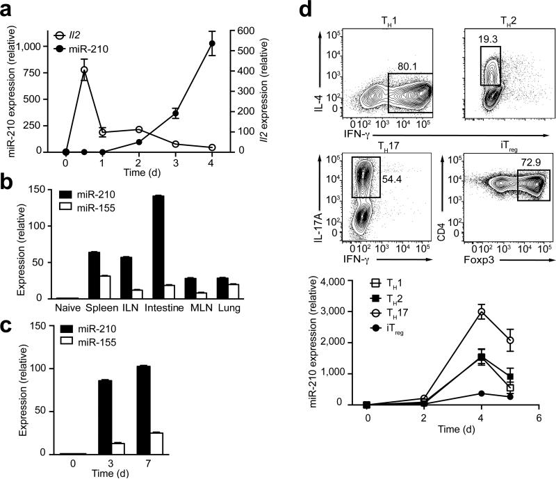
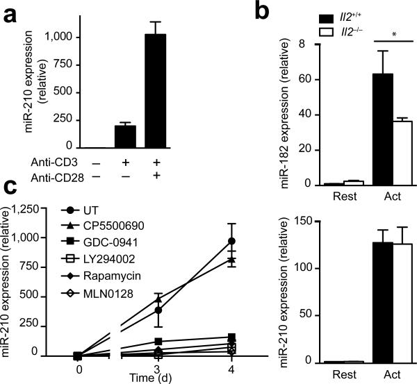
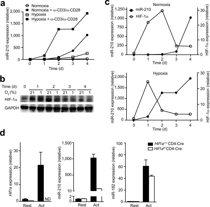
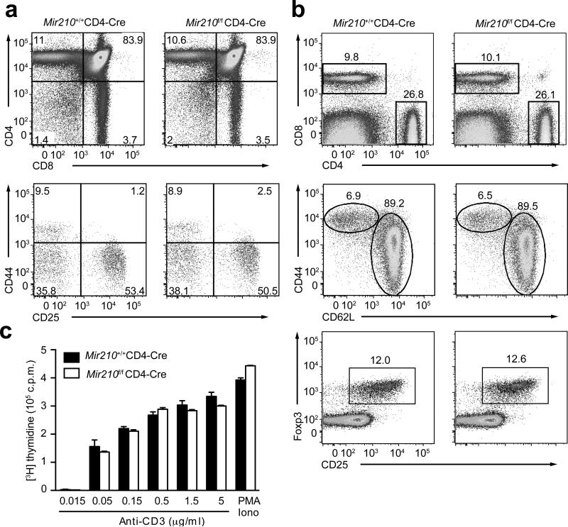
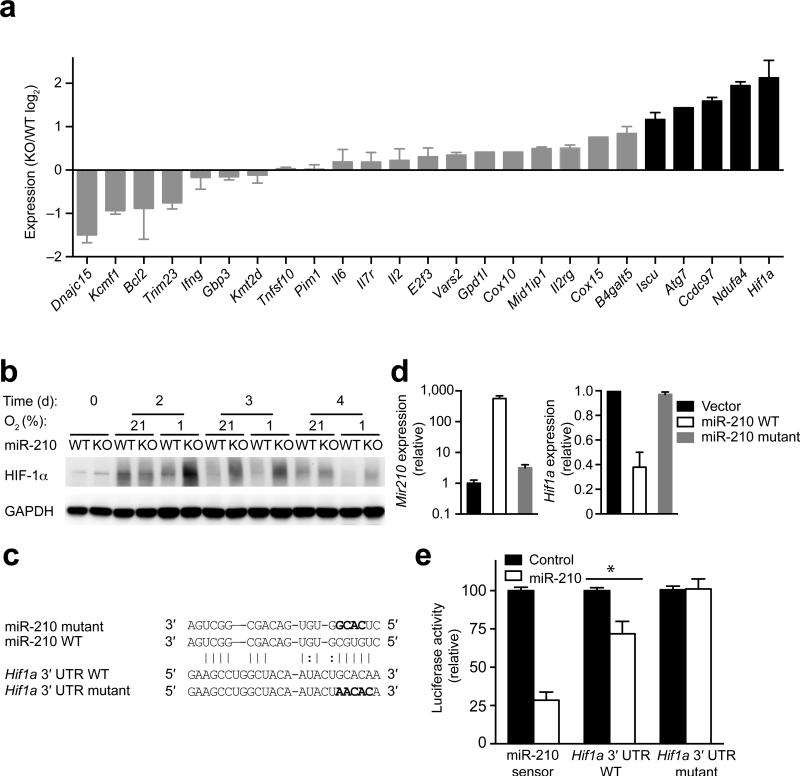
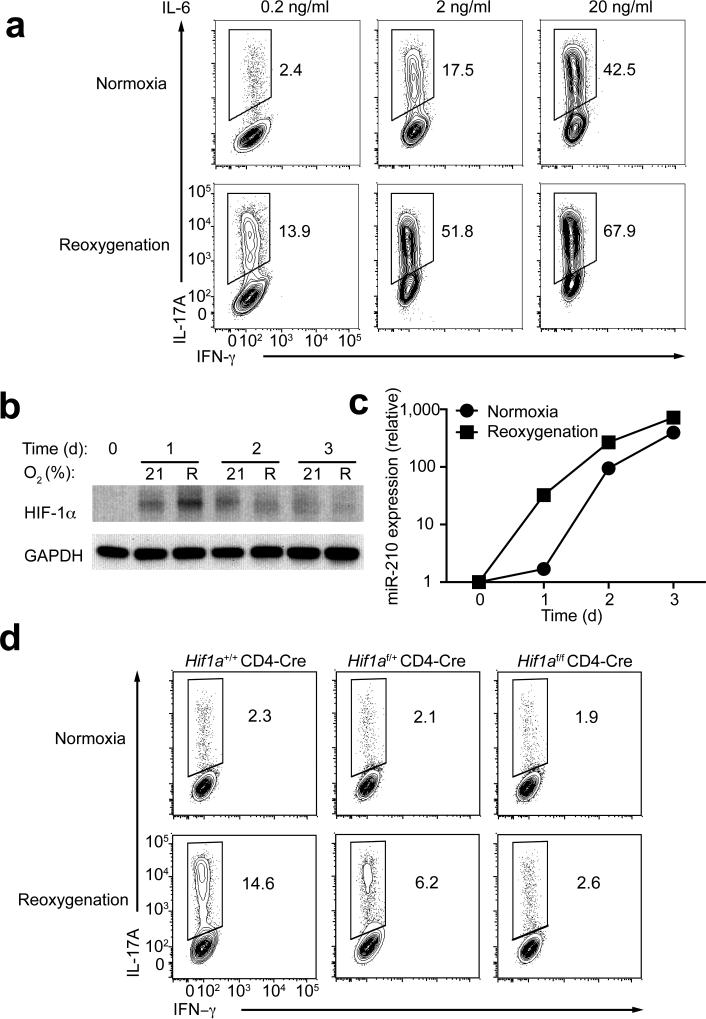
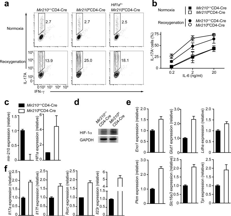
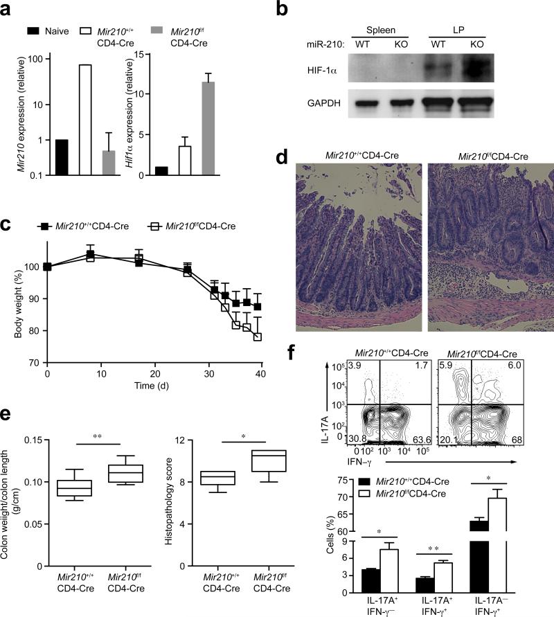
The microRNA miR-210 is a signature of hypoxia. We found robust increase in the abundance of miR-210 (>100-fold) in activated T cells, especially in the TH17 lineage of helper T cells. Hypoxia acted in synergy with stimulation via the T cell antigen receptor (TCR) and coreceptor CD28 to accelerate and increase Mir210 expression. Mir210 was directly regulated by HIF-1α, a key transcriptional regulator of TH17 polarization. Unexpectedly, we identified Hif1a as a target of miR-210, which suggested negative feedback by miR-210 in inhibiting HIF-1α expression. Deletion of Mir210 promoted TH17 differentiation under conditions of limited oxygen. In experimental colitis, miR-210 reduced the abundance of Hif1a transcripts and the proportion of cells that produced inflammatory cytokines and controlled disease severity. Our study identifies miR-210 as an important regulator of T cell differentiation in hypoxia, which can limit immunopathology.


Figures

Figure 1
Figure 1
Mir210 is induced after T cell activation and regulated during T cell differentiation. (a) The expression of miR-210 or Il2 in activated T cells was assessed by RT-PCR. The data were normalized by miR-210 expression in naive T cells (n=3 independent biological replicates per data point). (b) Homeostatically expanded CD4+ T cells were sorted from various tissues 3 weeks after adoptive transfer. The expression of miR-210 and miR-155 were assessed by RT-PCR. (c) miR-210 and miR-155 expression within in vivo activated OTII CD4+ T cells were determined by RT-PCR. (d) After 4 d polarizing naive CD4+ T cells towards the TH1, TH2, TH17 or iTreg lineage, cells with selective expression of IFN-γ, IL-4, IL-17A or Foxp3 were assessed by flow cytometry with the percentages of gated cells depicted (top). The time dependency of miR-210 expression in polarized T cells was measured by RT-PCR (bottom). Relative expression is normalized to sno202. Data are from one experiment representative of two (b,c) or three (d) independent experiments (mean and s.d. in ad).
Figure 2
Figure 2
CD28-costimulation controls Mir210 expression via the PI(3)K-mTOR pathway. (a) Naive CD4+ T cells were activated by anti-CD3 in the presence or absence of anti-CD28 for 4 d and miR-210 expression was analyzed by RT-PCR. (b) RT-PCR analysis of miR-182 and miR-210 expression in activated T cells from wild-type or Il2−/− DO11.10 Rag2−/− mice. (c) CD4+ naive T cells were stimulated in the presence of the kinase inhibitors CP550 690 (100 nM), GDC-0941 (1 μM), LY294002 (5 μM), Rapamycin (20 nM) or MLN0128 (200 nM). miR-210 expression was measured by RTPCR. Relative expression is normalized to sno202 (n=3 independent biological replicates per data point). Data are from one experiment representative of three (a) independent experiments (mean and s.d. in ac, * P<0.05, unpaired t-test in b, abbreviations: Rest, resting; Act, activated; UT, untreated).
Figure 3
Figure 3. HIF-1α is required for Mir210 induction
(a) Quantitative RT-PCR analysis of miR-210 abundance in unstimulated or anti-CD3– anti-CD28 stimulated CD4+ T cells. Cells were induced for a period of 4 d under normoxic (21% O2) or hypoxic (1% O2) conditions and RNA was harvested at the indicated time points. Relative miR-210 expression is normalized to its expression in naive T cells. (b) Immunoblot analysis with a HIF-1α-specific or GAPDH-specific antibody in total protein lysates of unstimulated or anti-CD3–anti-CD28 stimulated CD4+ T cells. Cells were induced for a period of 4 d under normoxic (21% O2) or hypoxic (1% O2) conditions and protein was harvested at the indicated time points. (c) Densitometric analysis of HIF-1α protein levels shown in (b) in comparison to miR-210 expression levels presented in (a). (d) Quantitative RT-PCR analysis of Hif1a, miR-210, and miR-182 transcripts in resting or stimulated wild-type (Hif1a+/+CD4-cre) or HIF-1α–deficient (Hif1af/fCD4-cre) CD4+ T cells. Relative expression is normalized to β2 (Hif1a) or sno202 (miR-210 and miR-182) and n=3 independent biological replicates per data point. Data are from one experiment representative of three (ac) independent experiments. (mean and s.d. in a,c,d; abbreviations: Rest, resting; Act, activated; ND not detectable).
Figure 4
Figure 4. miR-210–deficiency has no apparent effect on T cell development and proliferation
(a) Flow cytometry analysis of thymocytes (top panels) or gated double negative thymocytes (bottom panels) from 7 weeks old wild-type or Mir210–/– mice, stained with anti-CD4 and anti-CD8 or anti-CD44 and anti-CD25. Numbers adjacent to (or in) outlined areas indicate percent cells in each throughout. (b) The surface expression of CD4, CD8 (top), CD44, CD62L (middle) or CD25 and the intracellular expression of Foxp3 (bottom) in splenocytes derived from 7 weeks old wild-type or Mir210–/– mice were assessed by flow cytometry. (c) T cell proliferation was analyzed in triplicate samples by 3H-thymidine uptake 72 h after stimulation with either anti-CD3 or PMA plus ionomycin (mean and s.d.). Data are representative of three (ac) independent experiments.
Figure 5
Figure 5. miR-210 directly targets Hif1a
(a) RT-PCR analysis of the expression of predicted miR-210 target and control gene transcripts in anti-CD3–anti-CD28–stimulated wild-type or Mir210–/– CD4+ T cells. The genes with more than 2-fold upregulation were highlighted. (b) Naive CD4+ T cells from wild-type (WT) or Mir210–/– (KO) mice were activated by anti-CD3–anti-CD28 under normoxic (21% O2) or hypoxic (1% O2) conditions for 4 d and HIF-1α as well as GAPDH protein abundance was detected by immunoblot analysis. (c) Sequence alignment between miR-210 and the 3′ UTR of Hif1a. The mutated sequences in seed regions of miR-210 and Hif1a are highlighted in bold figures. (d) Mir210–/– CD4+ T cells were anti-CD3 plus anti-CD28 stimulated for 36 h, transduced with bicistronic retroviruses expressing GFP (Vector), GFP and wild-type pri-miR-210 (miR-210 WT) or GFP and mutated pri-miR-210 (miR-210 mutant) followed by RT-PCR analysis for Hif1a and miR-210 expression in GFP+, CD4+ T cells at 84 h. (e) Mir210–/– CD4+ T cells were cotransfected with luciferase reporters and miR-210 or control miRNA mimics. Luciferase activity was measured 24 h after transfection and was normalized to control transfected cells. (* P<0.05, n=4 independent biological replicates per data point t-test). Data are from one experiment representative of two (a,b) or three (d) independent experiments. (mean and s.d. in a,d,e).
Figure 6
Figure 6. Priming of naive T cells under reoxygenation increases miR-210 abundance
(a) Naive CD4+ T cells were polarized towards TH17 cells with titrated doses of IL-6 under normoxic or reoxygenation conditions (Supplementary Fig. 6a), followed by intracellular staining of IL-17A and IFN-γ. (b–c) Immunoblot analysis of HIF-1α and GAPDH and RT-PCR analysis of miR-210 in TH17 cells polarized under normoxic or reoxygenation conditions (0.2 ng/ml of IL-6). Relative miR-210 abundance was normalized to its expression in naive T cells. (d) Naive CD4+ T cells from Hif1a+/+CD4-Cre, Hif1a f/+CD4-Cre or Hif1af/fCD4-Cre mice were differentiated under TH17 skewing conditions with 0.2 ng/ml of IL-6 under normoxic or reoxygenation conditions, followed by intracellular staining of IL-17A and IFN-γ. Data are representative of two (b,c,d) or three (a) independent experiments. (mean and s.d. in c).
Figure 7
Figure 7. miR-210deficiency along with reoxygenation conditions markedly increases TH17 differentiation
(a) Naive CD4+ T cells from wild-type (Mir210+/+CD4-Cre), Mir210–/– (Mir210f/fCD4-Cre) or Hif1af/+Mir210f/fCD4-Cre mice were differentiated under TH17 skewing conditions (0.2 ng/ml of IL-6), followed by intracellular staining of IL-17A and IFN-γ. (b) Naive CD4+ T cells from wild-type (Mir210+/+CD4-Cre) or Mir210–/– (Mir210f/fCD4-Cre) mice were differentiated under TH17 skewing conditions with varying doses of IL-6 under normoxic or reoxygenetion conditions, followed by intracellular staining of IL-17A and IFN-γ. (c–d) Wild-type or Mir210–/– T cells were TH17 differentiated under reoxygenetion conditions as described in (a) for 3 d. Transcripts of miR-210 and Hif1a were detected by RT-PCR analysis; HIF-1α and GAPDH protein levels were detected by immunoblot analysis. (e–f) Quantitative RT-PCR analysis of HIF-1α target genes of the glycolytic pathway (Eno1, Glut1, Ldha, Pkm, Slc16a3 and Tpi) and TH17 signature genes (Il17a, Il17f, Rorc and Il23r) in wild-type or Mir210–/– CD4+ TH17 cells that were differentiated under reoxygenetion conditions. Relative expression is normalized to sno202 (miR-210) or β-2m (the rest of genes). Data are from one experiment representative of two (a right, d,e), three (c,f) or four (a left and middle, b) independent experiments. (mean and s.d. in b,c,e,f).
Figure 8
Figure 8. miR-210 in T cells ameliorates IBD disease in a CD4+ T cell transfer model of colitis
(a) Wild-type (Mir210+/+CD4-Cre) or Mir210–/– (Mir210f/fCD4-Cre) naive T cells were transferred into Rag2−/− recipient mice. Five weeks later, the colons from recipient mice were excised and lamina propria CD4+ T cells were isolated from the colon. Naive CD4+ T cells were included as a control. miR-210 abundance and Hif1a transcripts were analyzed by RT-PCR analysis. (b) HIF-1α as well as GAPDH protein abundance in CD4+ T cells isolated from spleen or lamina propria of Rag2−/− recipient mice described in (a) was detected by immunoblot analysis. (c) Change in body weight of Rag2−/− recipient mice given wild-type or Mir210–/– naive T cells (n=9 per group). (d) Histopathology of distal colon at 5.5 weeks after transfer. Original magnification, X100. (e) Ratio of colon weight vs colon length (n=8–9 per group) and histopathological score (n=6–8 per group) of each group are shown. (f) Expression of IL-17A and IFN-γ in CD4+ T cells isolated from lamina propria (n=3–4 mice per group). * P<0.05, ** P<0.001, unpaired t-test. Data are from one experiment representative of two (af) independent experiments.

Similar articles

Cited by

References

    1. Caldwell CC, et al. Differential effects of physiologically relevant hypoxic conditions on T lymphocyte development and effector functions. J. Immunol. 2001;167:6140–6149. - PubMed
    1. McNamee EN, Korns Johnson D, Homann D, Clambey ET. Hypoxia and hypoxia-inducible factors as regulators of T cell development, differentiation, and function. Immunol. Res. 2013;55:58–70. - PMC - PubMed
    1. Ye J. Emerging role of adipose tissue hypoxia in obesity and insulin resistance. Int. J. Obes. 2009;33:54–66. - PMC - PubMed
    1. Bedogni B, et al. The hypoxic microenvironment of the skin contributes to Akt-mediated melanocyte transformation. Cancer Cell. 2005;8:443–454. - PubMed
    1. Taylor CT, Colgan SP. Hypoxia and gastrointestinal disease. J. Mol. Med. 2007;85:1295–1300. - PubMed

Publication types

MeSH terms