

Pentosan polysulfate decreases myocardial expression of the extracellular matrix enzyme ADAMTS4 and improves cardiac function in vivo in rats subjected to pressure overload by aortic banding
- PMID: 24595230
- PMCID: PMC3940660
- DOI: 10.1371/journal.pone.0089621
Pentosan polysulfate decreases myocardial expression of the extracellular matrix enzyme ADAMTS4 and improves cardiac function in vivo in rats subjected to pressure overload by aortic banding
Abstract
Background: We hypothesized that cleavage of the extracellular matrix (ECM) proteoglycans versican and aggrecan by ADAMTS (a disintegrin and metalloprotease with thrombospondin motifs) proteases, which contributes to stress-induced ECM-reorganization in atherogenesis and osteoarthritis, also play a role in heart failure development.
Objectives: The primary objective was to identify alterations in expression of ADAMTS versicanases and aggrecanases during development of heart failure, while evaluation of the effects of in vivo modulation of relevant changes in ADAMTS activity constituted the secondary objective.
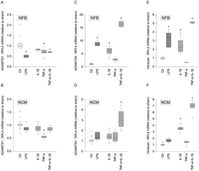
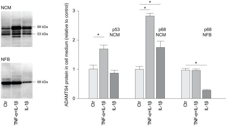
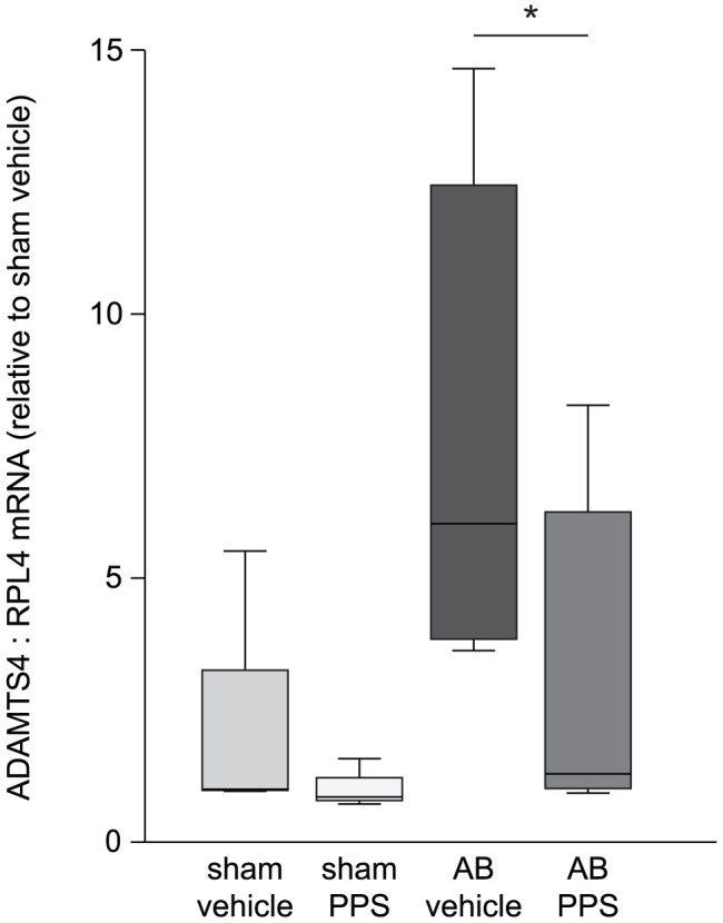
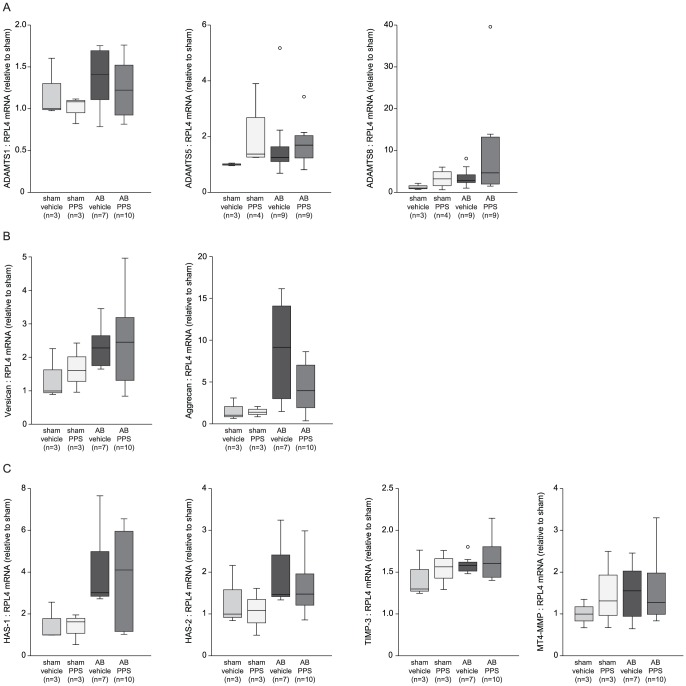
Methods: Myocardial levels of versican, aggrecan, and their ADAMTS cleaving proteases were examined in Wistar rats six weeks after aortic banding (AB), and versican and selected ADAMTS versicanases were further analyzed in neonatal cardiomyocytes (NCM) and cardiac fibroblasts (NFB) after stimulation by inflammatory mediators. Based on the initial findings, ADAMTS4 was selected the most promising therapeutic target. Thus, rats with AB were treated with pentosan polysulfate (PPS), a polysaccharide with known ADAMTS4-inhibitory properties, and effects on versican fragmentation, left ventricular function and geometry were evaluated.
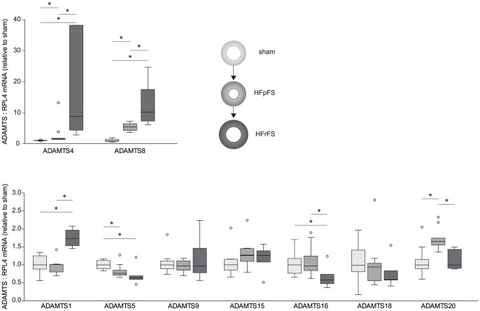
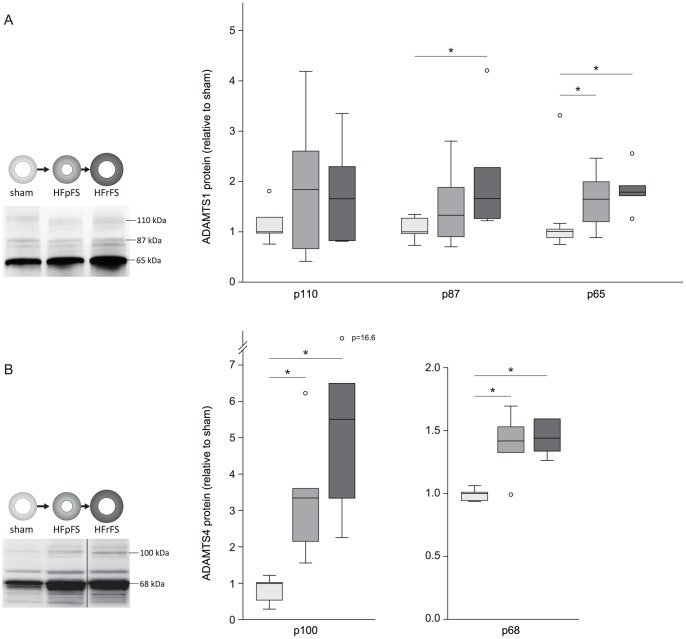
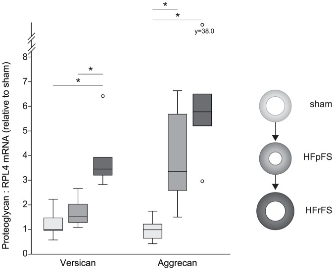
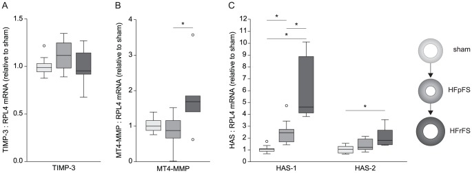
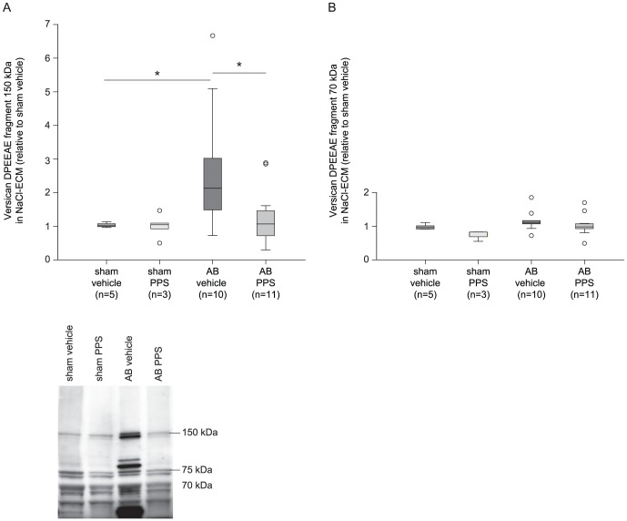
Results: We discovered that myocardial mRNA and protein levels of ADAMTS1 and -4, and mRNA levels of versican, aggrecan, and ADAMTS8 increased after AB, and TNF-α and IL-1β synergistically increased mRNA of versican and ADAMTS4 in NCM and NFB and secretion of ADAMTS4 from NCM. Furthermore, PPS-treatment improved systolic function, demonstrated by an improved fractional shortening (vehicle 48±3% versus PPS 60±1%, p<0.01) after AB. Following PPS-treatment, we observed an ∼80% reduction in myocardial ADAMTS4 mRNA (p = 0.03), and ∼50% reduction in the extracellular amount of the p150 versican fragments (p = 0.05), suggesting reduced versicanase activity.
Conclusions: Our findings suggest that AB induces an increase in myocardial ADAMTS4 versicanase activity, and that PPS-treatment improved systolic function in the pressure-overloaded heart, holding promise as a novel therapeutic agent in heart failure.
Conflict of interest statement
Figures
Similar articles
-
Distribution and processing of a disintegrin and metalloproteinase with thrombospondin motifs-4, aggrecan, versican, and hyaluronan in equine digital laminae.Am J Vet Res. 2012 Jul;73(7):1035-46. doi: 10.2460/ajvr.73.7.1035. Am J Vet Res. 2012. PMID: 22738056 Free PMC article.
-
ADAMTS4 and ADAMTS5 knockout mice are protected from versican but not aggrecan or brevican proteolysis during spinal cord injury.Biomed Res Int. 2014;2014:693746. doi: 10.1155/2014/693746. Epub 2014 Jul 3. Biomed Res Int. 2014. PMID: 25101296 Free PMC article.
-
Calcium pentosan polysulfate directly inhibits enzymatic activity of ADAMTS4 (aggrecanase-1) in osteoarthritic chondrocytes.FEBS Lett. 2008 Aug 20;582(19):2945-9. doi: 10.1016/j.febslet.2008.07.036. Epub 2008 Jul 29. FEBS Lett. 2008. PMID: 18671975
-
[Research progress of a disintegrin and metalloproteinase with thrombospondin motif 4 and 5 in osteoarthritis].Zhongguo Xiu Fu Chong Jian Wai Ke Za Zhi. 2013 Sep;27(9):1080-4. Zhongguo Xiu Fu Chong Jian Wai Ke Za Zhi. 2013. PMID: 24279019 Review. Chinese.
-
The multiple, complex roles of versican and its proteolytic turnover by ADAMTS proteases during embryogenesis.Matrix Biol. 2014 Apr;35:34-41. doi: 10.1016/j.matbio.2014.01.005. Epub 2014 Jan 18. Matrix Biol. 2014. PMID: 24444773 Free PMC article. Review.
Cited by
-
Hypochlorous Acid and Chloramines Induce Specific Fragmentation and Cross-Linking of the G1-IGD-G2 Domains of Recombinant Human Aggrecan, and Inhibit ADAMTS1 Activity.Antioxidants (Basel). 2023 Feb 8;12(2):420. doi: 10.3390/antiox12020420. Antioxidants (Basel). 2023. PMID: 36829979 Free PMC article.
-
ADAMTS proteases in cardiovascular physiology and disease.Open Biol. 2020 Dec;10(12):200333. doi: 10.1098/rsob.200333. Epub 2020 Dec 23. Open Biol. 2020. PMID: 33352066 Free PMC article. Review.
-
A Disintegrin and Metalloproteinase with Thrombospondin Motifs 4 Regulates Pulmonary Vascular Hyperpermeability through Destruction of Glycocalyx in Acute Respiratory Distress Syndrome.Int J Mol Sci. 2023 Nov 12;24(22):16230. doi: 10.3390/ijms242216230. Int J Mol Sci. 2023. PMID: 38003418 Free PMC article.
-
Cardiac Molecular Analysis Reveals Aging-Associated Metabolic Alterations Promoting Glycosaminoglycans Accumulation via Hexosamine Biosynthetic Pathway.Adv Sci (Weinh). 2024 Oct;11(38):e2309211. doi: 10.1002/advs.202309211. Epub 2024 Aug 9. Adv Sci (Weinh). 2024. PMID: 39119859 Free PMC article.
-
Hitting the Target! Challenges and Opportunities for TGF-β Inhibition for the Treatment of Cardiac fibrosis.Pharmaceuticals (Basel). 2024 Feb 20;17(3):267. doi: 10.3390/ph17030267. Pharmaceuticals (Basel). 2024. PMID: 38543053 Free PMC article. Review.
References
-
- Dorman G, Kocsis-Szommer K, Spadoni C, Ferdinandy P (2007) MMP inhibitors in cardiac diseases: an update. Recent Pat Cardiovasc Drug Discov 2: 186–194. - PubMed
-
- Hudson MP, Armstrong PW, Ruzyllo W, Brum J, Cusmano L, et al. (2006) Effects of Selective Matrix Metalloproteinase Inhibitor (PG-116800) to Prevent Ventricular Remodeling After Myocardial Infarction: Results of the PREMIER (Prevention of Myocardial Infarction Early Remodeling) Trial. Journal of the American College of Cardiology 48: 15–20. - PubMed
-
- Strand ME, Herum KM, Rana ZA, Skrbic B, Askevold ET, et al. (2013) Innate immune signaling induces expression and shedding of the heparan sulfate proteoglycan syndecan-4 in cardiac fibroblasts and myocytes, affecting inflammation in the pressure-overloaded heart. FEBS J 280: 2228–2247. - PubMed
Publication types
MeSH terms
Substances
Grants and funding
LinkOut - more resources
Full Text Sources
Other Literature Sources
Miscellaneous
