. 2014 Apr 11;289(15):10748-10760.
doi: 10.1074/jbc.M113.528380. Epub 2014 Feb 25.

Phosphoregulation of the ceramide transport protein CERT at serine 315 in the interaction with VAMP-associated protein (VAP) for inter-organelle trafficking of ceramide in mammalian cells

Affiliations

Phosphoregulation of the ceramide transport protein CERT at serine 315 in the interaction with VAMP-associated protein (VAP) for inter-organelle trafficking of ceramide in mammalian cells

Keigo Kumagai et al. J Biol Chem. .

Abstract

The ceramide transport protein CERT mediates the inter-organelle transport of ceramide for the synthesis of sphingomyelin, presumably through endoplasmic reticulum (ER)-Golgi membrane contact sites. CERT has a short peptide motif named FFAT, which associates with the ER-resident membrane protein VAP. We show that the phosphorylation of CERT at serine 315, which is adjacent to the FFAT motif, markedly enhanced the interaction of CERT with VAP. The phosphomimetic CERT S315E mutant exhibited higher activity to support the ER-to-Golgi transport of ceramide than the wild-type control in a semi-intact cell system, and this enhanced activity was abrogated when its FFAT motif was deleted. The level of phosphorylation of CERT at Ser-315 increased in HeLa cells treated with a sphingolipid biosynthesis inhibitor or exogenous sphingomyelinase. Expression of CERT S315E induced intracellular punctate structures, to which CERT and VAP were co-localized, and the occurrence of the structure was dependent on both phosphatidylinositol 4-monophosphate binding and VAP binding activities of CERT. Phosphorylation of another region (named a serine-rich motif) in CERT is known to down-regulate the activity of CERT. Analysis of various CERT mutant constructs showed that the de-phosphorylation of the serine-rich motif and the phosphorylation of Ser-315 likely have the additive contribution to enhance the activity of CERT. These results demonstrate that the phosphorylation of CERT at the FFAT motif-adjacent serine affected its affinity for VAP, which may regulate the inter-organelle trafficking of ceramide in response to the perturbation of cellular sphingomyelin and/or other sphingolipids.

Keywords: Endoplasmic Reticulum (ER); Golgi; Lipid Transport; Lipid-binding Protein; Sphingolipid.


Figures

FIGURE 1.
FIGURE 1.
Enhancement of the CERT-VAP interaction by phosphomimetic mutation at Ser-315 of CERT. A, schematic view of the structure of human CERT and the amino acid sequence containing Ser-315 near the FFAT motif (GenBankTM accession number NP_112729). The core region of the FFAT motif is highlighted by a gray box, and the asterisk represents the position of Ser-315. B, HeLa-S3 cells were transfected with an expression plasmid encoding HA-CERT WT, HA-CERT S315A, or an empty vector and cultured for 48 h before harvesting. Cell lysates were prepared with previously described lysis buffer (29) and subjected to SDS-PAGE followed by Western blotting (WB) with the anti-HA antibody (α-HA) and anti-Ser(P)-315 antibody (α-pS315). The experiments were performed at least three times, and similar results were obtained. Typical blot patterns are shown. C, amino acid sequences (312–331) around the FFAT motif of various human CERT constructs are shown. The core region of the FFAT motif is highlighted by a gray box; an asterisk represents the position of Ser-315, and the mutated residues are in bold letters. D, digitonin extracts were prepared from CHO-K1 cells co-expressing the indicated HA-CERT constructs and FLAG-VAP-A (FL-VAP-A). FLAG-VAP-A was immunoprecipitated (IP) from extracts with the anti-FLAG antibody (α-FL) and analyzed by Western blotting with the indicated antibodies. Experiments were performed at least three times, and similar results were obtained. E, Triton X-100 extracts were prepared from HeLa-S3 cells co-expressing the indicated HA-CERT constructs and FLAG-VAP-A. FLAG-VAP-A was immunoprecipitated from the extracts and analyzed by Western blotting with the indicated antibodies.
FIGURE 2.
FIGURE 2.
Phosphorylation state of wild-type CERT at Ser-315. A, Triton X-100 extracts were prepared from HeLa-S3 cells co-expressing the indicated HA-CERT constructs and FLAG-VAP-A (FL-VAP-A). HA-CERT or FLAG-VAP-A was immunoprecipitated (IP) from the extracts with the indicated antibody. The abundance of HA-CERT proteins in the immunoprecipitated fractions was quantified by Western blotting (WB) with the anti-HA antibody, and these fractions were then diluted with sample buffer to adjust the loading amounts of HA-CERT for comparisons between immunoprecipitated fractions with anti-HA and anti-FLAG antibodies. Loaded volumes of anti-HA and anti-FLAG immunoprecipitated fractions were equivalent to 0.04 and 4 μl of the original fraction, respectively. CERT was detected by Western blotting with anti-HA antibody or anti-Ser(P)-315 antiserum. Experiments were performed at least twice, and similar results were obtained. B, HeLa-S3 cells stably expressing HA-CERT WT were treated with 5 μm ISP-1, mock-treated (dimethyl sulfoxide, DMSO), or nontreated (NT) for 24 h in serum-free DMEM. Triton X-100 extracts were prepared from the cells, and HA-CERT WT was immunoprecipitated from extracts with the anti-HA antibody, and precipitated CERT was detected by Western blotting with the anti-HA antibody and anti-Ser(P)-315 antiserum. Experiments were performed at least twice, and similar results were obtained. C, HeLa-S3 cells stably expressing HA-CERT WT were treated with 30 milliunits/ml B. cereus sphingomyelinase (bSMase), mock-treated (40% glycerol), or nontreated (NT) for 2 h in serum-free DMEM. HA-CERT WT was immunoprecipitated and analyzed by Western blotting as described in B. Experiments were performed twice, and similar results were obtained.
FIGURE 3.
FIGURE 3.
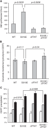
Effects of the phosphomimetic mutation of CERT at Ser-315 on CERT functions. A, CERT WT and CERT mutants expressed in E. coli BL21 (DE3) cells were purified. The purified CERT proteins (20 fmol) were used to measure the activity of mediating the ER-to-Golgi trafficking of ceramide in semi-intact LY-A cells as described previously (18). The data shown are the means ± S.D. from three experiments. Significance of changes was analyzed by Student's t test, and p values are shown. B, inter-membrane ceramide transfer activity of purified CERT proteins was measured in a cell-free system using artificial phospholipid vesicles. The data shown are the means ± S.D. from three experiments. Significance of changes was analyzed by Student's t test, and p values are shown. C, purified CERT proteins were examined on their binding to liposomes containing various amounts of PtdIns(4)P. The percentage of the amount of co-precipitated CERT relative to the total was estimated. Without PtdIns(4)P is shown by white bars; 0.1% PtdIns(4)P is shown by light gray bars; and 0.3% PtdIns(4)P is shown by dark gray bars. The data shown are the means ± S.D. from three experiments. Significance of changes was analyzed by Student's t test, and p values are shown.
FIGURE 4.
FIGURE 4.
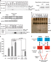
Functional interactions between the SRM and Ser(P)-315 in CERT. A, HeLa-S3 cells were transfected with an expression plasmid encoding HA-CERT WT, HA-CERT S315A, or an empty vector and cultured for 48 h before harvesting. Cell lysates were prepared by sonication with buffer A supplemented with 0.2% SDS and subjected to SDS-PAGE followed by Western blotting (WB) with the anti-HA antibody (α-HA) and anti-Ser(P)-315 antibody (α-pS315). The area enclosed with dotted line (CERT WT and CERT S315E) is enlarged in the right panel to show the doublet appearance of CERT more clearly. The upper band represents the hyper-phosphorylated form, and the lower band represents the hypo-phosphorylated or de-phosphorylated form of CERT at the SRM (14, 31). Experiments were performed at least three times, and similar results were obtained. Typical blot patterns are shown. IP, immunoprecipitated. B, amino acid sequences (312–335) around the FFAT motif of various human CERT constructs are shown. The core region of the FFAT motif is enclosed by a dotted line, and the asterisk represents the position of Ser-315, and the mutated residues are in bold letters. C, HA-tagged CERT WT and mutated CERT transiently expressed in HeLa-S3 cells were purified, subjected to SDS-PAGE, and analyzed by silver staining. CERT (70 kDa) and the putative endogenous VAP protein (marked by Band X) are indicated. The ladder bands in the high molecular range are most likely oligomers of CERT (41). D, purified proteins described in C were analyzed by Western blotting with the anti-HA antibody (α-HA) and anti-VAP-A antibody (α-pS315). E, purified CERT proteins (20 fmol) described in C were used to measure the ER-to-Golgi trafficking activity of ceramide in semi-intact LY-A cells as described previously (18). The data shown are the means ± S.D. from three experiments. The significance of changes was analyzed by the Student's t test, and p values are shown. F, hypothetical model of the functional regulation of CERT by the phosphorylation of the SRM and Ser-315 is depicted as follows: phosphorylated state (P), non-phosphorylated state (non-P), less-active form (blue boxes), and active form (red boxes). Typical mutations mimicking these states are shown in parentheses. The phosphorylation states of one region may mutually affect those of the other region. The de-phosphorylation of the SRM and the phosphorylation of Ser-315 additively enhance the activity of CERT to transport ceramide from the ER to the Golgi apparatus for the synthesis of SM.
FIGURE 5.
FIGURE 5.
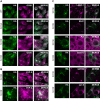
Effects of Ser-315 phosphorylation on the intracellular distribution of CERT. A, HeLa-S3 cells stably expressing HA-CERT WT or HA-CERT mutations were double-labeled with the anti-HA antibody (green) and anti-GS28 antibody (magenta) by indirect immunostaining and were then observed by confocal microscopy. Experiments were performed at least three times, and similar results were obtained. Typical staining patterns are shown. B, HeLa-S3 cells stably expressing HA-CERT S315E were double-labeled with the anti-HA antibody (green) and anti-VAP-A antibody (magenta) by indirect immunostaining and observed by confocal microscopy (upper panel). A high magnification of the selected region (boxes with dotted line in upper panel) is also shown (lower panel). C, HeLa-S3 cells stably expressing HA-CERT S315E were double-labeled with the anti-HA antibody (green) and the indicated antibodies (magenta) by indirect immunostaining and observed by confocal microscopy. The antibodies used were as follows: a late endosomal/lysosomal marker, LAMP-1; an early endosomal marker, EEA1; an ER marker, protein-disulfide isomerase, PDI; a membrane-associated ER marker, Sec61β; a Golgi marker, GM130; a trans-Golgi network marker, syntaxin-6 (syn-6). The bar indicates 10 μm.
FIGURE 6.
FIGURE 6.
Dependence of the punctate formation of CERT S315E on its PtdIns(4)P binding and VAP binding activities. A, HeLa-S3 cells were transiently transfected with HA-CERT S315E, HA-CERT S315E/G67E, and HA-CERT S315E/ΔFFAT constructs and were then cultured for 24 h. Cells were double-labeled with the anti-HA antibody (green) and anti-VAP-A antibody (magenta) by indirect immunostaining and observed by confocal microscopy. The bars indicate 10 μm. B, HeLa-S3 cells stably expressing HA-CERT S315E were transfected with siRNA against VAP-A and incubated for 24 h. The cells were double-labeled with the anti-HA antibody (green) and anti-VAP-A antibody (magenta) by indirect immunostaining and observed by confocal laser-scanning microscope (Axio Observer Z1; Carl Zeiss) equipped with an LSM 700 ZEN system (Carl Zeiss) using a Plan-Apochromat 63×/1.4 Oil differential interference contrast objective lens. The bar indicates 10 μm.

Similar articles

Cited by

References

    1. Pewzner-Jung Y., Ben-Dor S., Futerman A. H. (2006) When do Lasses (longevity assurance genes) become CerS (ceramide synthases)? Insights into the regulation of ceramide synthesis. J. Biol. Chem. 281, 25001–25005 - PubMed
    1. Mullen T. D., Hannun Y. A., Obeid L. M. (2012) Ceramide synthases at the centre of sphingolipid metabolism and biology. Biochem. J. 441, 789–802 - PMC - PubMed
    1. Hanada K. (2013) Co-evolution of sphingomyelin and the ceramide transport protein CERT. Biochim. Biophys. Acta, in press, 10.1016/j.bbalip/2013.06.006 - DOI - PubMed
    1. van Meer G., Voelker D. R., Feigenson G. W. (2008) Membrane lipids: Where they are and how they behave. Nat. Rev. Mol. Cell Biol. 9, 112–124 - PMC - PubMed
    1. Holthuis J. C., Levine T. P. (2005) Lipid traffic: Floppy drives and a superhighway. Nat. Rev. Mol. Cell Biol. 6, 209–220 - PubMed

Publication types

MeSH terms

LinkOut - more resources