. 2014 Feb 15;28(4):328-41.
doi: 10.1101/gad.233247.113.

SOX9: a stem cell transcriptional regulator of secreted niche signaling factors

Affiliations

SOX9: a stem cell transcriptional regulator of secreted niche signaling factors

Meelis Kadaja et al. Genes Dev. .

Abstract

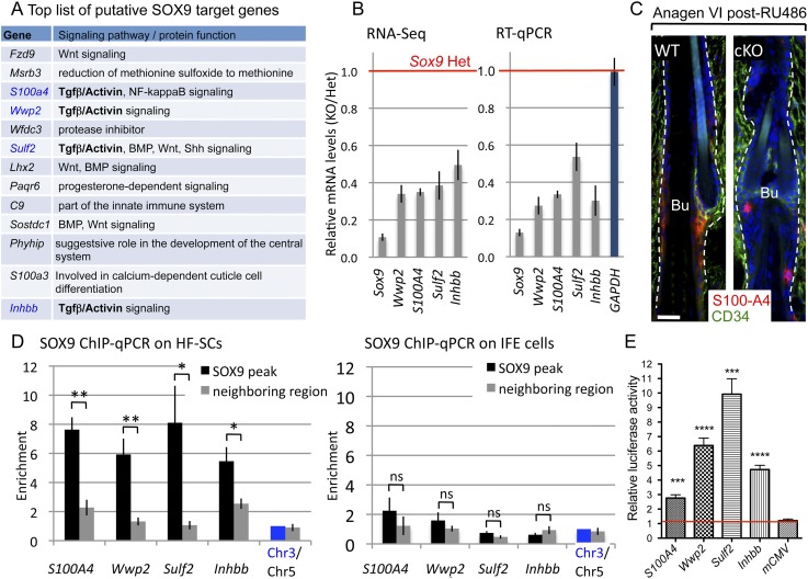
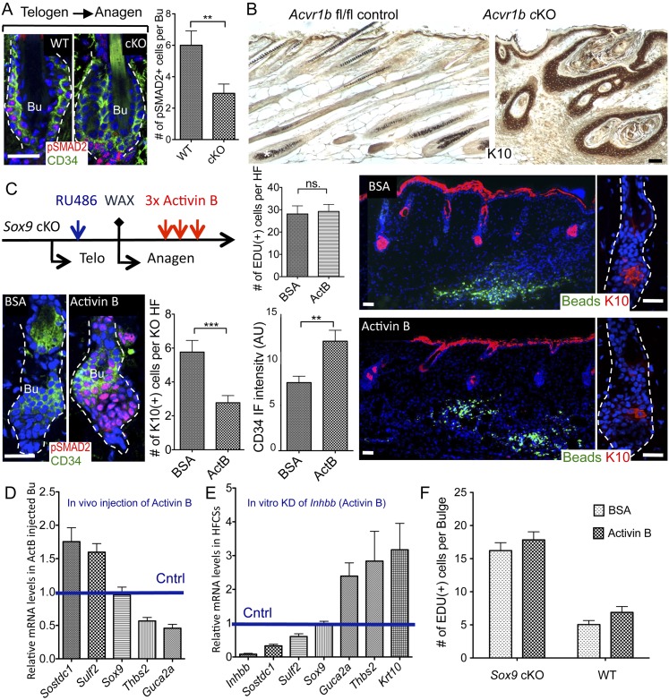
Hair follicles (HFs) undergo cyclical periods of growth, which are fueled by stem cells (SCs) at the base of the resting follicle. HF-SC formation occurs during HF development and requires transcription factor SOX9. Whether and how SOX9 functions in HF-SC maintenance remain unknown. By conditionally targeting Sox9 in adult HF-SCs, we show that SOX9 is essential for maintaining them. SOX9-deficient HF-SCs still transition from quiescence to proliferation and launch the subsequent hair cycle. However, once activated, bulge HF-SCs begin to differentiate into epidermal cells, which naturally lack SOX9. In addition, as HF-SC numbers dwindle, outer root sheath production is not sustained, and HF downgrowth arrests prematurely. Probing the mechanism, we used RNA sequencing (RNA-seq) to identify SOX9-dependent transcriptional changes and chromatin immunoprecipitation (ChIP) and deep sequencing (ChIP-seq) to identify SOX9-bound genes in HF-SCs. Intriguingly, a large cohort of SOX9-sensitive targets encode extracellular factors, most notably enhancers of Activin/pSMAD2 signaling. Moreover, compromising Activin signaling recapitulates SOX9-dependent defects, and Activin partially rescues them. Overall, our findings reveal roles for SOX9 in regulating adult HF-SC maintenance and suppressing epidermal differentiation in the niche. In addition, our studies expose a role for SCs in coordinating their own behavior in part through non-cell-autonomous signaling within the niche.

Keywords: Activin; SOX9; hair follicle; skin; stem cells.


Figures

Figure 1.
Figure 1.
Loss of SOX9 in adult HF-SCs causes deficient hair coat recovery. (A) Immunofluorescence of SOX9 and CD34 in the telogen HFs. (B) Strategy to knock out Sox9 in adult HF-SCs. (C) YFP and SOX9 immunofluorescence in telogen cKO HFs 2 wk after targeting. SOX9 is gone in HF-SCs, but K6+ inner bulge cells and YFP cells in the CD34 layer (arrows) still express SOX9. (D) Hair coat recovery of Sox9 wild-type (WT), Het, and cKO mice after RU486 treatment at the beginning of second telogen (postnatal days 60–74 [P60–P74]). Untreated Sox9fl/fl × K15-CrePGR × R26YFP mice were also used as a control. Bars, 30 μm. The white dashed line denotes the epidermal–dermal border, and the blue channel is DAPI staining. (Ana) Anagen, (Bu) bulge, (Telo) telogen.
Figure 2.
Figure 2.
Sox9 ablation in adult HF-SCs results in bulge deformation and an anagen-dependent failure to return to quiescence. (A) Semithin sections of Sox9 wild-type (WT) and cKO HFs in the first anagen phase of the hair cycle following RU486 treatment in previous telogen. Note that (1) cKO HFs are shorter, and (2) IRS/hair shaft perturbations are subsequent to those of the bulge. (B) Anti-SOX9 immunofluorescence. (C) EdU incorporation (24-h pulse) in HF-SCs in first (full) Anagen VI following RU486. (D,E) K6 in the companion layer (Cp), hair keratins (AE13) in the hair shaft (HS) cortex and cuticle, and trichohyalin (AE15) in the IRS and HS medulla are still expressed in Sox9 cKO HFs. (F) Quantifications of EdU incorporation and total matrix cell number of Anagen VI HFs post-RU486. N = 3 mice for each genotype. (G) Quantification of activated caspase 3 in the matrix of Anagen VI HFs post-RU486. N = 3 mice for each genotype. (H) Quantifications of EdU incorporation in the bulge at the indicated time points. N = 4 mice for each genotype. Data are mean ± SEM. (****) P < 0.0001. Bars, 30 μm. White dashed lines denote the epidermal–dermal border, and the blue channel is DAPI staining. Asterisk indicates the nonspecific staining of SOX9 Ab. (Amp) Arrector pili muscle; (Bu) bulge; (DP) dermal papilla; (IRS) inner root sheath; (ORS) outer root sheath; (SG) sebaceous gland; (Mx) matrix; (ns) not significant.
Figure 3.
Figure 3.
Sox9 cKO HF-SCs differentiate into IFE cells. (A) Ultrastructure of the SC niche of wild-type (WT) and Sox9 cKO bulge of Anagen VI HFs after RU486 treatment. The new bulge (Bu), deformed at this stage, is marked by red pseudocolor; dark brown labels the old bulge, and light brown indicates the sebaceous gland. The boxed area, magnified at right, reveals keratinized pearls and keratohyalin granules (arrows) within the cKO bulge. (B) K10. (C) Immunostaining for filaggrin (FIL). K10 reveals atypical labeling of epidermal markers in the cKO bulge. (D) Immunolabeling shows that the K6+ inner bulge layer is not targeted and remains intact during the first anagen following Sox9 ablation in HF-SCs. Dashed lines denote the epidermal–dermal border. (E) Representative images of K10 immunostainings of telogen end-phase HFs treated with RU486 3 mo earlier at telogen start. Quantifications are at right. N = 3 mice for each genotype. Data are mean ± SEM. (****) P < 0.0001. (F) Depletion of SOX9 in adult HF-SCs leads eventually to the formation of K10-positive cysts. Blue channel is DAPI staining. (Cntrl) Control. Bars, 30 μm.
Figure 4.
Figure 4.
Changes in gene expression in HF-SCs lacking SOX9. (A) Transcriptional profile of SOX9-deficient HF-SCs is shifted away from the normal HF-SC “signature” toward the basal IFE “signature.” The top diagram shows overlap of genes differentially expressed by ≥1.5× in Sox9 cKO versus Het HF-SCs (Sox9 RNA-seq), and genes were enriched ≥2× in telogen HF-SCs versus basal IFE (HF-SC signature genes). The bottom diagram shows overlap of Sox9 RNA-seq and genes enriched ≥2× in IFE versus telogen HF-SCs (IFE signature genes). (B) Table shows examples of genes ≥1.5-fold down-regulated (green) or ≥1.5-fold up-regulated (red) in Sox9 RNA-seq. (C) Gene ontology (GO) analyses of genes differentially expressed by ≥1.5× or ≥2× in Sox9 cKO versus Het HF-SCs denotes the “extracellular region” category as the most significantly enriched compared with the 7.6% frequency in the Mouse Genome Informatics (MGI) database. (D) K10 and YFP immunofluorescence of telogen Sox9 cKO bulge prior to hair cycling. (Blue) DAPI. At right, the percentage of K10+ cells within YFP+ and YFP HF-SCs of the mosaic Sox9 cKO bulge (YFP is in 60%–80% of HF-SCs) and wild-type (WT) bulge. N = 3 mice for each genotype. Data are mean ± SEM. (****) P < 0.0001. Bar, 30 μm.
Figure 5.
Figure 5.
SOX9 functions primarily as a transcriptional activator in adult HF-SCs. (A) Distribution of SOX9 peaks between promoters (−2 kb to +2 kb of transcription start sites) and “enhancers” (−50 kb of transcription start sites, gene body, +5 kb downstream from the gene) of SOX9-bound genes. (B) Diagram showing overlap of in vivo SOX9-bound targets (ChIP-seq) and mRNAs differentially expressed (RNA-seq) in Sox9 cKO versus Het HF-SCs. Overlaps represent the number of genes bound by SOX9 and whose expression is up-regulated or down-regulated by ≥1.5-fold in Sox9 cKO. Overlaps with HF-SC signature and IFE signature genes are also shown. (C) ChIP-seq profiles of the S100A4 genomic locus in wild-type (WT) telogen HF-SCs. Input represents the sequencing profile of genomic DNA used in ChIP pull-downs. Black arrows indicate locations of DNA fragments enriched in SOX9 ChIP. The SOX9 ChIP-seq profile is also compared with published data of histone modifications in telogen HF-SCs (Lien et al. 2011): H3K4me3 Ab pulls down promoter regions, H3K79me2 marks actively transcribed DNA, and the H3K27me3 label is enriched in polycomb-repressed chromatin. Below is an RNA-seq profile of the S100A4 locus in Sox9 cKO and Het HF-SCs. The locations of RNA-seq fragments match with exons in the S100A4 gene. All tracks are set to the same scale. The lengths of genes and direction of transcription are indicated by blue arrows. (D) SOX9-binding motifs (from left): the most enriched motif within SOX9 peaks in HF-SCs, the motif enriched most within SOX9 peaks associated with differentially expressed genes in Sox9 cKO versus Het, and the previously reported SOX9 in vitro motif (Mertin et al. 1999). Below, relative luciferase activities driven by SOX9 motifs were used as an enhancer. Assays were performed in 293FT cells (SOX9-negative) in the presence of a SOX9 expression vector or empty vector. All results were normalized to luciferase activity when firefly luciferase reporter with minimal CMV was used and to the Renilla luciferase control reporter driven by the HSV TK promoter. Experiments for each construct were performed in triplicate in duplicate wells. Data are mean ± SEM. (ns) Not significant; (*) P < 0.05.
Figure 6.
Figure 6.
SOX9 is a positive regulator of genes involved in Activin/TGFβ signaling. (A) Top list of putative SOX9 targets that were highly expressed in adult HF-SCs (FPKM >5 in Het RNA-seq), strongly influenced by SOX9 loss (down-regulated by twofold or more in cKO versus Het), and associated with a strong SOX9 peak in ChIP-seq (peak height ≥13). (B) RT-qPCR verification of RNA-seq data. Independent FACS-purified samples were analyzed for putative SOX9 target genes involved in TGFβ/Activin signaling. For RT-qPCR, all results were normalized to Ppib expression; GAPDH was used as an internal control. Data are mean ± SEM. N = 4 mice per genotype. (C) S100A4 immunofluorescence in Sox9 cKO and wild-type (WT) HFs in the first full anagen after targeting. White lines denote the epidermal–dermal border. (Blue) DAPI. Bar, 30 μm. (D) Independent SOX9 ChIP-qPCR on FACS-purified HF-SCs (SOX9-positive) and IFE (SOX9-negative) cells. For each gene, primers were designed to amplify region where the SOX9 peak was detected (black bars) and neighbor regions 4–8 kb away from the SOX9 peak (gray bars). Note that enrichment of DNA fragments was specific to HF-SCs and regions underneath the peaks, validating the specificity of SOX9 Ab and binding. Results were normalized to the intergenic Chr3 region; the intergenic Chr5 region was used as a control. Experiments were done in triplicate. Data are mean ± SEM. (*) P < 0.05; (**) P < 0.01. (E) Relative luciferase activities driven by sequences (300–600 bp) encompassing SOX9 ChIP-seq binding sites. Results are presented as the enhancement of luciferase activity in the presence of a SOX9 expression vector compared with an empty expression vector. All results are normalized to the luciferase activity of Renilla luciferase control reporter vector. Experiments for each construct were performed in four replicates in duplicate wells. Data are mean ± SEM. (***) P < 0.001; (****) P < 0.0001.
Figure 7.
Figure 7.
SOX9 maintains the identity of HF-SCs through Activin signaling. (A) pSMAD2 immunofluorescence of HFs during the first telogen–anagen transition after RU486. Quantifications are at right. N = 3 mice per genotype. Data are mean ± SEM. (**) P < 0.01. (B) K10 immunohistochemistry of cKO Acvr1b mice and fl/fl control (wild-type [WT]) mice. Asterisk indicates melanin. (C) Intradermally injected Activin B inhibits the trans-differentiation of HF-SCs to IFE caused by SOX9 loss. Mice treated with RU486 in second telogen were depilated to synchronize hair cycles and expedite the phenotype. Recombinant Activin B and BSA were injected intradermally into the same mouse at two different locations over the 3 d. Immunostaining for pSMAD2 and CD34 in Activin B and BSA-treated HFs are shown at left; K10 is shown at right. Quantifications of K10+ cells, EdU+ cells per HF, and the total fluorescence intensity of the CD34 label are shown in the middle. N = 4 mice. Data are mean ± SEM. (**) P < 0.01; (***) P < 0.001. White lines denote the epidermal–dermal border. (Blue) DAPI. Bar, 30 μm. (D) RT-qPCR quantification of mRNA levels of secreted factors in Activin B-treated bulge. HF-SCs were FACS-purified from Activin B and BSA injection sites independently from two Sox9 cKO mice. Data are mean ± SEM. (E) RT-qPCR quantification of mRNA levels of secreted factors in cultured wild-type HF-SCs, where Activin B. expression has been reduced by Inhbb shRNA. Data are mean ± SEM based on four independent experiments. (F) Quantifications of EdU+ cells in wild-type and Sox9 cKO bulge at Anagen IV, after treatment with BSA or Activin B. N = 2 mice. Data are mean ± SEM.

Similar articles

Cited by

References

    1. Akiyama H, Chaboissier M-C, Martin JF, Schedl A, de Crombrugghe B 2002. The transcription factor Sox9 has essential roles in successive steps of the chondrocyte differentiation pathway and is required for expression of Sox5 and Sox6. Genes Dev 16: 2813–2828 - PMC - PubMed
    1. Bailey TL, Boden M, Buske FA, Frith M, Grant CE, Clementi L, Ren J, Li WW, Noble WS 2009. MEME suite: Tools for motif discovery and searching. Nucleic Acids Res 37: W202–W208. - PMC - PubMed
    1. Bhandari RK, Haque MM, Skinner MK 2012. Global genome analysis of the downstream binding targets of testis determining factor SRY and SOX9. PLoS ONE 7: e43380. - PMC - PubMed
    1. Blanpain C, Lowry WE, Geoghegan A, Polak L, Fuchs E 2004. Self-renewal, multipotency, and the existence of two cell populations within an epithelial stem cell niche. Cell 118: 635–648 - PubMed
    1. Botchkarev VA, Botchkareva NV, Roth W, Nakamura M, Chen LH, Herzog W, Lindner G, McMahon JA, Peters C, Lauster R, et al. 1999. Noggin is a mesenchymally derived stimulator of hair-follicle induction. Nat Cell Biol 1: 158–164 - PubMed

Publication types