. 2014 Feb 13;156(4):678-90.
doi: 10.1016/j.cell.2014.01.009.

Erk1/2 activity promotes chromatin features and RNAPII phosphorylation at developmental promoters in mouse ESCs

Affiliations

Erk1/2 activity promotes chromatin features and RNAPII phosphorylation at developmental promoters in mouse ESCs

Wee-Wei Tee et al. Cell. .

Abstract

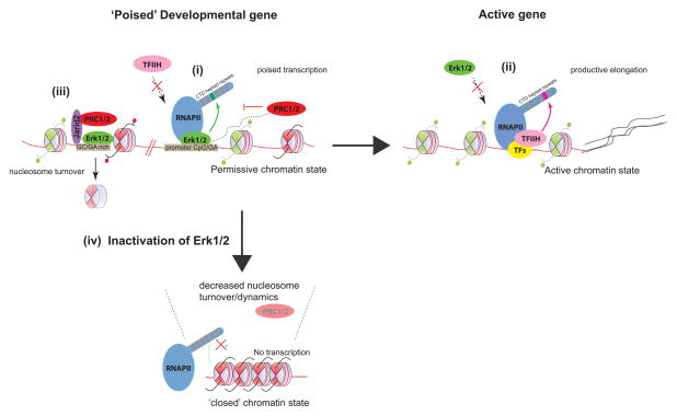
Erk1/2 activation contributes to mouse ES cell pluripotency. We found a direct role of Erk1/2 in modulating chromatin features required for regulated developmental gene expression. Erk2 binds to specific DNA sequence motifs typically accessed by Jarid2 and PRC2. Negating Erk1/2 activation leads to increased nucleosome occupancy and decreased occupancy of PRC2 and poised RNAPII at Erk2-PRC2-targeted developmental genes. Surprisingly, Erk2-PRC2-targeted genes are specifically devoid of TFIIH, known to phosphorylate RNA polymerase II (RNAPII) at serine-5, giving rise to its initiated form. Erk2 interacts with and phosphorylates RNAPII at its serine 5 residue, which is consistent with the presence of poised RNAPII as a function of Erk1/2 activation. These findings underscore a key role for Erk1/2 activation in promoting the primed status of developmental genes in mouse ES cells and suggest that the transcription complex at developmental genes is different than the complexes formed at other genes, offering alternative pathways of regulation.


Figures

Figure 1
Figure 1. Loss of PRC2 Components Abrogates Erk1/2 Activation
(A) Western blot comparing phospho-Erk1/2 (pErk1/2) and phospho-MEK1/2 (pMEK1/2) levels in Eed null ESCs and in Jarid2 stable knockdown (KD) ESCs. (B) Western blot comparing pErk1/2 and pMEK1/2 as a function of time of Ezh2 knockdown.
Figure 2
Figure 2. Erk1/2 Regulates PRC2 Occupancy on Developmental Genes in ESCs
(A) Western blot for Erk1/2 and pErk1/2 in ESC lines developed toward the generation of Erk1/2 mutant ESCs. 1 – 6 denotes different shRNA constructs against Erk1. (B) Heatmap representations of normalized read density of H3K27me3, H3K4me3, Jarid2 and total RNAPII corresponding to ~ 29000 annotated TSS (UCSC database) in wild type (WT) and Erk1/2 mutant ESCs. The normalized reads coverage against library size was calculated in the distance of ±10 kb to TSS with 200 bp bin. Heatmaps were ranked according to H3K27me3 enrichment in WT ESCs. Color scale represents normalized read density. (C) Representative ChIP-seq tracks for H3K27me3, H3K4me3, Jarid2 and RNAPII on the HoxA gene cluster in WT and Erk1/2 mutant ESCs. The X-axis corresponds to genomic location, and the Y-axis to normalized ChIP-seq signal density. (D) Generation of Erk2 rescue ESCs in the Erk1/2 mutant ESCs. Exogenous Erk2 is fused to an HA epitope tag and a downstream IRES-GFP. Western blots showing the expression of Erk2 (top panel) and HA-Erk2 (middle panel) in the respective cell lines. (E) Average H3K27me3 profile for WT, Erk1/2 mutant and Erk2-rescue ESCs, centered on TSS. The average reads coverage of ~29000 TSS regions was calculated and normalized by total mapped reads. Histogram window size is ±3 kb with 10 bp bin size. (F) ChIP-QPCR for Ezh2 and Jarid2 in WT, Erk1/2 mutant and Erk2-rescue ESCs. Results are presented as a percentage of input material. Bar represents mean of 2 replicates. See also Figures S1, S2 and Table S1.
Figure 3
Figure 3. Erk2 Binds to Polycomb-regulated developmental genes
(A) ChIP-seq gene tracks showing occupancy of Erk2 on PRC2 target genes. The X-axis corresponds to genomic location, and the Y-axis corresponds to normalized ChIP-seq signal density. (B) Average Erk2 profile for WT and MEK1/2 inhibited (PD0325901 and ‘2i’) ESCs, centered on TSS. The average reads coverage of ~29000 TSS regions was calculated and normalized by total mapped reads. Histogram window size is ±10 kb with 10 bp bin size. (C) Heatmaps of H3K27me3 and Jarid2 normalized reads distribution on all 2296 Erk2 enriched regions. H3K27me3 and Jarid2 are present in the majority of Erk2 enriched regions in wild type (WT) ESCs, but are depleted in Erk1/2 mutant cells. Data matrix was constructed with Erk2 peak regions centered by peak summit with ±5 kb extension on both sides and a bin size of 50 bp, as described in Extended Experimental Procedures. (D) Venn diagram of the overlap among target genes of Erk2, Jarid2 and H3K27me3 in WT ESCs. In this case, only respective target genes with at least one peak within 3 kb of the TSS were considered. A smaller list comprising Jarid2 and H3K27me3 target genes was generated by first annotating Jarid2 and H3K27me3 peaks to Erk2, as described in Extended Experimental Procedures. (E) GO term analysis of all Erk2 targets. (F) Motif analysis of all Erk2 enriched regions using MEME. Two distinct motifs were identified (GA- and GC-rich). (G) Left, Gel shift assay comparing recombinant Erk2, either wild type (WT), mutant in DNA-binding activity (DBM) or kinase activity (KDM) in the presence of probe containing a biotinylated GA-motif derived from the Pou3f2 locus. Top Right, Gel shift titrations and binding curve of WT- and KDM- Erk2. Increasing amounts of Erk2 proteins were used (0 – 10 μg) in the presence of the Pou3f2 GA-rich DNA probe. Bottom Right, Graph showing the effects of increasing amounts of different cold probes (0 – 250x) in affecting the binding between wild type Erk2 and the GA-rich DNA probe. ‘wt’ denotes wild type probe, while ‘mt’ denotes mutant GA -> GT probe. See also Figures S3, S4, Table S2 and Table S3.
Figure 4
Figure 4. Erk1/2 regulates nucleosome occupancy
Comparison of histone H3-ChIP QPCR for select Erk2-PRC2 bound developmental genes amongst wild type (WT) ESCs, two independent Erk2-rescue lines (rescue 1 and rescue 2), WT ESCs treated with MEK1/2 inhibitor (PD03), or with both MEK1/2 and GSK3 inhibitors (2i). Oct4 and Sox2 represent active genes, and the remaining represent Erk2-PRC2 developmental genes. Results are presented as a percentage of input material. Bar represents s.d of two replicates. (*) denotes p-value of <0.05, (#) denotes p-value <0.2 and (n.s) denotes not-significant. A two-tailed student t-test was performed. See also Figure S5.
Figure 5
Figure 5. Interplay Between Erk2 and TFIIH Determines Transcriptional States
(A) Representative ChIP-seq gene tracks showing loss of RNAPII(S5P) on developmental genes (HoxA cluster and Cebpa), but not on the active gene, Nanog. ‘WT’ denotes wild type ESCs and ‘Mutant’ denotes Erk1/2 mutant ESCs. The gene tracks for H3K27me3, Jarid2 and RNAPII are also shown. The X-axis represents genomic locus and the Y-axis corresponds to normalized read density. (B) Average normalized profiles of RNAPII(S5P), in wild type and Erk1/2 mutant cells, for different cohorts of genes as designated. Reads were centered at either ±500 bp or ±10 kb to the TSS. ‘High expressed’ and ‘silent’ genes represent the top 2000 most active and bottom 2000 least expressed genes in mouse ESCs respectively, as described in Extended Experimental Procedures. The ‘Erk2-PRC2’ group refers to the cohort of genes bound by Erk2 and H3K27me3, as classified in Figure 3D. A 10 bp bin was used in the plot generation. (C) Left, In vitro kinase assay using Erk2 and RNAPII-CTD as substrate. Right, Endogenous co-immunoprecipitation using HA antibody and extracts of wild type ESCs (control) or ESCs stably expressing HA-tagged Erk2. (D) Top, Ercc3 is only present on active genes (such as Klf2 and histone gene cluster) in ESCs, but not on Erk2-PRC2 bound developmental genes such as Gata6. (E) Average profile of Ercc3 in wild type and Erk1/2 mutant ESCs, based on gene expression levels (similar to the classification used in Figure 5B). A 10 bp bin was used in the generation of the plots. (F) ChIP-QPCR for RNAPII(S5P) comparing Ercc3-bound and Erk2-bound targets following Triptolide treatment (1 μM for 30 min). Bar represents s.d of two replicates. (G) ChIP-QPCR for RNAPII(S5P) showing restoration of RNAPII(S5P) on developmental genes upon wild type (WT) Erk2 expression, but not in the case of the kinase domain mutant (KDM). Bar represents s.d of two replicates. (H) ChIP-QPCR for Ezh2 showing restoration of Ezh2 on developmental genes upon wild type (WT) Erk2 expression but not in the case of kinase domain mutant (KDM). Bar represents s.d of two replicates. See also Figure S6.
Figure 6
Figure 6. Dynamics of Erk2 Binding During Differentiation
(A) ChIP-QPCR for Erk2 showing loss of Erk2 binding on Erk2-PRC2 targets upon retinoic acid differentiation (day 0 to day 6). Results are presented as a percentage of input material. Bar represents s.d of two replicates. (B) Top, ChIP-seq tracks showing the kinetics of Erk2 binding and RNAPII occupancy during mouse ES to Epiblast stem cell (EpiSC) differentiation (day 1 to day 3). Bottom, mRNA expression levels of the target genes specified, normalized to Gapdh. Bar represents s.d of two replicates. (C) Scatter plot of Erk2 and RNAPII occupancy at Erk2 target genes during ES to EpiSC conversion (day 1 to day 3). X and Y-axes indicate the log2 ratio (day1/day3) of normalized ChIP-seq read density for Erk2 and RNAPII (total) mapping within ±3 kb of each TSS. Black dots correspond to the TSS of high confidence Erk2 target genes, and grey dots represent all other TSS. Correlation coefficient = −.34 See also Figure S7.
Figure 7
Figure 7. Model
In the presence of basal Erk1/2 activity, lineage specific promoters exist in a permissive chromatin state. Erk2 can access and bind to underlying sequences, including specific DNA motifs that are rich in GA dinucleotides, and/or to promoter CpG islands. (i) At promoters, binding of Erk2 may potentially antagonize TFIIH recruitment. Through its own kinase activity, Erk2 phosphorylates Ser-5 in a particular RNAPII CTD heptad repeat, thus establishing a stalled/poised form of RNAPII that permits regulated transcription of promoter derived ncRNAs or nascent transcripts (represented as black dashed lines). This event may trigger the subsequent recruitment of PRC1/2 complexes to buffer/fine-tune gene expression. (ii) Upon gene activation, Erk2 is displaced, and TFIIH must be recruited, alongside other transcription factors (TFs) and cofactors to promote robust gene activation (mRNA represented by black solid lines). TFIIH may target a CTD heptad repeat distinct from that of Erk2. (iii) Erk2 also binds to non-promoter regions, and may help potentiate Jarid2/PRC2 targeting and/or spreading through an initial wave of nucleosome eviction. (iv) In the absence of Erk1/2, developmental promoters may assume a more ‘inert’ chromatin state, with increased nucleosome stability and reduced propensity for permissive transcription. PRC2 may thereby be rendered dispensable on these developmental genes.

Comment in

Similar articles

Cited by

References

    1. Akhtar MS, Heidemann M, Tietjen JR, Zhang DW, Chapman RD, Eick D, Ansari AZ. TFIIH kinase places bivalent marks on the carboxy-terminal domain of RNA polymerase II. Molecular cell. 2009;34:387–393. - PMC - PubMed
    1. Badeaux AI, Shi Y. Emerging roles for chromatin as a signal integration and storage platform. Nature reviews Molecular cell biology. 2013;14:211–224. - PMC - PubMed
    1. Bellier S, Dubois MF, Nishida E, Almouzni G, Bensaude O. Phosphorylation of the RNA polymerase II largest subunit during Xenopus laevis oocyte maturation. Molecular and cellular biology. 1997;17:1434–1440. - PMC - PubMed
    1. Bernstein BE, Mikkelsen TS, Xie X, Kamal M, Huebert DJ, Cuff J, Fry B, Meissner A, Wernig M, Plath K, et al. A bivalent chromatin structure marks key developmental genes in embryonic stem cells. Cell. 2006;125:315–326. - PubMed
    1. Bonnet F, Vigneron M, Bensaude O, Dubois MF. Transcription-independent phosphorylation of the RNA polymerase II C-terminal domain (CTD) involves ERK kinases (MEK1/2) Nucleic acids research. 1999;27:4399–4404. - PMC - PubMed

MeSH terms

Associated data