Review
. 2014 Feb:24:132-40.
doi: 10.1016/j.sbi.2014.01.011. Epub 2014 Feb 11.

The eukaryotic RNA exosome

Affiliations
Review

The eukaryotic RNA exosome

Kurt Januszyk et al. Curr Opin Struct Biol. 2014 Feb.

Abstract

The eukaryotic RNA exosome is an essential multi-subunit ribonuclease complex that contributes to the degradation or processing of nearly every class of RNA in both the nucleus and cytoplasm. Its nine-subunit core shares structural similarity to phosphorolytic exoribonucleases such as bacterial PNPase. PNPase and the RNA exosome core feature a central channel that can accommodate single stranded RNA although unlike PNPase, the RNA exosome core is devoid of ribonuclease activity. Instead, the core associates with Rrp44, an endoribonuclease and processive 3'→5' exoribonuclease, and Rrp6, a distributive 3'→5' exoribonuclease. Recent biochemical and structural studies suggest that the exosome core is essential because it coordinates Rrp44 and Rrp6 recruitment, RNA can pass through the central channel, and the association with the core modulates Rrp44 and Rrp6 activities.


Figures

Figure 1
Figure 1. Exosome function in the eukaryotic cell
A) Budding yeast. Nuclear and cytoplasmic forms of the exosome have been detected that use the exosome core (Exo9). Co-factors and co-factor complexes that are either functionally or directly associated with the S. cerevisiae exosome are depicted in either the cytoplasm (Exo10) or nucleus (Exo11). B) Human. Evidence for three different forms of the exosome existed: cytoplasmic, nuclear, and nucleolar. Each associates with different classes of co-factors. Figure prepared by Abigail C. Wasmuth.
Figure 2
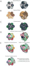
Figure 2. Conserved architecture of exosome core from bacteria, archaea, and eukaryotes
Structures of complexes reveal a six-component ring architecture with or without phosphorolytic active sites (shown as red dots). The left panel shows the cartoon representation, and the right panel shows the x-ray structure surface representation. A) RNase PH. The Aquifex aeolicus RNase PH structure (PDB ID = 1UDN) forms a homohexamer of PH subunits (colored dark grey and light grey). B) PNPase. The S. antibioticus PNPase structure (PDB ID = 1E3P) forms a homotrimer. Each of PNPase protomers are colored differently: light yellow, dark yellow, and light brown to emphasize the homotrimer of RNase PH 1-like (PH 1) and RNase PH 2-like (PH 2) domains. C) Archaeal exosome. The S. solfataricus archaeal exosome (PDB ID = 2JE6) is shown with Rrp41 subunits (blue) and Rrp42 subunits (green). Rrp41 and Rrp42 form a heterodimer; the resulting heterodimer can trimerize, culminating in a six-component ring. Either Csl4 or Rrp4 (shown in grey) form a trimeric cap above the ring. The surface representation of the structure depicts the Rrp4-bound form. D) Eukaryotic exosome: 9-subunit core. Human subunits are labeled and color-coded and include the PH-like ring subunits Mtr3 (orange), Rrp42 (red), Rrp41 (purple), Rrp45 (blue), Rrp46 (green) and Rrp43 (yellow); the S1/KH domain proteins Csl4 (light blue), Rrp4 (green) and Rrp40 (pink). E) Cytoplasmic, nuclear and nucleolar exosome architectures. The non-catalytic exosome core interacts with additional hydrolytic enzymes to form: the 10 subunit cytoplasmic exosome (panel 1, Exo9 + Rrp44), the 11 subunit nuclear exosome (panel 2, Exo9 + Rrp44 + Rrp6), and the 10 subunit nucleolar exosome (panel 3, Exo9 + Rrp6). The S1/KH protein ring is shown on the top of the PH-like ring with Rrp44 shown below the PH-like ring to reflect structural models of the complex. Rrp6 is shown on the other side of the complex below the PH-like ring, although there is no definitive structural data for this complex. The exoribonuclease active sites are depicted with red circles in Rrp44 and Rrp6, and the endoribonuclease active site of Rrp44 is shown as a yellow circle. Graphics generated with Pymol [59].
Figure 3
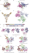
Figure 3. Structures of ‘exosome’ domains and exosome associated exoribonucleases
Residues in red indicate phosphate binding regions and residues in yellow highlight RNA binding surfaces. RNA is shown as green ribbon. Structures depicted in cartoons with helices as tubes and β-strands as arrows. A) C. crescentus PNPase RNase PH 1/RNase PH 2 domain binding interface (PDB ID = 4AM3). Phosphate binding residues include: H405, S440, and S441. RNA binding interface residues: R93, R97, R100, and R401. A second RNA binding site includes residues F77, F78, K79, and R80. B) S. cerevisiae exosome Rrp41/Rrp45 domain binding interface (PDB ID = 4IFD). Rrp41 RNA binding interface residues are R3, K62, S63, T67, R95, and R119. Rrp45 RNA binding site includes residues Y68, R71, R86, R106, R113, and R114. C) C. crescentus PNPase KH/S1 protein ring + RNA (PDB ID = 4AM3). Ribbon and surface diagram of the cap of PNPase bound to RNA. Solely the KH and RNA are represented (because no electron density was detected fro the S1 domains). A circle indicates the central pore. D) S. cerevisiae exosome KH/S1 protein ring + RNA (PDB ID = 4IFD). Ribbon and surface diagram of the budding yeast S1/KH proteins shown from the “top” with subunits labeled and color coded as in previous figures with Csl4 (light blue), Rrp4 (green), and Rrp40 (pink). Note that the S1 domains from each S1/KH protein face the central channel for the eukaryotic exosome complexes, while the KH domains face the channel for PNPase. E) Domain structure of the Rrp44 and Rrp6. Rrp44 contains five domains: a PIN (PIlus N terminal) domain with a Cysteine-Rich sequence (CR3), two Cold Shock Domains (CSD1 and CSD2), a Ribo Nuclease Binding (RNB) domain, and an S1 domain. The hydrolytic endoribonucleolytic active site is located within the PIN domain (yellow circle), and the processive 3′ to 5′ hydrolytic exoribonucleolytic active site is in the RNB domain (red circle). Rrp6 contains three domains: PMC2NT, EXO (EXOribonuclease domain), and HRDC (Homology to RNase D domain C-terminal). A second putative HRDC domain (HRDC2) may also exist similar to one detected in RNase D. The 3′ to 5′ distributive hydrolytic exoribonucleolytic active site is located within the EXO domain (red circle). F) Structures of Rrp6 and RNase D. Structures depicted in cartoons with helices as tubes and β-strands as arrows. Left panel depicts a cartoon ribbon representation of budding yeast Rrp6 catalytic domain structure (PDB ID = 2HBK). Middle panel depicts a cartoon ribbon representation of human Rrp6 catalytic domain structure (PDB 3SAF). Left panel depicts a cartoon ribbon representation of RNase D catalytic domain structure (PDB ID = 1YT3). For all three panels, domains are colored and labeled as in the domain schematic. The EXO active sites are colored red and shown in stick representation; the magnesium ions are shown as green spheres. G) Structure of RNase II + RNA. E. coli RNase II bound to RNA (PDB ID = 2IX1). H) Structure of yeast Rrp44 without PIN + RNA. Budding yeast Rrp44 bound to RNA (PDB ID = 2VNU). I) Structure of yeast Rrp44 from trimer. Structure of full-length budding yeast Rrp44 in complex with Rrp41 and Rrp45 (PDB ID = 2WP8) J) Structure of yeast Rrp44 + RNA from Exo1144/6CTD. Structure of full-length yeast Rrp44 in the yeast core exosome and the caboxy terminus of Rrp6 (PDB ID = 4IFD) For G–I, the domains are labeled and color-coded as in the schematic. The exoribonuclease active site residues are colored red in stick and surface representation. RNA is shown as a green ribbon with the 5′ end labeled; the 3′ end is buried in the exoribonuclease active site. The endoribonuclease active site residues are colored yellow in stick and surface representation. Red and yellow arrows are drawn in H–I on two helices to represent the 100–120° rigid body rotation conformational change of Rrp44 within Exo1144/6CTD.
Figure 4
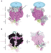
Figure 4. The 10 component exosome with CT of Rrp6
A) Overall architecture of the Exo1144/6CTD. Orthogonal view of the budding yeast 10-subunit exosome in complex with the carboxy terminus of Rrp6 (PDB ID = 4IFD). The S1/KH cap is shown in light gray, the six-membered ring is shown in dark pink, the Rrp6 CT is shown as a light blue cartoon, the location of the EXO and HRDC are currently unknown but they are predicted to be above the cap (light blue ellipse), and each of the Rrp44 domains are colored as discussed in Fig 3. B) RNA ingress into Rrp44 exo active site. The RNA ingress via the central channel is highlighted in two different views: Left, the central channel is viewed by cutting the core in half, and right, the RNA ingress is viewed by making the core transparent.

Similar articles

Cited by

References

    1. Porrua O, Libri D. RNA quality control in the nucleus: the Angels’ share of RNA. Biochim Biophys Acta. 2013;1829:604–611. - PubMed
    1. Schneider C, Tollervey D. Threading the barrel of the RNA exosome. Trends Biochem Sci. 2013 - PMC - PubMed
    1. Chlebowski A, Lubas M, Jensen TH, Dziembowski A. RNA decay machines: the exosome. Biochim Biophys Acta. 2013;1829:552–560. - PubMed
    1. Symmons MF, Jones GH, Luisi BF. A duplicated fold is the structural basis for polynucleotide phosphorylase catalytic activity, processivity, and regulation. Structure. 2000;8:1215–1226. - PubMed
    1. Liu Q, Greimann JC, Lima CD. Reconstitution, activities, and structure of the eukaryotic RNA exosome. Cell. 2006;127:1223–1237. - PubMed

Publication types

LinkOut - more resources