. 2014 Feb 20;506(7488):328-33.
doi: 10.1038/nature13038. Epub 2014 Feb 12.

Identification of pre-leukaemic haematopoietic stem cells in acute leukaemia

Collaborators, Affiliations

Identification of pre-leukaemic haematopoietic stem cells in acute leukaemia

Liran I Shlush et al. Nature. .

Erratum in

  • Nature. 2014 Apr 17;508(7496):420. Yousif, Fouad [added]

Abstract

In acute myeloid leukaemia (AML), the cell of origin, nature and biological consequences of initiating lesions, and order of subsequent mutations remain poorly understood, as AML is typically diagnosed without observation of a pre-leukaemic phase. Here, highly purified haematopoietic stem cells (HSCs), progenitor and mature cell fractions from the blood of AML patients were found to contain recurrent DNMT3A mutations (DNMT3A(mut)) at high allele frequency, but without coincident NPM1 mutations (NPM1c) present in AML blasts. DNMT3A(mut)-bearing HSCs showed a multilineage repopulation advantage over non-mutated HSCs in xenografts, establishing their identity as pre-leukaemic HSCs. Pre-leukaemic HSCs were found in remission samples, indicating that they survive chemotherapy. Therefore DNMT3A(mut) arises early in AML evolution, probably in HSCs, leading to a clonally expanded pool of pre-leukaemic HSCs from which AML evolves. Our findings provide a paradigm for the detection and treatment of pre-leukaemic clones before the acquisition of additional genetic lesions engenders greater therapeutic resistance.


Figures

Extended Data Figure 1
Extended Data Figure 1. FLT3-ITD is a late event in patients carrying DNMT3A mutation
PCR analysis of FLT3-ITD in stem/progenitor, mature lymphoid and blast (CD45dim CD33+) cell populations from patient no. 13 (a) and no. 14 (b). FLT3-ITD was present in the blasts from both patients, and also in MLPs from patient no. 14. In contrast, DNMT3Amut without FLT3-ITD was detected in multiple non-blast cell populations (see Extended Data Fig. 2). HSC, haematopoietic stem cell; MPP, multipotent progenitor; CMP, common myeloid progenitor; MLP, multilymphoid progenitor; GMP, granulocyte monocyte progenitor; NK, natural killer cells.
Extended Data Figure 2
Extended Data Figure 2. Frequent occurrence of DNMT3A mutation without NPM1 mutation in stem/progenitor and mature lymphoid cells in AML patients at diagnosis
a, Summary of the allele frequency (%) of DNMT3A and NPM1 mutations in stem/progenitor, mature lymphoid, and blast (CD45dim CD33+) cell populations from 11 AML patient peripheral blood samples obtained at diagnosis, as determined by droplet digital PCR (ddPCR). Phenotypically normal cell populations were isolated by fluorescence activated cell sorting according to the strategy depicted in Fig. 2a. Mutant allele frequency ~50% is consistent with a heterozygous cell population. Departures from 50% mutant allele frequency may be stochastic, related to clonal heterogeneity, or due to the presence of copy number variations, for example loss of the wild type allele (loss of heterozygosity) or amplification of the mutant allele. NA, no cell population detected; HSC, haematopoietic stem cell; MPP, multipotent progenitor; CMP, common myeloid progenitor; MEP, megakaryocyte erythroid progenitor; MLP, multilymphoid progenitor; GMP, granulocyte monocyte progenitor; NK, natural killer cells. Blank boxes indicate no DNMT3A or NPM1 mutation detected. b, Representative plots showing ddPCR analysis of DNMT3Amut and NPM1c allele frequency in sorted cell populations from patient no. 11. The mutant allele frequency (%) is indicated on each plot.
Extended Data Figure 3
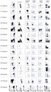
Extended Data Figure 3. Phenotypically normal stem/progenitor and mature cell populations are present in AML patient samples at diagnosis, remission and relapse
Flow cytometric analysis showing the gating strategy used to isolate phenotypically normal stem/progenitor and mature lymphoid cell populations from AML patient samples. Diagnosis and relapse samples are from peripheral blood; remission samples are from bone marrow.
Extended Data Figure 4
Extended Data Figure 4. Cells bearing mutations in DNMT3A but not NPM1 are present at diagnosis in AML patients and persist at remission and relapse
Allele frequency of DNMT3A and NPM1 mutations of patients no. 28, 35, 55, and 57 in stem/progenitor, mature and blast (CD45dim CD33+) cell populations, as determined by droplet digital PCR (ddPCR). Cells were isolated from diagnosis (blue), early remission (white), relapse (red) or late remission (yellow) samples. At remission, CD33+ myeloid cells were also analysed. HSC, haematopoietic stem cell; MPP, multipotent progenitor; MLP, multilymphoid progenitor; CMP, common myeloid progenitor; GMP, granulocyte monocyte progenitor; MEP, megakaryocyte erythroid progenitor; NK, natural killer cells.
Extended Data Figure 5
Extended Data Figure 5. PreL-HSCs in the peripheral blood of AML patients generate multilineage human grafts in immunodeficient mice
Summary of results of limiting dilution experiments to assess frequency of pre-leukaemic HSCs generating multilineage grafts after xenotransplantation. Cohorts of NSG mice were transplanted intrafemorally with varying numbers of peripheral blood mononuclear cells from diagnostic samples of AML patient no. 11 (a) and no. 55 (b) and analysed after 8 or 16 weeks by flow cytometry. Engraftment was defined as >0.1% human CD45+ cells in the injected right femur. Shown is the number of mice with multilineage human grafts containing both CD33+ myeloid cells and CD33CD19+ cells. The frequency of pre-leukaemic HSCs was calculated using the ELDA platform.
Extended Data Figure 6
Extended Data Figure 6. Frequent generation of non-leukaemic multilineage human grafts following xenotransplantation of peripheral blood cells from AML patients
Summary of xenograft characteristics in 123 sublethally irradiated NSG mice transplanted intrafemorally with mononuclear peripheral blood cells from 20 AML patients at diagnosis and analysed after 8 weeks by flow cytometry. The proportion of myeloid (CD33+) and B-lymphoid (CD33CD19+) cells in the human (CD45+) graft is shown. Leukaemic (AML) engraftment is characterized by a dominant myeloid (CD45dimCD33+) graft, whereas non-leukaemic multilineage grafts contain both lymphoid (predominantly CD33CD19+ B cells) and myeloid (CD33+) cells. No leukaemic or multilineage graft could be detected in 65/123 mice (53%) in this cohort. Red box indicates AML grafts (27 mice, 22%); blue box indicates multilineage grafts (31 mice, 25%).
Figure 1
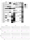
Figure 1. Recurrent somatic DNMT3a mutations are common in T-cells from AML patients
a, Summary of the allele frequency (%) of missense and frameshift somatic single nucleotide variants (sSNV) in AML-related genes assessed by deep targeted sequencing (read depth 250×) in AML blasts and T cells from the peripheral blood of 12 AML patients. The sSNV numbers indicated at the top of the table correspond to the numbers in Supplementary Table 3. Somatic mutations in DNMT3a (*, R882H; †, R137C) were found in both T-cells and AML blasts in Patients #9, 11 and 12. Patient #12 also had a low frequency IDH2 mutation (‡, R140L) in T cells. b, Frequency (%) of DNMT3amut and NPM1c in freshly isolated CD33+ blasts (AML) and matched T-cell controls from 17 patients with normal karyotype AML, as determined by droplet digital PCR. For a and b, the length of the bars is proportional to the mutant allele frequency (the scale bar under the first column applies to all columns).
Figure 2
Figure 2. DNMT3a mutation precedes NPM1 mutation in human AML and is present in stem/progenitor cells at diagnosis and remission
a, Flow cytometric analysis showing the gating strategy used to isolate phenotypically normal stem and progenitor cell populations from AML patient samples. Plots show analysis of samples from Patient #11: diagnosis (day 0, peripheral blood mononuclear cells), remission (day 62, CD34+ enriched bone marrow) and relapse (day 379, peripheral blood mononuclear cells). b, Allele frequency of DNMT3a and NPM1 mutations in stem/progenitor, mature lymphoid and blast (CD45dim CD33+) cell populations, as indicated, isolated from diagnosis (blue), remission (white) and relapse (red) samples of Patient #11 as determined by droplet digital PCR (ddPCR). At remission, CD33+ myeloid cells were also analyzed. c, Summary of the occurrence of DNMT3amut and NPM1c in isolated stem/progenitor, mature and blast cell populations from 11 AML patient peripheral blood samples as determined by ddPCR. White, DNMT3amut or NPM1c not detected; gray, DNMT3amut alone; black, DNMT3amut + NPM1c. NA, no population detected; HSC, hematopoietic stem cell; MPP, multipotent progenitor; MLP, multilymphoid progenitor; CMP, common myeloid progenitor; GMP, granulocyte monocyte progenitor; MEP, megakaryocyte erythroid progenitor; NK, natural killer cells. d, Graphic representation of DNMT3amut allele frequency in sorted cell populations isolated from diagnosis (0 months), early (3) and late (36) remission samples of Patient #57.
Figure 3
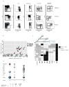
Figure 3. Pre-leukemic HSC bearing DNMT3amut generate multilineage engraftment and have a competitive advantage in xenograft repopulation assays
a, Representative flow cytometric analysis of engrafted human cells harvested from NSG mouse bone marrow (BM) 16 weeks after intrafemoral (i.f.) transplantation of peripheral blood mononuclear cells (PB MNC) from diagnosis and relapse samples of Patient #11. b, Analysis of human graft composition in NSG mouse BM 16 weeks after i.f. transplantation of PB MNC from the diagnosis sample of Patient #11 across a range of cells doses. The percentage of human (CD45+) B (CD19+) and myeloid (CD33+) cells was determined by flow cytometry. Mutant allele frequency (%) in the human graft was determined by droplet digital PCR (ddPCR) analysis of sorted human cells. The length of the bars is proportional to the mutant allele frequency (the scale bar under the first column applies to all columns). c, Summary of DNMT3amut allele frequency in the human graft from mice analyzed by ddPCR 8 and 16 weeks after transplantation of PB MNC from Patient #11, compared to isolated hematopoietic stem cell/multipotent progenitors (HSC/MPP) from the patient’s PB at diagnosis. *, P<0.05. Bars indicate mean and standard deviation.
Figure 4
Figure 4. Identification of preL-HSC with IDH2 mutation
a, Summary of the occurrence of mutations in NPM1, DNMT3a, and IDH1/2 determined by Sanger sequencing, in AML patient peripheral blood samples (n=25) that generated a non-leukemic multilineage graft after transplantation into immune-deficient mice. b, Representation of the proportion (%) of AML patient samples with DNMT3a and/or IHD1/2 mutations among samples that generated a non-leukemic multilineage graft in xenotransplanted mice. c, IDH2 and NPM1 mutant allele frequency (%) in stem/progenitor, mature lymphoid and blast (CD45dimCD33+) cell populations isolated from the peripheral blood of Patients #52, 64 and 77 at diagnosis, as determined by droplet digital PCR (ddPCR). Blank boxes indicate no mutation detected.

Comment in

  • Cancer: Persistence of leukaemic ancestors.
    Potter NE, Greaves M. Potter NE, et al. Nature. 2014 Feb 20;506(7488):300-1. doi: 10.1038/nature13056. Epub 2014 Feb 12. Nature. 2014. PMID: 24522525 No abstract available.
  • Leukaemia: a pre-leukaemic reservoir.
    Seton-Rogers S. Seton-Rogers S. Nat Rev Cancer. 2014 Apr;14(4):212. doi: 10.1038/nrc3706. Epub 2014 Mar 6. Nat Rev Cancer. 2014. PMID: 24599218 No abstract available.
  • On the origin of leukemic species.
    Vasanthakumar A, Godley LA. Vasanthakumar A, et al. Cell Stem Cell. 2014 Apr 3;14(4):421-2. doi: 10.1016/j.stem.2014.03.008. Cell Stem Cell. 2014. PMID: 24702991

Similar articles

Cited by

References

    1. Fialkow PJ, et al. Clonal development, stem-cell differentiation, and clinical remissions in acute nonlymphocytic leukemia. N Engl J Med. 1987;317:468–73. - PubMed
    1. McCulloch EA, Howatson AF, Buick RN, Minden MD, Izaguirre CA. Acute myeloblastic leukemia considered as a clonal hemopathy. Blood Cells. 1979;5:261–82. - PubMed
    1. Vogelstein B, Fearon ER, Hamilton SR, Feinberg AP. Use of restriction fragment length polymorphisms to determine the clonal origin of human tumors. Science. 1985;227:642–5. - PubMed
    1. Kandoth C, et al. Mutational landscape and significance across 12 major cancer types. Nature. 2013;502:333–9. - PMC - PubMed
    1. Greaves M, Maley CC. Clonal evolution in cancer. Nature. 2012;481:306–13. - PMC - PubMed

Publication types

MeSH terms