

Flavivirus NS1 structures reveal surfaces for associations with membranes and the immune system
- PMID: 24505133
- PMCID: PMC4263348
- DOI: 10.1126/science.1247749
Flavivirus NS1 structures reveal surfaces for associations with membranes and the immune system
Abstract
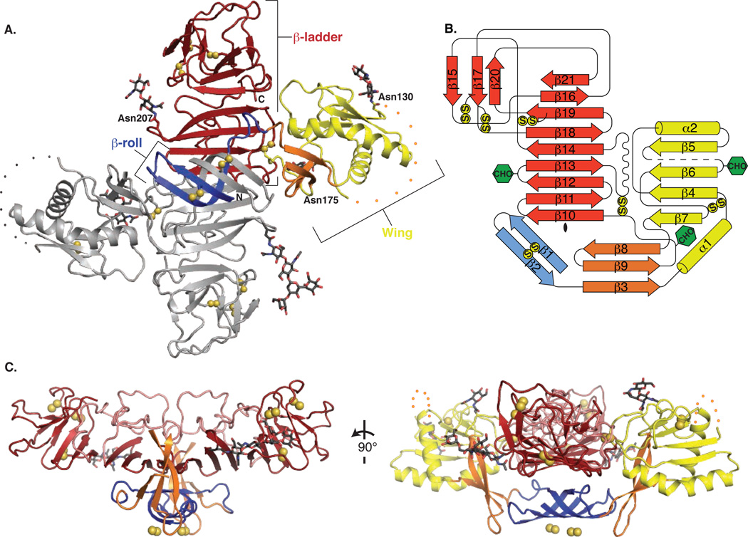
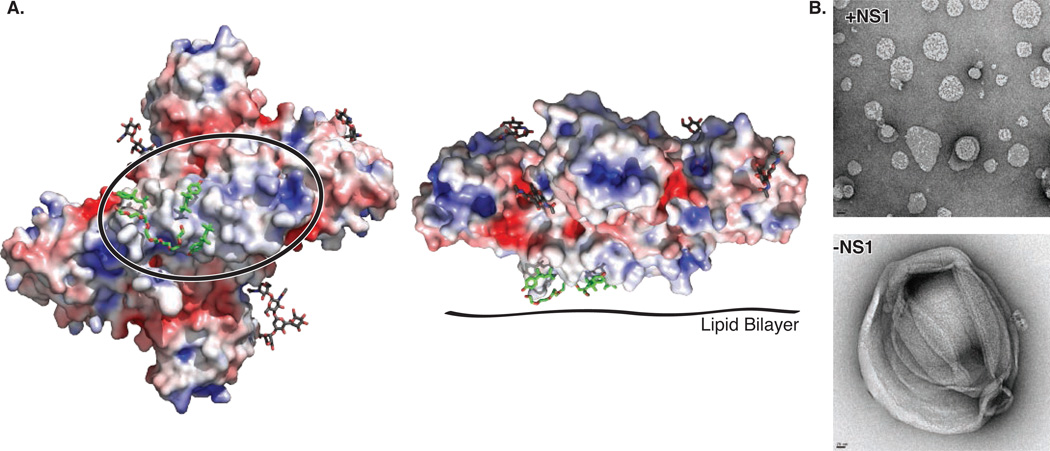
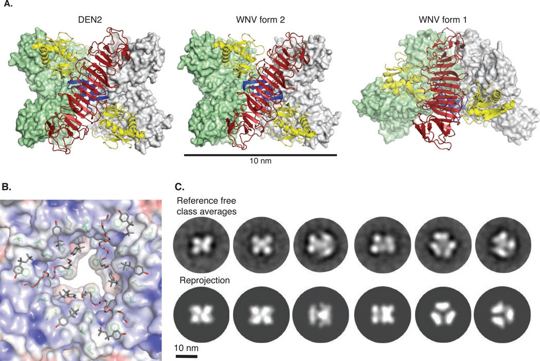
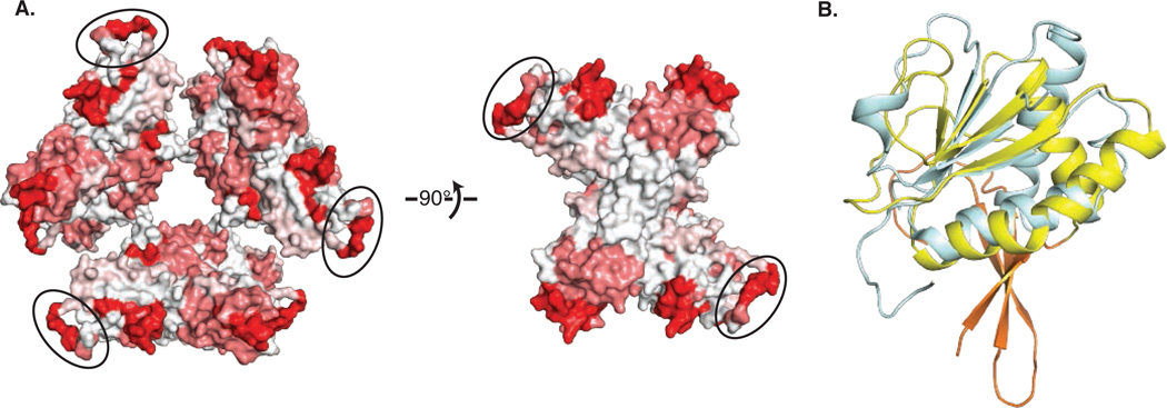
Flaviviruses, the human pathogens responsible for dengue fever, West Nile fever, tick-borne encephalitis, and yellow fever, are endemic in tropical and temperate parts of the world. The flavivirus nonstructural protein 1 (NS1) functions in genome replication as an intracellular dimer and in immune system evasion as a secreted hexamer. We report crystal structures for full-length, glycosylated NS1 from West Nile and dengue viruses. The NS1 hexamer in crystal structures is similar to a solution hexamer visualized by single-particle electron microscopy. Recombinant NS1 binds to lipid bilayers and remodels large liposomes into lipoprotein nanoparticles. The NS1 structures reveal distinct domains for membrane association of the dimer and interactions with the immune system and are a basis for elucidating the molecular mechanism of NS1 function.
Figures
Comment in
-
Structural biology. Unraveling a flavivirus enigma.Science. 2014 Feb 21;343(6173):849-50. doi: 10.1126/science.1251249. Science. 2014. PMID: 24558152 No abstract available.
Similar articles
-
Flavivirus NS1: a multifaceted enigmatic viral protein.Virol J. 2016 Jul 29;13:131. doi: 10.1186/s12985-016-0590-7. Virol J. 2016. PMID: 27473856 Free PMC article. Review.
-
The step-by-step assembly mechanism of secreted flavivirus NS1 tetramer and hexamer captured at atomic resolution.Sci Adv. 2024 May 3;10(18):eadm8275. doi: 10.1126/sciadv.adm8275. Epub 2024 May 1. Sci Adv. 2024. PMID: 38691607 Free PMC article.
-
Structure-guided insights on the role of NS1 in flavivirus infection.Bioessays. 2015 May;37(5):489-94. doi: 10.1002/bies.201400182. Epub 2015 Mar 11. Bioessays. 2015. PMID: 25761098 Free PMC article.
-
Zika virus NS1 structure reveals diversity of electrostatic surfaces among flaviviruses.Nat Struct Mol Biol. 2016 May;23(5):456-8. doi: 10.1038/nsmb.3213. Epub 2016 Apr 18. Nat Struct Mol Biol. 2016. PMID: 27088990
-
Structural biology of flavivirus NS1 protein and its antibody complexes.Antiviral Res. 2024 Jul;227:105915. doi: 10.1016/j.antiviral.2024.105915. Epub 2024 May 20. Antiviral Res. 2024. PMID: 38777094 Review.
Cited by
-
ApoA1 Neutralizes Proinflammatory Effects of Dengue Virus NS1 Protein and Modulates Viral Immune Evasion.J Virol. 2021 Jun 10;95(13):e0197420. doi: 10.1128/JVI.01974-20. Epub 2021 Jun 10. J Virol. 2021. PMID: 33827950 Free PMC article.
-
Dengue NS1 interaction with lipids alters its pathogenic effects on monocyte derived macrophages.J Biomed Sci. 2024 Sep 4;31(1):86. doi: 10.1186/s12929-024-01077-8. J Biomed Sci. 2024. PMID: 39232783 Free PMC article.
-
Development of a Model System for Tick-Borne Flavivirus Persistence in HEK 293T Cells.mBio. 2015 Jun 4;6(3):e00614. doi: 10.1128/mBio.00614-15. mBio. 2015. PMID: 26045539 Free PMC article.
-
Cross-Reactive Antibodies to the NS1 Protein of Omsk Hemorrhagic Fever Virus Are Absent in the Sera of Patients with Tick-Borne Encephalitis.Viruses. 2024 Jun 27;16(7):1032. doi: 10.3390/v16071032. Viruses. 2024. PMID: 39066195 Free PMC article.
-
The Role of NS1 Protein in the Diagnosis of Flavivirus Infections.Viruses. 2023 Feb 19;15(2):572. doi: 10.3390/v15020572. Viruses. 2023. PMID: 36851784 Free PMC article. Review.
References
-
- Lindenbach BD, Rice CM. Molecular biology of flaviviruses. Adv Virus Res. 2003;59:23–61. - PubMed
-
- Suthar MS, Diamond MS, Gale M., Jr West Nile virus infection and immunity. Nat Rev Microbiol. 2013;11:115–128. - PubMed
-
- Muller DA, Young PR. The flavivirus NS1 protein: molecular and structural biology, immunology, role in pathogenesis and application as a diagnostic biomarker. Antiviral Res. 2013;98:192–208. - PubMed
Publication types
MeSH terms
Substances
Associated data
- Actions
- Actions
- Actions
Grants and funding
LinkOut - more resources
Full Text Sources
Other Literature Sources
Medical
Research Materials
