

γδ T-cell-deficient mice show alterations in mucin expression, glycosylation, and goblet cells but maintain an intact mucus layer
- PMID: 24503767
- PMCID: PMC3962592
- DOI: 10.1152/ajpgi.00218.2013
γδ T-cell-deficient mice show alterations in mucin expression, glycosylation, and goblet cells but maintain an intact mucus layer
Abstract
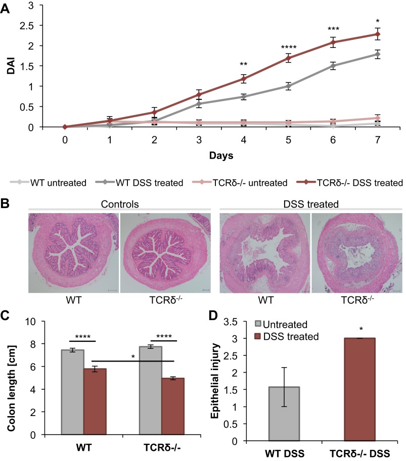
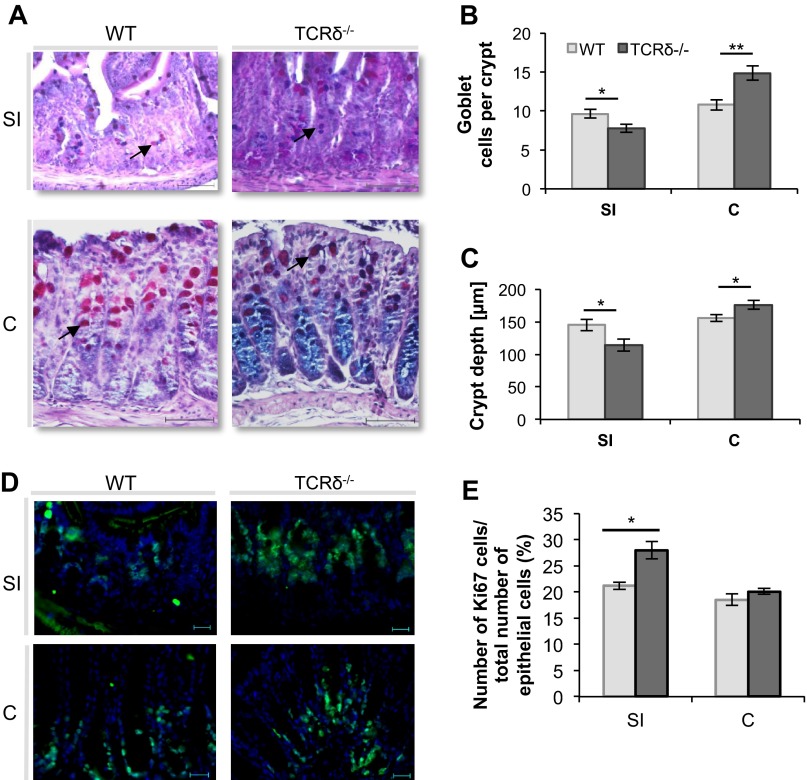
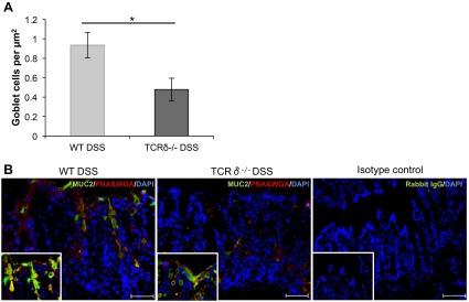
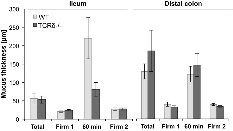
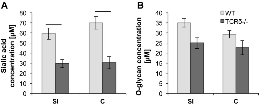
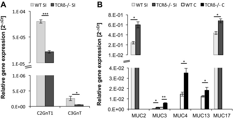
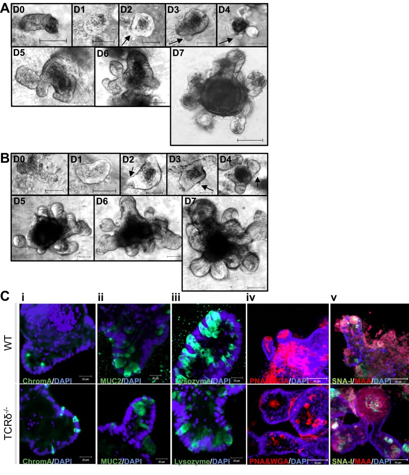
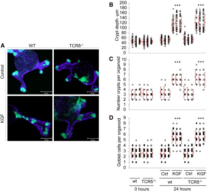
Intestinal homeostasis is maintained by a hierarchy of immune defenses acting in concert to minimize contact between luminal microorganisms and the intestinal epithelial cell surface. The intestinal mucus layer, covering the gastrointestinal tract epithelial cells, contributes to mucosal homeostasis by limiting bacterial invasion. In this study, we used γδ T-cell-deficient (TCRδ(-/-)) mice to examine whether and how γδ T-cells modulate the properties of the intestinal mucus layer. Increased susceptibility of TCRδ(-/-) mice to dextran sodium sulfate (DSS)-induced colitis is associated with a reduced number of goblet cells. Alterations in the number of goblet cells and crypt lengths were observed in the small intestine and colon of TCRδ(-/-) mice compared with C57BL/6 wild-type (WT) mice. Addition of keratinocyte growth factor to small intestinal organoid cultures from TCRδ(-/-) mice showed a marked increase in crypt growth and in both goblet cell number and redistribution along the crypts. There was no apparent difference in the thickness or organization of the mucus layer between TCRδ(-/-) and WT mice, as measured in vivo. However, γδ T-cell deficiency led to reduced sialylated mucins in association with increased gene expression of gel-secreting Muc2 and membrane-bound mucins, including Muc13 and Muc17. Collectively, these data provide evidence that γδ T cells play an important role in the maintenance of mucosal homeostasis by regulating mucin expression and promoting goblet cell function in the small intestine.
Keywords: T cell receptor-γδ; intestinal intraepithelial lymphocytes; mouse mucin.
Figures
Similar articles
-
Mice deficient in intestinal γδ intraepithelial lymphocytes display an altered intestinal O-glycan profile compared with wild-type littermates.Glycobiology. 2015 Jan;25(1):42-54. doi: 10.1093/glycob/cwu088. Epub 2014 Sep 3. Glycobiology. 2015. PMID: 25187161
-
Study of mucin turnover in the small intestine by in vivo labeling.Sci Rep. 2018 Apr 10;8(1):5760. doi: 10.1038/s41598-018-24148-x. Sci Rep. 2018. PMID: 29636525 Free PMC article.
-
γδ T cells play a protective role during infection with Nippostrongylus brasiliensis by promoting goblet cell function in the small intestine.Immunology. 2011 Dec;134(4):448-58. doi: 10.1111/j.1365-2567.2011.03503.x. Immunology. 2011. PMID: 22044210 Free PMC article.
-
Intestinal goblet cells and mucins in health and disease: recent insights and progress.Curr Gastroenterol Rep. 2010 Oct;12(5):319-30. doi: 10.1007/s11894-010-0131-2. Curr Gastroenterol Rep. 2010. PMID: 20703838 Free PMC article. Review.
-
The relationship between intestinal goblet cells and the immune response.Biosci Rep. 2020 Oct 30;40(10):BSR20201471. doi: 10.1042/BSR20201471. Biosci Rep. 2020. PMID: 33017020 Free PMC article. Review.
Cited by
-
γδ T Cells Control Gut Pathology in a Chronic Inflammatory Model of Colorectal Cancer.Cell Mol Gastroenterol Hepatol. 2021;12(3):1163-1165.e8. doi: 10.1016/j.jcmgh.2021.05.002. Epub 2021 May 11. Cell Mol Gastroenterol Hepatol. 2021. PMID: 33989816 Free PMC article. No abstract available.
-
Innate-like lymphocytes in intestinal infections.Curr Opin Infect Dis. 2015 Oct;28(5):457-63. doi: 10.1097/QCO.0000000000000189. Curr Opin Infect Dis. 2015. PMID: 26270655 Free PMC article. Review.
-
Butyrate modulates bacterial adherence on LS174T human colorectal cells by stimulating mucin secretion and MAPK signaling pathway.Nutr Res Pract. 2015 Aug;9(4):343-9. doi: 10.4162/nrp.2015.9.4.343. Epub 2015 Jul 17. Nutr Res Pract. 2015. PMID: 26244071 Free PMC article.
-
Intestinal Mucosal Wound Healing and Barrier Integrity in IBD-Crosstalk and Trafficking of Cellular Players.Front Med (Lausanne). 2021 Mar 23;8:643973. doi: 10.3389/fmed.2021.643973. eCollection 2021. Front Med (Lausanne). 2021. PMID: 33834033 Free PMC article. Review.
-
Impaired Barrier Function and Immunity in the Colon of Aldo-Keto Reductase 1B8 Deficient Mice.Front Cell Dev Biol. 2021 Feb 12;9:632805. doi: 10.3389/fcell.2021.632805. eCollection 2021. Front Cell Dev Biol. 2021. PMID: 33644071 Free PMC article.
References
-
- Atuma C, Strugala V, Allen A, Holm L. The adherent gastrointestinal mucus gel layer: thickness and physical state in vivo. Am J Physiol Gastrointest Liver Physiol 280: G922–G929, 2001 - PubMed
-
- Beagley KW, Fujihashi K, Lagoo AS, Lagoo-Deenadaylan S, Black CA, Murray AM, Sharmanov AT, Yamamoto M, McGhee JR, Elson CO, Kiyono H. Differences in intraepithelial lymphocyte T cell subsets isolated from murine small versus large intestine. J Immunol 154: 5611–5619, 1995 - PubMed
-
- Boismenu R, Havran WL. Modulation of epithelial cell growth by intraepithelial gamma delta T cells. Science 266: 1253–1255, 1994 - PubMed
Publication types
MeSH terms
Substances
Grants and funding
- BBS/E/F/00044452/BB_/Biotechnology and Biological Sciences Research Council/United Kingdom
- BB/J004529/1/BB_/Biotechnology and Biological Sciences Research Council/United Kingdom
- IFR/08/01/BB_/Biotechnology and Biological Sciences Research Council/United Kingdom
- BB/F016816/1/BB_/Biotechnology and Biological Sciences Research Council/United Kingdom
LinkOut - more resources
Full Text Sources
Other Literature Sources
Molecular Biology Databases
Research Materials
Miscellaneous