Migration of neutrophils targeting amyloid plaques in Alzheimer's disease mouse model
- PMID: 24485508
- PMCID: PMC4248665
- DOI: 10.1016/j.neurobiolaging.2014.01.003
Migration of neutrophils targeting amyloid plaques in Alzheimer's disease mouse model
Abstract
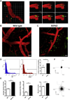
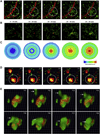
Immune responses in the brain are thought to play a role in disorders of the central nervous system, but an understanding of the process underlying how immune cells get into the brain and their fate there remains unclear. In this study, we used a 2-photon microscopy to reveal that neutrophils infiltrate brain and migrate toward amyloid plaques in a mouse model of Alzheimer's disease. These findings suggest a new molecular process underlying the pathophysiology of Alzheimer's disease.
Keywords: Alzheimer's disease; Amyloid plaques; Immune cells; Neutrophils.
Copyright © 2014 Elsevier Inc. All rights reserved.
Conflict of interest statement
The authors declare that they have no conflicts of interest, financial or otherwise, that are related to the present work.
Figures


Similar articles
-
T cells specifically targeted to amyloid plaques enhance plaque clearance in a mouse model of Alzheimer's disease.PLoS One. 2010 May 26;5(5):e10830. doi: 10.1371/journal.pone.0010830. PLoS One. 2010. PMID: 20520819 Free PMC article.
-
IFN-γ Production by amyloid β-specific Th1 cells promotes microglial activation and increases plaque burden in a mouse model of Alzheimer's disease.J Immunol. 2013 Mar 1;190(5):2241-51. doi: 10.4049/jimmunol.1200947. Epub 2013 Jan 30. J Immunol. 2013. PMID: 23365075
-
Microglia limit the expansion of β-amyloid plaques in a mouse model of Alzheimer's disease.Mol Neurodegener. 2017 Jun 12;12(1):47. doi: 10.1186/s13024-017-0188-6. Mol Neurodegener. 2017. PMID: 28606182 Free PMC article.
-
The neuroinflammatory response in plaques and amyloid angiopathy in Alzheimer's disease: therapeutic implications.Curr Drug Targets CNS Neurol Disord. 2005 Jun;4(3):223-33. doi: 10.2174/1568007054038229. Curr Drug Targets CNS Neurol Disord. 2005. PMID: 15975026 Review.
-
Immunological approaches for amyloid-beta clearance toward treatment for Alzheimer's disease.Rejuvenation Res. 2008 Apr;11(2):349-57. doi: 10.1089/rej.2008.0689. Rejuvenation Res. 2008. PMID: 18341423 Review.
Cited by
-
Blood cells and endothelial barrier function.Tissue Barriers. 2015 Apr 3;3(1-2):e978720. doi: 10.4161/21688370.2014.978720. eCollection 2015. Tissue Barriers. 2015. PMID: 25838983 Free PMC article.
-
Inflammatory pathway analytes predicting rapid cognitive decline in MCI stage of Alzheimer's disease.Ann Clin Transl Neurol. 2020 Jul;7(7):1225-1239. doi: 10.1002/acn3.51109. Epub 2020 Jul 7. Ann Clin Transl Neurol. 2020. PMID: 32634865 Free PMC article.
-
Neuroinflammation in Alzheimer's disease: chemokines produced by astrocytes and chemokine receptors.Int J Clin Exp Pathol. 2014 Dec 1;7(12):8342-55. eCollection 2014. Int J Clin Exp Pathol. 2014. PMID: 25674199 Free PMC article. Review.
-
Transcriptome analysis reveals potential marker genes for diagnosis of Alzheimer's disease and vascular dementia.Front Genet. 2022 Nov 24;13:1038585. doi: 10.3389/fgene.2022.1038585. eCollection 2022. Front Genet. 2022. PMID: 36506318 Free PMC article.
-
Ultrathin Dual-Scale Nano- and Microporous Membranes for Vascular Transmigration Models.Small. 2019 Feb;15(6):e1804111. doi: 10.1002/smll.201804111. Epub 2019 Jan 11. Small. 2019. PMID: 30632319 Free PMC article.
References
-
- Fiala M, Cribbs DH, Rosenthal M, Bernard G. Phagocytosis of amyloid-beta and inflammation: two faces of innate immunity in Alzheimer’s disease. J. Alzheimer’s Dis. 2007;11:457–463. - PubMed
-
- Fortin CF, McDonald PP, Lesur O, Fulop T., Jr Aging and neutrophils: there is still much to do. Rejuvenation Res. 2008;11:873–882. http://dx.doi.org/10.1089/rej.2008.0750. - DOI - PubMed
-
- Friese MA, Steinle A, Weller M. The innate immune response in the central nervous system and its role in glioma immune surveillance. Onkologie. 2004;27:487–491. http://dx.doi.org/10.1159/000080371. - DOI - PubMed
-
- Gate D, Rezai-Zadeh K, Jodry D, Rentsendorj A, Town T. Macrophages in Alzheimer’s disease: the blood-borne identity. J. Neural Transm. 2010;117:961–970. http://dx.doi.org/10.1007/s00702-010-0422-7. - DOI - PMC - PubMed
Publication types
MeSH terms
Grants and funding
LinkOut - more resources
Full Text Sources
Other Literature Sources
Medical
Molecular Biology Databases

