p300 acetyltransferase regulates androgen receptor degradation and PTEN-deficient prostate tumorigenesis
- PMID: 24480624
- PMCID: PMC3971883
- DOI: 10.1158/0008-5472.CAN-13-2485
p300 acetyltransferase regulates androgen receptor degradation and PTEN-deficient prostate tumorigenesis
Abstract
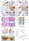
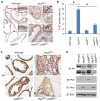
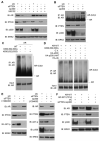
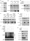
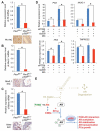
Overexpression of the histone acetyltransferase p300 is implicated in the proliferation and progression of prostate cancer, but evidence of a causal role is lacking. In this study, we provide genetic evidence that this generic transcriptional coactivator functions as a positive modifier of prostate tumorigenesis. In a mouse model of PTEN deletion-induced prostate cancer, genetic ablation of p300 attenuated expression of the androgen receptor (AR). This finding was confirmed in human prostate cancer cells in which PTEN expression was abolished by RNA interference-mediated attenuation. These results were consistent with clinical evidence that the expression of p300 and AR correlates positively in human prostate cancer specimens. Mechanistically, PTEN inactivation increased AR phosphorylation at serine 81 (Ser81) to promote p300 binding and acetylation of AR, thereby precluding its polyubiquitination and degradation. In support of these findings, in PTEN-deficient prostate cancer in the mouse, we found that p300 was crucial for AR target gene expression. Taken together, our work identifies p300 as a molecular determinant of AR degradation and highlights p300 as a candidate target to manage prostate cancer, especially in cases marked by PTEN loss.
©2014 AACR.
Figures

Similar articles
-
The pace of prostatic intraepithelial neoplasia development is determined by the timing of Pten tumor suppressor gene excision.PLoS One. 2008;3(12):e3940. doi: 10.1371/journal.pone.0003940. Epub 2008 Dec 15. PLoS One. 2008. PMID: 19081794 Free PMC article.
-
Activation of p300 histone acetyltransferase activity and acetylation of the androgen receptor by bombesin in prostate cancer cells.Oncogene. 2006 Mar 30;25(14):2011-21. doi: 10.1038/sj.onc.1209231. Oncogene. 2006. PMID: 16434977
-
Reciprocal feedback regulation of PI3K and androgen receptor signaling in PTEN-deficient prostate cancer.Cancer Cell. 2011 May 17;19(5):575-86. doi: 10.1016/j.ccr.2011.04.008. Cancer Cell. 2011. PMID: 21575859 Free PMC article.
-
P300 acetyltransferase regulates fatty acid synthase expression, lipid metabolism and prostate cancer growth.Oncotarget. 2016 Mar 22;7(12):15135-49. doi: 10.18632/oncotarget.7715. Oncotarget. 2016. PMID: 26934656 Free PMC article.
-
CBP/p300: Critical Co-Activators for Nuclear Steroid Hormone Receptors and Emerging Therapeutic Targets in Prostate and Breast Cancers.Cancers (Basel). 2021 Jun 8;13(12):2872. doi: 10.3390/cancers13122872. Cancers (Basel). 2021. PMID: 34201346 Free PMC article. Review.
Cited by
-
N-terminal domain of androgen receptor is a major therapeutic barrier and potential pharmacological target for treating castration resistant prostate cancer: a comprehensive review.Front Pharmacol. 2024 Sep 18;15:1451957. doi: 10.3389/fphar.2024.1451957. eCollection 2024. Front Pharmacol. 2024. PMID: 39359255 Free PMC article. Review.
-
AR coactivators, CBP/p300, are critical mediators of DNA repair in prostate cancer.Oncogene. 2024 Oct;43(43):3197-3213. doi: 10.1038/s41388-024-03148-4. Epub 2024 Sep 13. Oncogene. 2024. PMID: 39266679 Free PMC article.
-
Prostate cancers with distinct transcriptional programs in Black and White men.Genome Med. 2024 Jul 23;16(1):92. doi: 10.1186/s13073-024-01361-0. Genome Med. 2024. PMID: 39044302 Free PMC article.
-
Discovery of a peptide proteolysis-targeting chimera (PROTAC) drug of p300 for prostate cancer therapy.EBioMedicine. 2024 Jul;105:105212. doi: 10.1016/j.ebiom.2024.105212. Epub 2024 Jul 1. EBioMedicine. 2024. PMID: 38954976 Free PMC article.
-
Epigenetic developmental mechanisms underlying sex differences in cancer.J Clin Invest. 2024 Jul 1;134(13):e180071. doi: 10.1172/JCI180071. J Clin Invest. 2024. PMID: 38949020 Free PMC article. Review.
References
-
- Debes JD, Tindall DJ. Mechanisms of androgen-refractory prostate cancer. N Engl J Med. 2004;351:1488–90. - PubMed
-
- Isaacs JT. Antagonistic effect of androgen on prostatic cell death. Prostate. 1984;5:545–57. - PubMed
-
- Scher HI, Sawyers CL. Biology of progressive, castration-resistant prostate cancer: directed therapies targeting the androgen-receptor signaling axis. J Clin Oncol. 2005;23:8253–61. - PubMed
-
- Scher HI, Fizazi K, Saad F, Taplin ME, Sternberg CN, Miller K, et al. Increased survival with enzalutamide in prostate cancer after chemotherapy. N Engl J Med. 2012;367:1187–97. - PubMed
Publication types
MeSH terms
Substances
Grants and funding
LinkOut - more resources
Full Text Sources
Other Literature Sources
Medical
Molecular Biology Databases
Research Materials
Miscellaneous

