. 2014:5:3071.
doi: 10.1038/ncomms4071.

Generation of folliculogenic human epithelial stem cells from induced pluripotent stem cells

Affiliations

Generation of folliculogenic human epithelial stem cells from induced pluripotent stem cells

Ruifeng Yang et al. Nat Commun. 2014.

Abstract

Epithelial stem cells (EpSCs) in the hair follicle bulge are required for hair follicle growth and cycling. The isolation and propagation of human EpSCs for tissue engineering purposes remains a challenge. Here we develop a strategy to differentiate human iPSCs (hiPSCs) into CD200(+)/ITGA6(+) EpSCs that can reconstitute the epithelial components of the hair follicle and interfollicular epidermis. The hiPSC-derived CD200(+)/ITGA6(+) cells show a similar gene expression signature as EpSCs directly isolated from human hair follicles. Human iPSC-derived CD200(+)/ITGA6(+) cells are capable of generating all hair follicle lineages including the hair shaft, and the inner and outer root sheaths in skin reconstitution assays. The regenerated hair follicles possess a KRT15(+) stem cell population and produce hair shafts expressing hair-specific keratins. These results suggest an approach for generating large numbers of human EpSCs for tissue engineering and new treatments for hair loss, wound healing and other degenerative skin disorders.


Figures
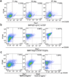
Figure 1
Figure 1. Generation of human EpSCs from hiPSCs
a. Flow cytometric ananlysis of the percentage of CD200+/ITGA6+ cell population at day 11, 18 and 25 after hiPSCs were induced with BMP4 and without EGF. b. Flow cytometric analysis of the percentage of CD200+/ITGA6+ cell population at day 11, 18 and 25 after hiPSCs were induced with BMP4 and EGF, while EGF was added one day after induction with BMP4. c. Flow cytometric analysis of the percentage of CD200+/ITGA6+ cell population at day 11, 18 and 25 after hiPSCs were induced with BMP4 and EGF, while EGF was added two days after induction with BMP4.
Figure 2
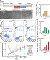
Figure 2. Staged differentiation of hiPSCs into human EpSCs
a. An outline of the protocol used to differentiate hiPSCs to EpSCs and then mature keratinocytes. b. Morphologies of hiPSCs, hiPSC-derived EpSCs (hiPSC-EpSCs, obtained at day 18 after differentiation) and hiPSC-derived mature keratinocytes (hiPSC-keratinocytes, obtained at day 45 after differentiation). Scale bar, 100 µm. c-e. Flow cytometric analysis of CD200+/ITGA6+, KRT15+ and KRT14+ cells at day 0 and 18 during the differentiation. f-h. Quantitation of CD200+/ITGA6+, KRT15+ and KRT14+ cells by flow cytometric analysis. Data shown are mean ± SD of cell percentage from three independent experiments. i. qPCR analysis of OCT3/4, NANOG, KRT5, KRT8, KRT14, KRT15, LamB3, involucrin and filaggrin expression in hiPSC-derived cells at different stages of differentiation. Samples collected at day 0, day 11, day 18, day 25 and day 30 after differentiation were used for qRT-PCR analysis. Data shown are mean ± SD of the expression from three independent experiments.
Figure 3
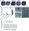
Figure 3. Colony formation capacity of hiPSC-derived EpSCs
a. Colony formation assays. Representative dishes of hiPSC-derived mature keratinocytes (hiPSC-keratinocytes), hiPSC-derived CD200+/ITGA6+ cells (hiPSC-EpSCs), passaged normal keratinocytes from adult skin (passage 3), and hiPSC-derived CD200/ITGA6+ cells (hiPSC-CD200/ITGA6+), cultured for 3 weeks on 3T3 fibroblast feeder cells. The dishes were stained with H&E. Representative images from three independent experiments. b. qPCR analysis of pluripotent stem cell markers in the hiPSC-derived CD200+/ITGA6+, CD200+/ITGA6 and CD200/ITGA6+ cells. The pluripotent stem cell markers used are OCT3/4, NANOG and REX1. Data shown are mean ± SD of the expression from three independent experiments. c. qPCR analysis of the keratinocyte specific genes in hiPSC-derived CD200+/ITGA6+ and CD200+/ITGA6 cells. Data shown are mean ± SD of the expression from three independent experiments. d. Morphology of colonies formed by CD200+/ITGA6+ and CD200+/ITGA6 cells. Scale bar in upper panel, 100um; scale bar in lower panel, 20 um.
Figure 4
Figure 4. Molecular characterization of hiPSC-derived EpSCs
a. qPCR analysis of known EpSC markers, including LGR5, LGR6, CD200, KRT15, ITGA6, TCF4, FZD2, DKK3, CTNNB1, LEF1 and LHX2 in hiPSC-EpSCs compared to control CD200+/ITGA6+ cells isolated from fetal hair follicles (hEpSCs) and parental hiPSCs. Data shown are mean ± SD of the expression from three independent experiments. b-e. Immunocytochemical analysis of KRT15, KRT1, KRT10 and ITGB1 in hiPSC-EpSCs and hEpSCs culture. Secondary antibodies are conjugated with FITC. Antibody against ITGB1 is conjugated with PE. Scale bar, 50 um. f. Hierarchical clustering among the three cell populations analyzed. g. Heat-map of genes differentially expressed in RNA-microarray analysis performed on hiPSCs, hiPSC-EpSCs and hEpSCs. h,i. Scatter plots show that epithelial markers are expressed in hiPSC-EpSCs, whereas iPSCs markers are silenced.
Figure 5
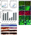
Figure 5. Folliculogeneic capacity of EpSCs derived from hiPSC tested in two different types of reconstitution assays
a. hiPSC-derived CD200+/ITGA6+/SSEA3 cells form hair follicles in a patch reconstitution assay. hiPSC-derived CD200+/ITGA6+/SSEA3 cells were combined with mouse neonatal dermal cells and injected into the dermis of an immunodeficient mouse. After 3 weeks, hair follicles and hair follicle like structures were observed at the site of injection photographed from the underside of the skin. Dotted short lines outline a hair follicle. An arrowhead points to the pigmented bulb region of a hair follicle. Representative image from seven independent experiments. Scale bar, 500 µm. b. H&E staining of an epidermal cyst with attached hair follicles formed from hiPSC-EpSCs. An arrowhead points to a hair follicle. Scale bar, 200 µm. c. Human specific Alu probe staining (green nuclei) confirms human origin of follicular epithelium and epidermal cyst lining which were generated by hiPSC-derived EpSCs. An arrowhead points to a hair follicle. Scale bar, 200 µm. d, e. In situ hybridization using pan-centromeric probes specific for human (red) and mouse (green) respectively show human origin of follicular epithelium (d) and epidermal lining (e). Scale bar, 30 µm. f. Hair follicles form from hiPSC-derived CD200+/ITGA6+/SSEA3 cells and mixed with mouse neonatal dermal cells in a reconstitution assay using a silicone chamber. hiPSC-EpSCs and mouse neonatal dermal fibroblasts were mixed together and placed in a chamber transplanted onto the back skin of a nude mouse. After three weeks, skin and hair follicles formed in the chamber. Pigmented human-like hair shafts were thicker than the surrounding mouse hair shafts. Arrowheads point to the pigmented hair shafts. Scale bar, 1 mm. g. H&E staining of a reconstituted hair follicle (H&E stain). An arrowhead points to the hair shaft. Scale bar, 150 um. h. A human-like multilayered epidermis was formed (H&E stain). Scale bar, 100 um. i,j. Human specific Alu probe staining of human hair follicles (i) and epidermis (j). Human cells were stained by human specific Alu probe labeled with FITC. An arrowhead points to the hair shaft (i). Scale bar, 150 um (i), 100 um (j). Representative images from three independent experiments.
Figure 6
Figure 6. Characterization of hair follicles and interfollicular epidermis formed by hiPSC-EpSCs
a, b. Reconstitution of stem cell niche in hair follicles by EpSCs derived from hiPSCs. CD200+/ITGA6+/SSEA3 cells derived from hiPSCs were combined with mouse neonatal dermal cells and injected into the dermis of an immunodeficient mouse. Neonatal foreskin keratinocytes were used as a positive control. Immunostaining of reconstituted hair follicles formed by neonatal foreskin keratinocytes or hiPSC-EpSCs was performed using antibodies against KRT15 (a) or KRT14 (b), respectively. Scale bar, 100 µm. hiPSC-hair follicle represents the hair follicle derived from hiPSC-EpSCs. FK-hair follicle represents the hair follicle formed by neonatal foreskin keratinocytes. c-e. Immunostaining of hair follicles formed by hiPSC-EpSCs using hair differentiation markers AE13 (c), AE15 (d) and K75 (e). Arrowheads point to the positive areas. Fetal scalp tissue was sectioned and stained with antibodies against AE13, AE15 and K75, respectively as positive controls. AE13 marks hair follicle cortex and AE15 marks inner root sheath and medulla. K75 marks companion layer of a hair follicle. Scale bar, 100 µm. f,g. Immunostaining of interfollicular epidermal lining of cyst formed by hiPSC-EpSCs was performed using antibodies against KRT10 (f) or Involucrin (g). Scale bar, 50 µm.
Figure 7
Figure 7. Differentiation of hiPSCs into mature keratinocytes
a. Flow cytometric analysis of the hiPSC-derived mature keratinocytes. hiPSC-derived CD200+/ITGA6+ and CD200/ITGA6+ cells were cultured for additional 25 days and flow cytometric analysis was performed to detect the number of KRT14+ cells. b. qPCR analysis of keratinocyte markers. Keratinocyte markers, including KRT5, KRT8, KRT14, KRT15, KRT19, Involucrin and Filaggrin, were analyzed by qPCR in mature keratinocytes derived from hiPSCs (hiPSC-keratinocytes) and normal skin-derived keratinocytes(Normal skin-keratinocytes). Data shown are mean ± SD of the expression from three independent experiments. c-g. Immunocytochemical stains of keratinocytes markers, including KRT14, p63, ITGA6, pan-cytokeratin, ITGB4 and E-cadherin (E-CAD) in hiPSC-derived mature keratinocytes (hiPSC-keratinocytes) and normal skin keratinocytes, Scale bar, 20 um. h. 3D skin equivalents using hiPSC-derived mature keratinocytes. Neonatal foreskin-derived keratinocytes (foreskin-keratinocytes) were used as a control. H&E stain of the 3D skin equivalents showed multilayered epidermis. The epidermis expressed pan-cytokeratin (Pan-CK), KRT5 and KRT14. Scale bar, 30 um.

Similar articles

Cited by

References

    1. Cherry AB, Daley GQ. Reprogrammed cells for disease modeling and regenerative medicine. Annu Rev Med. 2013;64:277–290. - PMC - PubMed
    1. Cherry AB, Daley GQ. Reprogramming cellular identity for regenerative medicine. Cell. 2012;148:1110–1122. - PMC - PubMed
    1. Morris RJ, et al. Capturing and profiling adult hair follicle stem cells. Nat Biotechnol. 2004;22:411–417. - PubMed
    1. Ito M, et al. Wnt-dependent de novo hair follicle regeneration in adult mouse skin after wounding. Nature. 2007;447:316–320. - PubMed
    1. Cotsarelis G, Sun TT, Lavker RM. Label-retaining cells reside in the bulge area of pilosebaceous unit: implications for follicular stem cells, hair cycle, and skin carcinogenesis. Cell. 1990;61:1329–1337. - PubMed

Publication types

Associated data