. 2014 Apr;33(4):429-37.
doi: 10.1016/j.healun.2013.11.004. Epub 2013 Nov 28.

T-regulatory cell treatment prevents chronic rejection of heart allografts in a murine mixed chimerism model

Affiliations

T-regulatory cell treatment prevents chronic rejection of heart allografts in a murine mixed chimerism model

Nina Pilat et al. J Heart Lung Transplant. 2014 Apr.

Abstract

Background: The mixed chimerism approach induces donor-specific tolerance in both pre-clinical models and clinical pilot trials. However, chronic rejection of heart allografts and acute rejection of skin allografts were observed in some chimeric animals despite persistent hematopoietic chimerism and tolerance toward donor antigens in vitro. We tested whether additional cell therapy with regulatory T cells (Tregs) is able to induce full immunologic tolerance and prevent chronic rejection.

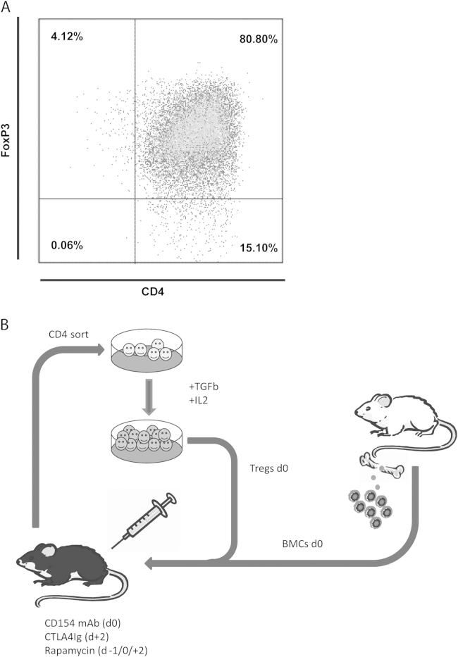
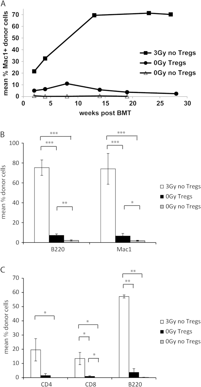
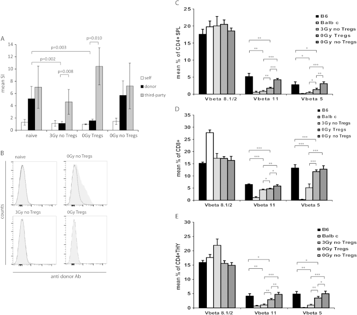
Methods: We recently developed a murine "Treg bone marrow (BM) transplantation (BMT) protocol" that is devoid of cytoreductive recipient pre-treatment. The protocol consists of a moderate dose of fully mismatched allogeneic donor BM under costimulation blockade, together with polyclonal recipient Tregs and rapamycin. Control groups received BMT under non-myeloablative irradiation and costimulation blockade without Treg therapy. Multilineage chimerism was followed by flow cytometry, and tolerance was assessed by donor-specific skin and heart allografts.

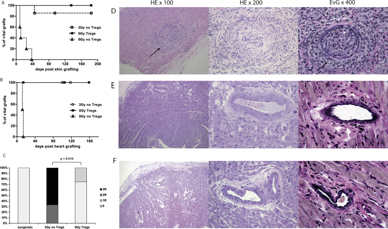
Results: Durable multilineage chimerism and long-term donor skin and heart allograft survival were successfully achieved with both protocols. Notably, histologic examination of heart allografts at the end of follow-up revealed that chronic rejection is prevented only in chimeras induced with the Treg protocol.

Conclusions: In a mouse model of mixed chimerism, additional Treg treatment at the time of BMT prevents chronic rejection of heart allografts. As the Treg-chimerism protocol also obviates the need for cytoreductive recipient treatment it improves both efficacy and safety over previous non-myeloablative mixed chimerism regimens. These results may significantly impact the development of protocols for tolerance induction in cardiac transplantation.

Keywords: chronic rejection; costimulation blockade; heart transplantation; mixed chimerism; regulatory T cells; tolerance.


Figures

Figure 1
Figure 1
Efficient expression of FoxP3 in TGF-β–induced Tregs enhances BM engrafment in a murine mixed chimerism model. (A) Representative FACS blot depicting FoxP3 expression among CD4 T cells after in vitro cultivation in the presence of TGF-β. (B) Schematic drawing of the non-cytotoxic BMT protocol using Tregs. Recipient-type CD4 T cells were separated by magnetic bead sorting and cultivated in the presence of TGF-β in vitro. Tregs were infused with fully mismatched allogeneic donor BM under the cover of costimulation blockade and rapamycin.
Figure 2
Figure 2
Treg treatment induces low but persistent levels of hematopoietic chimerism without the need for recipient irradiation. (a) Groups of B6 mice received fully mismatched BALB/c BM cells (20 × 106), combined costimulation blockade with CTLA4Ig and MR1, and either pre-treatment with 3-Gy TBI (3 Gy, 6/6 chimeras), polyclonal recipient-type Tregs with short-term rapymycin (5/5 chimeras) or rapaymycin alone (0/5 chimeras). Long-term donor (H-2Dd) chimerism among leukocytes of the myeloid (Mac1+) lineage was assessed by flow-cytometric analysis of peripheral blood at multiple time-points. (B, C) Multilineage chimerism in lymphoid tissue (BM, spleen) was assessed at the end of follow-up (~7 months post-BMT). (B) Mean levels of B-cell and myloid chimerism within BM are shown for irradiated BMT recipients (3 Gy, open bars, n = 6), Treg-treated BMT recipients (0-Gy Tregs, filled bars, n = 4) and the unirradiated control group (0-Gy control, shaded bars, n = 5). (C) Mean levels of CD4 T-cell, CD8 T-cell and B-cell chimerism among splenocytes are shown for irradiated BMT recipients (3 Gy, open bars, n = 6), Treg-treated BMT recipients (0-Gy Tregs, filled bars, n = 4) and the unirradiated control group (0-Gy control, shaded bars, n = 5). ***p < 0.0005, **p < 0.005 and *p < 0.05 (Student’s t-test). Error bars indicate standard deviation. Data represent multiple experiments.
Figure 3
Figure 3
Treg treatment induces full donor-specific tolerance and prevents chronic rejection. (A) Tolerance was assessed by grafting fully mismatched skin allografts. Donor-specific BALB/c skin survived indefinitely in most chimeras induced by irradiation (3 Gy, n = 7, open circles) and all chimeras treated with Tregs (0-Gy Tregs, n = 5, filled squares), whereas control mice uniformly rejected donor-specific grafts (0-Gy control, n = 5, open triangles). (B) Cardiac allograft survival was monitored by daily palpation (>100 days) in BMT recipients that were pre-conditioned with irradiation (3 Gy, n = 6, open circles), Tregs (0-Gy Tregs, n = 5, filled squares) and unirradiated control BMT recipients (0-Gy control, n = 5, open triangles). (C) Clinical ISHLT rejection score of cardiac allografts >100 days after transplantation into BMT recipients treated with 3-Gy irradiation or therapeutic Treg infusion (0-Gy Tregs; p = 0.010, Fisher’s exact test). (D–F) Representative features of cardiac histopathology harvested >100 days after transplantation of BALB/c hearts into (D) 3-Gy–irradiated chimeras, (E) Treg-treated chimeras or (F) syngeneic recipients. Compared with syngeneic allografts, 3-Gy–irradiated chimeras show areas with increased lymphocytic infiltrates (black arrow, left) and intimal proliferation (black arrows, right) and arterial occlusion (white arrow, right). Heart allografts were fixed and stained with HE (original magnifications: ×100 [left] and ×200 [middle]) and EvG (original magnification: ×400 [right]).
Figure 4
Figure 4
Chimeras induced by Treg treatment show donor-specific tolerance in vitro, although there is less central and peripheral deletion of donor-reactive T cells. (A) Donor reactivity was assessed in MLRs at the end of follow-up (7 months post-BMT). Chimeras induced with 3-Gy irradiation (n = 6) and Treg-treated BMT recipients (n = 4) showed specific hyporesponsiveness toward fully mismatched donor antigen in vitro compared with naive B6 mice (n = 7; p = 0.002 vs 3-Gy chimeras and p = 0.003 vs Treg chimeras). No donor-specific hyporesponsiveness was evident in control BMT recipients (n = 6) receiving BM, costimulation blockade and rapamycin only. Reactivity against third party was preserved in both groups (donor vs third-party: p = 0.008 for 3-Gy chimeras and p = 0.010 for Treg chimeras). SIs were calculated by dividing the mean count per million (cpm) from responses against recipient (open bars, B6), donor (filled bars, BALB/c) or third-party (shaded bars, C3H) stimulator cells by mean background cpm (i.e., cpm with no stimulator population). The p-values are shown for comparison between groups (Student’s t-test). Error bars indicate standard deviation. (B) Groups of mice grafted with allogeneic heart grafts were analyzed for presence of anti-donor antibodies in serum >5 months post-BMT. Chimeras induced with either irradiation (n = 6) or Tregs (n = 4) uniformly failed to develop detectable levels of anti-donor antibodies, whereas non-chimeric control mice (n = 5) developed substantial donor-specific antibody levels. The reactivity of sera with syngeneic (B6, dashed shaded line) and donor (BALB/c, solid gray line) thymocytes is shown by flow cytomery through indirect staining with anti-mouse IgG. Representative histograms are shown. (C) Chimeras induced by irradiation show significant deletion among donor-reactive T cells, as measured by percentages of Vβ11 and Vβ5 (but not Vβ8) CD4 T-cell splenocytes (SPL) (3-Gy chimeras vs naive B6: p = 0.0019 for Vβ11, p = 0.0081 for Vβ5; 3-Gy chimeras vs 0-Gy control: p < 0.0001 for Vβ11, p < 0.0001 for Vβ5). Deletion among Treg chimeras was evident (Treg chimeras vs naive B6: p = 0.0037 for Vβ11, p = 0.0311 for Vβ5; 0-Gy Treg chimeras vs 0-Gy Treg chimeras: p < 0.0001 for Vβ11, p = 0.0010 for Vβ5), but far less pronounced (3-Gy chimeras vs Treg chimeras: p = 0.0014 for Vβ11, p = 0.0140 for Vβ5). (D) Deletion of CD8-positive splenocytes (SPL) is shown in comparison to naive B6 controls (3-Gy chimeras: p < 0.0001 for Vβ11, p = 0.0001 for Vβ5; 0-Gy Treg chimeras: p = 0.0001 for Vβ11, p = not statistically significant [NS] for Vβ5) and in comparison to unirradiated BMT recipients without Tregs (3-Gy chimeras: p = 0.0016 for Vβ11, p < 0.0001 for Vβ5; 0-Gy Treg chimeras: p = 0.0077 for Vβ11, p = NS for Vβ5). Intrathymic deletion of CD8 cells was significantly more distinct in chimeras induced by irradiation (3 Gy vs 0-Gy Tregs: p = 0.0451 for Vβ11, p < 0.0001 for Vβ5). (E) Central deletion among single-positive CD4 thymocytes (THY) led to significantly reduced percentages of Vβ11- and Vβ5-expressing cells compared with naive B6 mice for BMT recipients receiving 3-Gy treatment (p = 0.0031 for Vβ11, p = 0.0020 for Vβ5) and BMT recipients receiving Tregs (p = 0.0495 for Vβ11, p = 0.0482 for Vβ5). Donor-reactive thymocytes in chimeras were also substantially reduced compared with control BMT recipients (3 Gy: p = 0.0001 for Vβ11, p = 0.0003 for Vβ5; 0-Gy Tregs: p = 0.0019 for Vβ11, p = 0.0204 for Vβ5), with significantly higher deletion in irradiated BMT (3-Gy vs 0-Gy Tregs: p = 0.0008 for Vβ11, p = 0.0012 for Vβ5). Deletion of donor-reactive T cells was assessed by multicolor flow cytometry in selected mice at the end of follow-up at ~7 months post-BMT (light-shaded bars: 3 Gy, n = 6; medium-shaded bars: 0-Gy Tregs, n = 4; dark-shaded bars: 0-Gy control, n = 5). Filled bars: naive B6 controls (n = 4); open bars: naive BALB/c controls (n = 4). ***p < 0.0005; **p < 0.005; and *p < 0.05 (Student’s t-test). Error bars indicate standard deviation.

Similar articles

Cited by

References

    1. Pilat N., Wekerle T. Transplantation tolerance through mixed chimerism. Nat Rev Nephrol. 2010;6:594–605. - PubMed
    1. Kawai T., Cosimi A.B., Spitzer T.R., et al. HLA-mismatched renal transplantation without maintenance immunosuppression. N Engl J Med. 2008;358:353–361. - PMC - PubMed
    1. Scandling J.D., Busque S., Dejbakhsh-Jones S., et al. Tolerance and chimerism after renal and hematopoietic-cell transplantation. N Engl J Med. 2008;358:362–368. - PubMed
    1. Scandling J.D., Busque S., Dejbakhsh-Jones S., et al. Tolerance and withdrawal of immunosuppressive drugs in patients given kidney and hematopoietic cell transplants. Am J Transplant. 2012;12:1133–1145. - PMC - PubMed
    1. Wekerle T., Sayegh M.H., Hill J., et al. Extrathymic T cell deletion and allogeneic stem cell engraftment induced with costimulatory blockade is followed by central T cell tolerance. J Exp Med. 1998;187:2037–2044. - PMC - PubMed

Publication types