. 2014 Jan 21;9(1):e85834.
doi: 10.1371/journal.pone.0085834. eCollection 2014.

Btk regulates macrophage polarization in response to lipopolysaccharide

Affiliations

Btk regulates macrophage polarization in response to lipopolysaccharide

Joan Ní Gabhann et al. PLoS One. .

Abstract

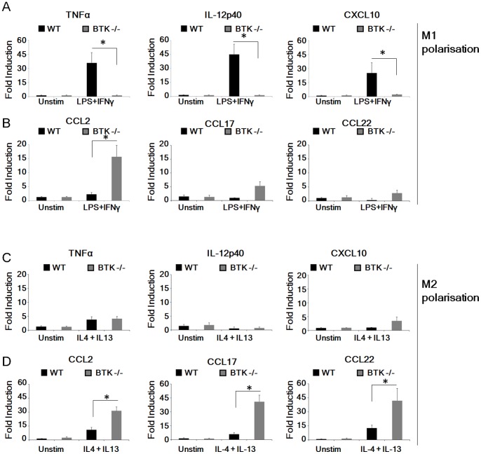
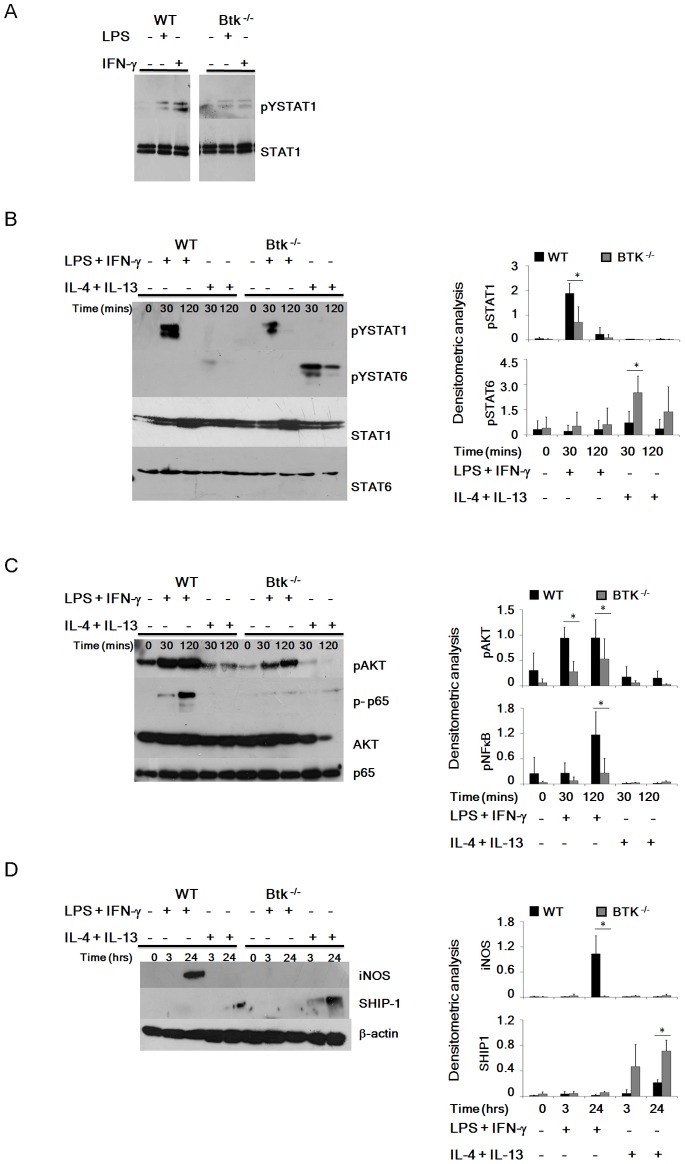
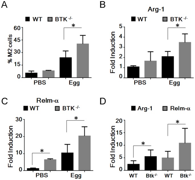
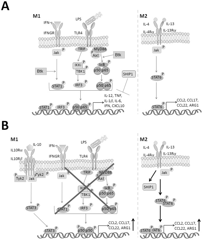
Bacterial Lipopolysaccharide (LPS) is a strong inducer of inflammation and does so by inducing polarization of macrophages to the classic inflammatory M1 population. Given the role of Btk as a critical signal transducer downstream of TLR4, we investigated its role in M1/M2 induction. In Btk deficient (Btk (-\-)) mice we observed markedly reduced recruitment of M1 macrophages following intraperitoneal administration of LPS. Ex vivo analysis demonstrated an impaired ability of Btk(-/-) macrophages to polarize into M1 macrophages, instead showing enhanced induction of immunosuppressive M2-associated markers in response to M1 polarizing stimuli, a finding accompanied by reduced phosphorylation of STAT1 and enhanced STAT6 phosphorylation. In addition to STAT activation, M1 and M2 polarizing signals modulate the expression of inflammatory genes via differential activation of transcription factors and regulatory proteins, including NF-κB and SHIP1. In keeping with a critical role for Btk in macrophage polarization, we observed reduced levels of NF-κB p65 and Akt phosphorylation, as well as reduced induction of the M1 associated marker iNOS in Btk(-/-) macrophages in response to M1 polarizing stimuli. Additionally enhanced expression of SHIP1, a key negative regulator of macrophage polarisation, was observed in Btk(-/-) macrophages in response to M2 polarizing stimuli. Employing classic models of allergic M2 inflammation, treatment of Btk (-/-) mice with either Schistosoma mansoni eggs or chitin resulted in increased recruitment of M2 macrophages and induction of M2-associated genes. This demonstrates an enhanced M2 skew in the absence of Btk, thus promoting the development of allergic inflammation.


Conflict of interest statement

Competing Interests: JAJ is an employee of the company Amgen. This does not alter the authors’ adherence to all the PLOS ONE policies on sharing data and materials.

Figures

Figure 1
Figure 1. Impaired TLR4-mediated induction of peritoneal M1 cells in Btk−/− mice in vivo.
(A–E) Peritoneal macrophages were harvested from WT or Btk−/− mice following i.p. LPS injection. (A) Representative plots demonstrate the gating strategies for macrophage discrimination where total peritoneal macrophages were defined following co-staining with CD11b APC-Cy7 and F4/80 PE-Cy7. (B–C) Macrophage subsets were further distinguished as F4/80intCD11bhi M1-like macrophages and F4/80hiCD11bhi M2-like macrophages (M1 and M2 gates, respectively). The relative percentage of M1 (B) and M2 (C) macrophages within the total macrophage gate were determined for WT or Btk−/− mice following i.p. LPS injection. (D) Expression of CD86 and MHC Class II was determined by flow cytometry. (E) MHC Class II expression was examined within the defined M1 and M2 gates following co-staining with F4/80 and CD11b. (F) Ly6C was determined by flow cytometry. In all cases data is presented as percent increased expression above background as determined by staining with the relevant isotype control (indicated by markers in panel (D). (G–H) Peritoneal macrophages harvested from WT or Btk−/− mice were treated ex vivo with LPS (100 ng/ml) for the indicated time course and the induction of M1- (G) and M2- (H) associated genes was determined by real time PCR (qPCR). Peritoneal macrophages were pooled after isolation, with each treatment group consisting of 3–4 animals, and all experiments were performed in triplicate. Student’s paired t test was performed comparing gene induction in Btk−/− peritoneal macrophages to WT cells at the indicated time points. Results shown are mean±SD from three independent experiments. * = p≤0.05.
Figure 2
Figure 2. Btk−/− macrophages have impaired ability to expand in vitro into M1 cells.
(A–D) Peritoneal macrophages were extracted from WT and Btk−/− mice and treated ex vivo with an M1 (LPS plus IFN-y) or an M2 (IL-4 plus IL-13) polarizing cocktail for 24 hr and induction of M1 (A–B) and M2- associated (C–D) genes determined by qPCR. In all cases peritoneal macrophages were pooled after isolation, with each treatment group consisting of 3–4 animals, and all experiments were performed in triplicate. Student’s paired t test was performed comparing gene induction in Btk−/− peritoneal macrophages to WT cells following treatment with polarizing cocktails as indicated. Results shown are mean±SD from three independent experiments. * = p≤0.05.
Figure 3
Figure 3. Altered phosphorylation of key signalling intermediaries in the absence of Btk.
(A) WT and Btk−/− BMDMs were treated ex vivo with LPS (100 ng/ml) or IFN-γ (100 U/ml) for 3 hours or 15 minutes, respectively, lysates were prepared and phosphorylated Y701-STAT1 levels determined by Western blot. WT and Btk−/− BMDMs were treated ex vivo with an M1 or M2 polarizing cocktail over the indicated time course, lysates were prepared and tyrosine phosphorylated STAT1 and STAT6 (B), serine phosphorylated AKT (upper panel) and NF-κB p65 (lower panel) (C) iNOS and SHIP-1 (D) levels were determined by Western blot. Results in each case are representative of three independent experiments. Densitometric analysis was performed and graphs represent phosphorylated protein levels relative to unphosphorylated proteins (B-C) or changes in total protein levels relative to β-actin (D) for WT and Btk−/− BMDMs. Student’s paired t test was performed comparing relative expression of phosphorylated or total proteins in Btk−/− BMDMs to WT BMDMs following treatment with polarizing cocktails as indicated. Results shown are mean±SD from three independent experiments. * = p≤0.05.
Figure 4
Figure 4. Increased in vivo M2 macrophage generation in the absence of Btk.
(A–C) WT and Btk−/− mice were injected i.v. with 5,000 live S.mansoni eggs. (A) The percentage of pulmonary M2 macrophages was evaluated by flow cytometry using F4/80 and CD11b co-staining. (B–C) Induction of M2-associated genes was determined by qPCR. (D) WT and Btk−/− mice were injected i.p. with approximately 800 ng Chitin. Peritoneal cells were collected by lavage after 48 hours and gene induction of M2-associated genes was determined by qPCR. In all cases peritoneal macrophages were pooled after isolation, treatment groups consisted of 3–4 animals, and experiments were performed in triplicate. Student’s paired t test was performed comparing gene induction following in vivo exposure to S.mansoni eggs or Chitin as indicated in WT and Btk−/− peritoneal macrophages. Results shown are mean±SD from three independent experiments. * = p≤0.05.
Figure 5
Figure 5. Proposed transcriptional regulation of macrophage polarization in the absence of Btk.
(A) This study has demonstrated that in response to LPS and IFN-γ Btk contributes to M1 polarizing of myeloid cells via promoting the phosphorylation of Akt and subsequently the p65 subunit of NFκB, in addition to enhancing to phosphorylation of STAT1. (B) In the absence of Btk exposure of myeloid cells to LPS and IFN-γ results in the preferential induction of M2 associated genes and preferentially recruitment of M2 cells in vivo. Previous studies in Btk−/− mice have observed increased levels IL-10 systemically following LPS treatment. IL-10 is known to activate STAT3 and there is some evidence to suggest that STAT3 may also play a role in promoting M2 macrophage polarization. Thus in the absence of Btk, in response to M1 polarizing stimuli increased IL-10 production together with reduced phosphorylation of key signaling intermediaries, combined with activation of p50 the inhibitory subunit of NF-κB could potentially account for the observed preferential skew towards an M2 phenotype. Additionally this study has shown that in response to IL-4 and IL-13 Btk−/− cells demonstrate an increased capacity to polarize towards an M2 phenotype as a result of enhanced STAT6 phosphorylation and increased SHIP1 expression.

Similar articles

Cited by

References

    1. Orme J, Mohan C (2012) Macrophage subpopulations in systemic lupus erythematosus. Discov Med 13: 151–158. - PubMed
    1. Shechter R, Schwartz M (2013) Harnessing monocyte-derived macrophages to control central nervous system pathologies: no longer ‘if’ but ‘how’. J Pathol 229: 332–346. - PubMed
    1. Hao NB, Lu MH, Fan YH, Cao YL, Zhang ZR, et al. (2012) Macrophages in tumor microenvironments and the progression of tumors. Clin Dev Immunol 2012: 948098. - PMC - PubMed
    1. Dasgupta P, Keegan AD (2012) Contribution of alternatively activated macrophages to allergic lung inflammation: a tale of mice and men. J Innate Immun 4: 478–488. - PMC - PubMed
    1. Sica A, Mantovani A (2012) Macrophage plasticity and polarization: in vivo veritas. J Clin Invest 122: 787–795. - PMC - PubMed

Publication types

MeSH terms

Grants and funding

This work was supported by the Health Research Board, Ireland (RP/2004/79) and P/2006/121 www.hrb.ie) and Science Foundation Ireland (Grant 08/IN.1/B2091 www.sfi.ie). The funders had no role in study design, data collection and analysis, decision to publish, or preparation of the manuscript.