. 2014 Jan;12(1):e1001773.
doi: 10.1371/journal.pbio.1001773. Epub 2014 Jan 21.

Conservation and divergence of regulatory strategies at Hox Loci and the origin of tetrapod digits

Affiliations

Conservation and divergence of regulatory strategies at Hox Loci and the origin of tetrapod digits

Joost M Woltering et al. PLoS Biol. 2014 Jan.

Abstract

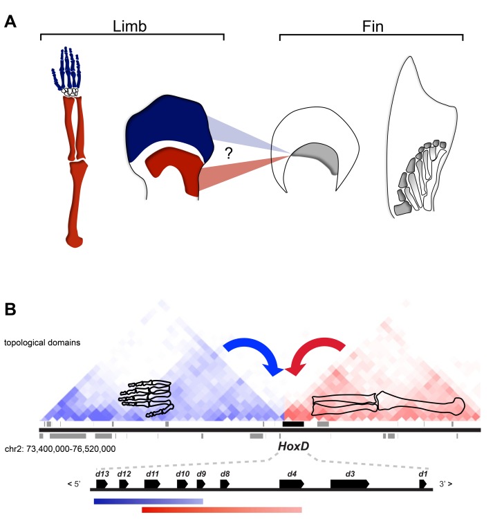
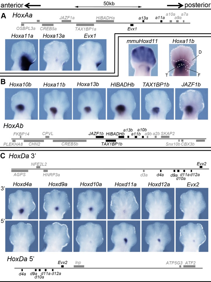
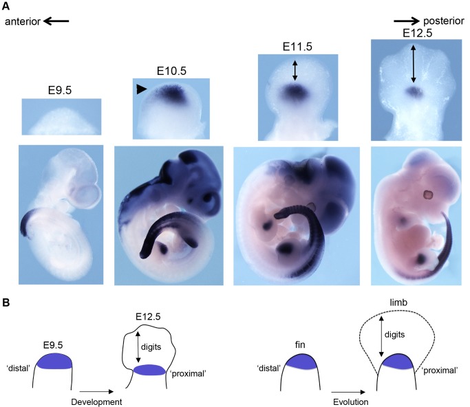
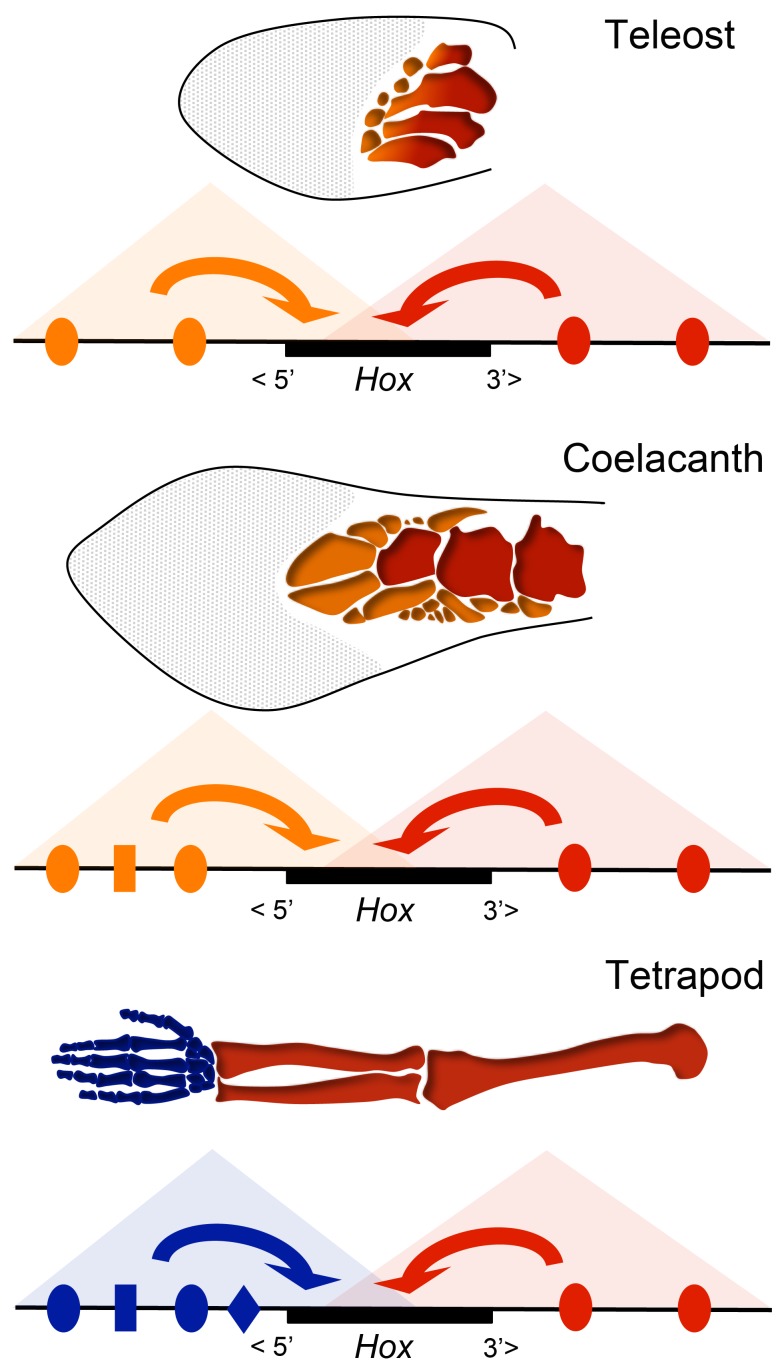
The evolution of tetrapod limbs from fish fins enabled the conquest of land by vertebrates and thus represents a key step in evolution. Despite the use of comparative gene expression analyses, critical aspects of this transformation remain controversial, in particular the origin of digits. Hoxa and Hoxd genes are essential for the specification of the different limb segments and their functional abrogation leads to large truncations of the appendages. Here we show that the selective transcription of mouse Hoxa genes in proximal and distal limbs is related to a bimodal higher order chromatin structure, similar to that reported for Hoxd genes, thus revealing a generic regulatory strategy implemented by both gene clusters during limb development. We found the same bimodal chromatin architecture in fish embryos, indicating that the regulatory mechanism used to pattern tetrapod limbs may predate the divergence between fish and tetrapods. However, when assessed in mice, both fish regulatory landscapes triggered transcription in proximal rather than distal limb territories, supporting an evolutionary scenario whereby digits arose as tetrapod novelties through genetic retrofitting of preexisting regulatory landscapes. We discuss the possibility to consider regulatory circuitries, rather than expression patterns, as essential parameters to define evolutionary synapomorphies.


Conflict of interest statement

The authors have declared that no competing interests exist.

Figures

Figure 1
Figure 1. Regulatory mechanisms and the homology conundrum between fins and limbs.
(A) The evolutionary changes that occurred during the transition from fins to limbs are mostly unresolved, in particular concerning the most distal segment of tetrapod limbs: the digits. Mammalian proximal and distal limb regions develop along with independent phases of Hoxd expression [indicated in red (arm) and blue (digits)] and fish fin buds have been probed for the existence of similar Hoxd expression patterns. A single expression domain of 5′ Hoxd genes along the distal fin margin (in grey) was interpreted either as corresponding to the distal phase in tetrapods or, alternatively, as homologous to the proximal phase ,,,,. Accordingly, radials (in grey) could be homologous with digits or this homology may not exist, in which case digits are tetrapod novelties. (B) Proximal (red) and distal (blue) Hoxd gene expression domains in the developing mouse limb are derived from enhancers located within distinct 3′- (red) and 5′- (blue) regulatory landscapes. The enhancer–promoter interaction profiles within these two landscapes were shown to precisely match two topological domains as determined by Hi-C using ES cells . This bimodal regulatory organization in tetrapods suggests distinct evolutionary trajectories for proximal and distal limbs. The presence or absence of such a modular regulatory strategy in fish would help clarify the origin of this mechanism and the homology between fins and limbs. The DNA domain shown is approximately 3 mb large.
Figure 2
Figure 2. Hoxa gene expression in limb buds.
(A) Expression of Hoxa4, Hoxa9, Hoxa10, Hoxa11, Hoxa11 antisense (Hoxa11as), and Hoxa13 in E12.5 limb buds. The Hoxa11as transcript originates from a promoter within the intron of Hoxa11 (upper panel) and is expressed like Hoxa13. (B) In control (WT) embryos, Hoxa11 and Hoxa13 are expressed in mutually exclusive domains, with Hoxa13 in the autopod and Hoxa11 in the distal zeugopod. In Hoxa13 homozygous mutants embryos, the Hoxa11 expression domain shifts into the proximal autopod, partly overlapping with Hoxa13. In Hoxa13 −/−/Hoxd13+/ double mutant animals, Hoxa11-expressing cells spread further distally. The Hoxa13 probe is within the 3′ UTR and thus detects Hoxa13 transcripts in mice carrying a loss of function for this gene. Although HOX13 proteins repress Hoxa11 transcription, this latter gene has the capacity to respond to global distal enhancers, much like its Hoxa9, Hoxa10, Hoxa11, and Hoxa13 neighbors (fl, forelimb; hl, hindlimb). The anterior-to-posterior polarity of the limb buds is indicated with arrows.
Figure 3
Figure 3. Interaction profiles of mouse Hoxa genes.
Circular chromosome conformation capture (4C) analysis of either distal (A) or proximal (B) E12.5 dissected limb bud (schematized in the left) or forebrain (C). The proximal or distal fates of these cells are illustrated by adult skeletons (left) with the same colors as in Figure 1. Dark grey squares indicate regions of local interactions excluded from the analysis. Four interaction profiles are shown after using Hoxa4, Hoxa9, Hoxa11, and Hoxa13 as viewpoints. The genomic orientation of HoxA is inverted with respect to HoxD. The percentage of contacts for each viewpoint is given, either in 5′ or in 3′ of the gene cluster. In both samples, Hoxa4 mostly interacts with the 3′ landscape, whereas Hoxa13 is biased toward the 5′ landscape. Both Hoxa9 and Hoxa11 change their bias from increased contacts in 3′, in the proximal limb bud sample (B), to contacts in 5′ in the distal sample (A), thus resembling Hoxd genes (Figure S2) . Note that the interaction profiles obtained when using either autopod, proximal limb, or brain (C) tissues are quite similar to one another, indicating a constitutive chromatin organization at the HoxA locus. The size of the displayed DNA interval is of ca. 3 Mb. (D and E) Summaries of the directional 4C signals using bar diagrams in the 3′ and 5′ flanking regions of both HoxA and HoxD clusters. The colored bars represent 100% of the signal for each of the three tissues (color code at the bottom) and for three genes in either the HoxD (D) or the HoxA (E) clusters. The position of each bar with respect to the central black line (0) represents the balance between the contacts scored either in 5′ (left in D; right in E) or in the 3′ (right in D; left in E) landscapes. The HoxA and HoxD clusters are shown in opposite orientation regarding 3′ and 5′ directions to reflect their inverse locations on chromosomes 2 and 6. The four displayed topological domains were extracted from the Hi-C ES cell dataset of Dixon et al. .
Figure 4
Figure 4. Zebrafish Hox clusters are partitioned into 3′ and 5′ interaction domains.
4C analysis of zebrafish whole embryos (5 dpf, including well-developed fin buds) using as viewpoints (left) several genes within the HoxAa (A and B), HoxAb (C and D), and HoxDa (E and F) gene clusters (for HoxAa, see also Figure S4). The HoxDa cluster has a reversed chromosomal orientation when compared to both HoxA clusters. The percentages of interactions between the viewpoints and either the 5′ or the 3′ landscapes are indicated above each profile. Bar diagrams in (B, D, and F) give a summary of the signal directionality per viewpoint in the 3′ and 5′ flanking regions (compare Figure 3C,D). The blue bars are as in Figure 3. Genes located at either extremity of their clusters display a strong bias toward the flanking landscape, such as Hoxa4a (B), Hoxd4a (F), Hoxa13a (B), or Hoxd13a (F). Genes located at more central positions in the clusters [e.g., Hoxa11a (B) or Hoxd11a, (F)] show more balanced interaction profiles, like for the mouse HoxA and HoxD clusters. Dark grey squares are regions of local interactions excluded from the analysis.
Figure 5
Figure 5. Regulatory potential of the fish HoxA and HoxD landscapes in mouse transgenic limbs.
(A) Scheme of the HoxAa BAC used for transgenesis in the mouse with the expression of the fish Hoxa11a, Hoxa13a, and Evx1 genes in mouse embryonic limbs. All genes assayed showed expression in a proximal domain, yet not in the presumptive digit domain. Note that Hoxa11a expression was not observed in forelimb buds. (B) Scheme of the HoxAb BAC (bottom) with the expression of several genes. The fish Hoxa10b, Hoxa11b, and Hoxa13b genes are expressed in a proximal domain, and transcripts are absent from the presumptive digit domain. Likewise, the 5′ flanking genes HIBADHb, TAX1BP1b, and JAZF1b respond to the same proximal regulation. A comparison with the endogenous Hoxd11 expression (mmuHoxd11) shows that limb expression of the transgenes is confined to the distal zeugopod and mesopod. (C) Two BAC clones containing either the entire 5′ (top) or 3′ (bottom) landscape flanking the HoxDa cluster with their corresponding expression patterns. Here again, expression is observed in a proximal domain but is absent from developing digits. In the various schemes, genes analyzed are shown in black. All samples are right hind limbs, dorsal views with anterior to the left, except for the endogenous mouse gene “mmuHoxd11” (B), which is a mirror image of the left hind limb of the limb bud stained for Hoxa11b to its right, in order to facilitate the comparison of transcription domains. The anterior-to-posterior polarity is indicated with arrows. (D, digits; F and T, distal parts of the femur and tibia, respectively).
Figure 6
Figure 6. The Tetraodon Hoxa13b expression domain in mice: from “distal” to “proximal.”
(A) In situ hybridization of a Tetraodon Hoxa13b probe using E9.5 to E12.5 fetuses transgenic for the Tetraodon HoxAb cluster. Top panels are dorsal views of forelimbs (anterior to the left), and bottom panels are whole mount pictures. Hoxa13b is expressed in limb buds and posterior trunk, whereas the staining in the head vesicles at E10.5 and E11.5 is a routinely observed artifact. At day E10.5, before the appearance of digits, expression initiates in the distal limb bud (arrowhead). In subsequent stages, however, this domain becomes increasingly “proximal” due to the distal expansion of the digit domain (arrows in E11.5 and E12.5 specimen). The distal expression of Hoxa13b at E10.5 is strikingly similar to the distal expression of Hoxa13b in the fish fin . The anterior-to-posterior polarity is indicated with arrows. (B) Scheme illustrating the difficulty in using relative parameters such as “proximal” or “distal” to assign homologies. Due to the developmental expansion of the autopod, the zeugopod domain becomes relatively more proximally positioned within the limb bud, along with time. During digit evolution, a similar process may have occurred and structures that are distal in the fin apparently shifted to a more proximal position in the limb, due to the distal growth of the autopod. The fish Hoxa13b expression in mouse limb buds (purple color in the left scheme) in fact illustrates that distal fish fin tissues correspond to proximal limb structures after the evolution into limbs (right scheme). The fin bud scheme only depicts the endoskeletal part of the fin and not the exoskeleton, which derives from a distinct developmental lineage.
Figure 7
Figure 7. Regulatory evolution and the fin-to-limb transition.
Fish and tetrapod HoxA and HoxD clusters are regulated by 3′ and 5′ regulatory landscapes, represented here as triangles due to their correspondence to topological domains ,. Enhancer (indicated with colored shapes) interactions within these domains (indicated by arrows) occur with the neighboring parts of the Hox clusters, resulting in a regulatory partition between 3′ and 5′ parts of the clusters. In fishes, this mechanism may be used for patterning the fin proximal (red) to distal (orange) (P-D) polarity, through the potential function of these two landscapes in slightly different fin domains. Variation in the regulatory balance between these 3′ and 5′ landscapes through the acquisition of novel enhancers potentially explains interspecies differences in P-D fin morphology, as for instance between zebrafish and species such as coelacanth, which possesses a more elaborate fin skeleton. Although these regulatory landscapes may underlie the P-D patterning of fin skeletons, they both elicit a proximal response when assessed in transgenic mice, and hence the fish 5′ landscape is indicated as “proximal” (orange). In tetrapods, the 5′ domain (blue) has acquired new enhancers or modified existing ones, thereby evolving a novel, more distal autopodial identity, perhaps as a response to preexisting signals emanating from the apical ectoderm.

Comment in

  • A footnote to the evolution of digits.
    Hoff M. Hoff M. PLoS Biol. 2014 Jan;12(1):e1001774. doi: 10.1371/journal.pbio.1001774. Epub 2014 Jan 21. PLoS Biol. 2014. PMID: 24465182 Free PMC article. No abstract available.

Similar articles

Cited by

References

    1. Woltering JM, Duboule D (2010) The origin of digits: expression patterns versus regulatory mechanisms. Dev Cell 18: 526–532. - PubMed
    1. Wagner GP, Chiu CH (2001) The tetrapod limb: a hypothesis on its origin. J Exp Zool 291: 226–240. - PubMed
    1. Gaffney ES (1979) Tetrapod monophyly: a phylogenetic analysis. Bull Carnegie Mus Nat Hist 13: 92–105.
    1. Holmgren N (1952) An embryological analysis of the mammalian carpus and its bearing upon the question of the origin of the tetrapod limb. Acta Zoologica 33: 1–115.
    1. Coates MI, Jeffery JE, Rut M (2002) Fins to limbs: what the fossils say. Evol Dev 4: 390–401. - PubMed

Publication types

Grants and funding

JMW was supported by a fellowship from EMBO. This work was carried out with funding from the University of Geneva, the EPFL Lausanne, the Swiss National Science Foundation, and the European Research Council (to DD). The funders had no role in study design, data collection and analysis, decision to publish, or preparation of the manuscript.

LinkOut - more resources