. 2014 Jan 22;81(2):366-78.
doi: 10.1016/j.neuron.2013.11.033.

Structural insights into competitive antagonism in NMDA receptors

Affiliations

Structural insights into competitive antagonism in NMDA receptors

Annie Jespersen et al. Neuron. .

Abstract

There has been a great level of enthusiasm to downregulate overactive N-methyl-D-aspartate (NMDA) receptors to protect neurons from excitotoxicity. NMDA receptors play pivotal roles in basic brain development and functions as well as in neurological disorders and diseases. However, mechanistic understanding of antagonism in NMDA receptors is limited due to complete lack of antagonist-bound structures for the L-glutamate-binding GluN2 subunits. Here, we report the crystal structures of GluN1/GluN2A NMDA receptor ligand-binding domain (LBD) heterodimers in complex with GluN1- and GluN2-targeting antagonists. The crystal structures reveal that the antagonists, D-(-)-2-amino-5-phosphonopentanoic acid (D-AP5) and 1-(phenanthrene-2-carbonyl)piperazine-2,3-dicarboxylic acid (PPDA), have discrete binding modes and mechanisms for opening of the bilobed architecture of GluN2A LBD compared to the agonist-bound form. The current study shows distinct ways by which the conformations of NMDA receptor LBDs may be controlled and coupled to receptor inhibition and provides possible strategies to develop therapeutic compounds with higher subtype-specificity.


Figures

Figure 1
Figure 1. Domain organization of NMDA receptor subunits and ligands
(A) NMDA receptor subunits are modular proteins composed of an amino terminal domain (ATD), a ligand-binding domain (LBD), a transmembrane domain (TMD), and a cytosolic domain (CTD). Their modular domains are oriented such that the N-terminus (NT) and the C-terminus (CT) are located at the extracellular and cytoplasmic regions, respectively. In this study, LBDs from GluN1 and GluN2A are isolated (scissors) by tethering two peptide fragments between TMD M1 and M3 by a Gly-Thr dipeptide linker (dashed line). (B) Ligands for GluN1 and GluN2 subunits. Glycine and 5,7-dichlorokynurenic acid (DCKA) are an agonist and an antagonist, respectively, for GluN1. l-glutamate and N-methyl-d-aspartate (NMDA) are agonists for GluN2 LBD. D-(−)-2-Amino-5-phosphonopentanoic acid (d-AP5) and 1-(Phenanthrene-2-carbonyl-)piperazine-2,3-dicarboxylic acid (PPDA) with distinct chemical structures both act as competitive antagonists at the GluN2 LBD.
Figure 2
Figure 2. Structures of GluN1/GluN2A LBDs in complex with various ligands
The four crystal structures of GluN1/GluN2A LBDs in complex with glycine (Gly; gray sphere) and l-glutamate (Glu; green sphere) (panel A), DCKA (blue sphere) and l-glutamate (panel B), glycine and d-AP5 (yellow sphere; panel C), and glycine and PPDA (cyan sphere; panel D). The GluN1 and GluN2A LBDs exist as a heterodimer in the asymmetric unit of the orthorhombic crystals obtained in this study (Table S1). The structures are oriented so that the N-terminal ends (NT) from both GluN1 and GluN2A face top of this figure. GluN1 and GluN2A LBDs are shaped like bilobed clamshells composed of upper (D1) and lower (D2) domains colored green and orange, respectively, in GluN1 and blue and magenta, respectively, in GluN2A. All of the ligands studied here bind at the D1-D2 interface. Sticks represent three disulfide bonds conserved among the NMDA receptor family members. The spheres at the bottom of the structures represent the Cα of the glycine residue in the Gly-Thr dipeptide linker, where loops leading to the M1 TMD and from the M3 TMD would be located in the full-length receptors.
Figure 3
Figure 3. Antagonist binding in GluN2A LBD
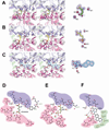
(A–C) Stereoview of the ligand binding sites located at the D1-D2 interface. Binding of l-glutamate (green sticks; panel A), the GluN2 antagonists involve residues from both D1 and D2 for both D-AP5 (cyan sticks; panel B), and PPDA (yellow sticks; panel C). Dashed lines represent polar interactions. The current crystallographic study shows unambiguous electron density of l-glutamate (A), d-AP5 (B) and PPDA (C) as well as critical water molecules for binding (red spheres; W1-3). The electron density (blue mesh) represents the Fo-Fc omit map contoured at 3σ. (D–F) Schematic representation of l-glutamate (D), d-AP5 (E), and (−)-PPDA (F) binding sites. The crystal structures around the ligand binding sites were analyzed by LIGPLOT v.4.5.3 (Wallace et al., 1995). Dotted lines represent polar interactions whereas red arcs with spokes represent hydrophobic interactions. Red spheres marked by “W” represent water molecules at the binding sites. Residues involved in binding of l-glutamate and/or d-AP5 have blue (from D1) and magenta (from D2) backgrounds. Residues uniquely involved in binding of (−)-PPDA have an emerald green background. Orientations of the D2 residues are different between the l-glutamate-bound, the d-AP5-bound, and the (−)-PPDA-bound GluN2A LBDs due to a large domain movement of D2 relative to D1.
Figure 4
Figure 4. GluN1/GluN2A NMDA receptors selectively bind (−)-PPDA over (+)-PPDA
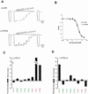
(A) Enantiomers of PPDA showing two chiral centers (stars) at the 2 and 3 positions of the piperazine ring. PPDA used in this crystallographic study is the enantiomeric mixture available commercially (TOCRIS). However, the electron density indicates the exclusive presence of (−)-(2S, 3R)-PPDA as shown in Figure 3C. (B) Inhibition of glycine/l-glutamate ion channel current by (+)-(2S, 3R)-PPDA (filled circle) or (−)-(2S, 3R)-PPDA (square) in the GluN1-1a/GluN2A NMDA receptors. The currents are formed by coapplication of 100 µM of glycine and 5 µM of l-glutamate and inhibited by various concentrations of PPDA. All of the recordings are done using the TEVC method. Error bars represent s.d.
Figure 5
Figure 5. Mutagenesis of the ligand-binding site
Residues surrounding the antagonist binding site are mutated and tested for inhibition of ion channel activities to validate physiological relevance of the crystal structures. (A) Typical dose-response inhibition pattern of the wild type GluN1/GluN2A NMDA receptor current assessed by TEVC. In this recording, currents formed by application of 100 µM of glycine and 5 µM l-glutamate are inhibited by various concentrations of d-AP5 (top panel) or (−)-PPDA (bottom panel). Note that there is no significant rundown of currents before and after the dose-response experiment. (B) All of the data points can be fit to a one-binding-site model of Hill equation. (C–D) Fold increase of Ki values for d-AP5 (C) and (−)-PPDA (D). Ki values were calculated by the Cheng-Prusoff equation using EC50 values for l-glutamate and IC50 values for the antagonists for every mutant (Table S3).
Figure 6
Figure 6. Comparison of GluN2A LBD and non-NMDAR LBDs
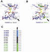
(A–B) Superposition of GluA2 (panel A) and GluK1 (panel B) LBDs onto the GluN2A LBD-D-AP5 structure. The LBD structures of GluA2 (PDB code: 1FTL) and GluK1 (PDB code: 2F34) are split into D1 and D2 domains and superposed onto the respective domains of GluN2A LBD-d-AP5 structure by the program LSQKAB (Kabsch, 1976). The GluN2A residues involved in binding of d-AP5 are shown in blue sticks whereas d-AP5 is shown in yellow sticks. Side chains of non-NMDA receptor residues that are involved in ligand-binding and are different from GluN2 subunits are shown in gray (GluA2) and green (GluK1). A notable difference is found at the position of GluN2A Tyr730 (arrows) where the equivalent residues in GluA2 and GluK1 are Leu704 (gray sticks) and Met722 (green sticks), respectively. (C) Sequence alignment of the glutamate binding iGluR subunits around GluN2A Tyr730 (arrow). Tyrosine is conserved in the NMDA subfamily (blue) whereas the equivalent residue in the AMPA (black) and kainate (green) subfamilies is either leucine or methionine.
Figure 7
Figure 7. (−)-PPDA binding on GluN2A LBD and non-NMDAR LBDs
(A–B) Superposition of GluA2 (panel A) and GluK1 LBDs (panel B) onto the GluN2A LBD-(−)-PPDA structure. The structural superposition is done in the same manner as in Figure 6. Side chains of non-NMDA receptor residues that are involved in ligand-binding and are different from GluN2 subunits are shown in gray (GluA2) and green (GluK1). In both GluA2 and GluK1 receptors, there appear to be no residue that may prevent (−)-PPDA from binding. (C) (−)-PPDA can robustly inhibit currents produced by GluN1-1a/GluN2A NMDA receptors, GluA2 AMPA receptors (Leu483Tyr, flip), and GluK1 kainate receptors (Tyr506Cys Leu768Cys). Here NMDA receptor and non-NMDA receptor currents are induced by 100 µM glycine/30 µM glutamate and 30 µM glutamate, respectively, and inhibited by application of 100 µM (−)-PPDA in the presence of agonists. All of the recordings were done using TEVC on Xenopus oocytes injected with cRNAs encoding GluN1/GluN2A NMDA receptor, GluA2 AMPA receptor mutant, and GluK1 kainate receptor mutant. The Ki values for (−)-PPDA inhibition are 7.85 µM and 1.17 µM for GluA2 (flip, Leu483Tyr) and GluK1 (Tyr506Cys Leu768Cys), respectively.
Figure 8
Figure 8. Binding of d-AP5 and (−)-PPDA induce different degree of domain movement
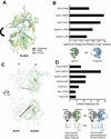
(A) Superposition of GluN1/GluN2A LBD structures in complex with l-glutamate (green), d-AP5 (yellow), and (−)-PPDA (cyan). The Cαs of D1 in GluN2A in d-AP5-bound or (−)-PPDA-bound forms and l-glutamate-bound form are superposed to observe the differences in the extent of domain opening caused by the GluN2-antagonist binding. d-AP5 and (−)-PPDA induce opening of the clamshell-like architecture through mobilization of Helix E and Helix G, respectively. Spheres at the bottom of the structures represent Cα of GluN2A Val662 next to the M3 transmembrane region. GluN1 LBD is omitted from this figure for clarity. (B) Analysis of domain opening of antagonist-bound LBD structures relative to the l-glutamate-bound LBD structures in GluA2 (PDB code: 1FTJ for l-glutamate vs. 1FTL for DNQX), GluK1 (PDB code: 2F36 for l-glutamate vs. 2F34 for UBP310) GluN1, and GluN2A. The rotation angles are measured using the program Dyndom (Hayward and Lee, 2002). Below the bar graph is the schematic presentation of domain opening resulting from binding of an antagonist. (C) The same superposition as “panel A” but showing GluN1 LBD in complex with glycine (light gray) viewed from the side (top panel) and from the bottom or the membrane plane (bottom panel). Spheres in GluN1 represent Cα of Ile664. Dashed lines show distances between GluN1 Ile664 and GluN2A Val662, which are plausible measures of the TMD movement. The linker distance for GluN1/GluN2A LBD-gly/glu is shown in red whereas that for GluN1/GluN2A LBD-gly/d-AP5 or GluN1/GluN2A LBD-gly/PPDA is in black. (D) Differences in the GluN1 Ile664 - GluN2A Val662 distances between antagonist-bound (black dash in panel C) and the agonist-bound (glycine and l-glutamate; red dash in panel C) structures (Δ-linker-distances). The inter-linker distances in the DCKA/PPDA and DCKA/d-AP5 forms (asterisks) are calculated using hypothetical models built by superposing the D1 of the DCKA/glu-bound GluN1/GluN2A LBD onto that of GluN1/GluN2A LBD structures bound to gly/d-AP5 or gly/(−)-PPDA. In GluA2, distances between the Cαs of Ile663 in the GluA2 LBD homodimers in the l-glutamate-bound (PDB code: 1FTJ) and DNQX-bound (PDB code: 1FTL) forms are measured. In GluK1, distances between the Cαs of Ile653 in the GluK1 LBD homodimers in l-glutamate-bound (PDB code: 2F36) and UBP310-bound (PDB code: 2F34) forms are measured. Below the bar graph is the schematic presentation to show that the antagonist-induced domain opening of LBD may result in a decrease in the inter-linker distance (arrows between two spheres), thereby causing the transmembrane ion channel to close.

Similar articles

Cited by

References

    1. Acker TM, Khatri A, Vance KM, Slabber C, Bacsa J, Snyder JP, Traynelis SF, Liotta DC. Structure-Activity Relationships and Pharmacophore Model of a Noncompetitive Pyrazoline Containing Class of GluN2C/GluN2D Selective Antagonists. J Med Chem. 2013;56:6434–6456. - PMC - PubMed
    1. Adams PD, Grosse-Kunstleve RW, Hung LW, Ioerger TR, McCoy AJ, Moriarty NW, Read RJ, Sacchettini JC, Sauter NK, Terwilliger TC. PHENIX: building new software for automated crystallographic structure determination. Acta Crystallogr D Biol Crystallogr. 2002;58:1948–1954. - PubMed
    1. Armstrong N, Gouaux E. Mechanisms for activation and antagonism of an AMPA-sensitive glutamate receptor: crystal structures of the GluR2 ligand binding core. Neuron. 2000;28:165–181. - PubMed
    1. Benveniste M, Mayer ML. Kinetic analysis of antagonist action at N-methyl-D-aspartic acid receptors. Two binding sites each for glutamate and glycine. Biophys J. 1991;59:560–573. - PMC - PubMed
    1. Bettini E, Sava A, Griffante C, Carignani C, Buson A, Capelli AM, Negri M, Andreetta F, Senar-Sancho SA, Guiral L, et al. Identification and characterization of novel NMDA receptor antagonists selective for NR2A- over NR2B-containing receptors. J Pharmacol Exp Ther. 2010;335:636–644. - PubMed

Publication types

MeSH terms

LinkOut - more resources