. 2014:5:3176.
doi: 10.1038/ncomms4176.

A role for sorting nexin 27 in AMPA receptor trafficking

Affiliations
Free PMC article

A role for sorting nexin 27 in AMPA receptor trafficking

Li Shen Loo et al. Nat Commun. 2014.
Free PMC article

Abstract

Sorting nexin 27 (SNX27), a PDZ domain-containing endosomal protein, was recently shown to modulate glutamate receptor recycling in Down's syndrome. However, the precise molecular role of SNX27 in GluA1 trafficking is unclear. Here we report that SNX27 is enriched in dendrites and spines, along with recycling endosomes. Significantly, the mobilization of SNX27 along with recycling endosomes into spines was observed. Mechanistically, SNX27 interacts with K-ras GTPase via the RA domain; and following chemical LTP stimuli, K-ras is recruited to SNX27-enriched endosomes through a Ca(2+)/CaM-dependent mechanism, which in turn drives the synaptic delivery of homomeric GluA1 receptors. Impairment of SNX27 prevents LTP and associated trafficking of AMPARs. These results demonstrate a role for SNX27 in neuronal plasticity, provide a molecular explanation for the K-ras signal during LTP and identify SNX27 as the PDZ-containing molecular linker that couples the plasticity stimuli to the delivery of postsynaptic cargo.


Figures
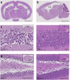
Figure 1
Figure 1. Histopathological changes in the SNX27−/− brain.
(a) Representative coronal brain section from SNX27−/− mouse stained with haematoxylin and eosin (H&E). There was dilatation of the lateral ventricle (LV), periventricular neuropil dissociation (arrow) with a rim of cortical tissue at the periphery (asterisk). Scale bar, 2 mm (b) Representative saggital sections from SNX27−/− brain stained with H&E. Scale bar, 3 mm. (c) Hippocampal CA1 region from SNX27−/− and WT mice (left and right panels, respectively). Inset shows region where the images were obtained. Scale bar, 40 μm. (d) Hippocampal dentate gyrus(DG) from SNX27−/− and WT mice (left and right panels respectively). Notable features include vacuolation of pyramidal neurons, thinning of dentate gyrus layer and cytoplasmic vacuolation. Inset shows region where the images were obtained. Scale bar, 100 μm.
Figure 2
Figure 2. Spine distribution of SNX27 in neurons.
(a) Cultured hippocampal neurons (17 div) transfected with GFP-tagged SNX27 and mCherry-PSD95 to identify spines. SNX27 was distributed on the base of spines and within dendritic spines. Scale bar, 4 μm. (b) Cultured hippocampal neurons (17 div) transfected with GFP-tagged SNX27. Neurons were then labelled with presynaptic marker, synaptophysin. SNX27 labelling is closely opposed to that of synaptophysin (arrows). Scale bar, 1 μm.
Figure 3
Figure 3. Loss of spine distribution in the truncated mutants.
Cultured hippocampal neurons (17 div) transfected with GFP-tagged full-length SNX27 or SNX27 deletion mutants. mCherry-PSD95 was also expressed to identify spines. Fluorescence signal was quantified along the length (dashed line) of the image. For full-length SNX27, there is overlap of the red and green peaks indicating colocalization of the corresponding spots, whereas for the mutants, the green peaks are much lower than the red peak indicating minimal localization in the spines. Scale bar, 2 μm.
Figure 4
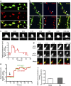
Figure 4. Mobilization of SNX27 into spines along with recycling endosomes.
(a) Colocalization of SNX27 with early endosomes marked by Rab5. Scale bar, 1 μm. (b) Recycling endosomes were labelled with transferrin receptor and Rab 11. Colocalization of SNX27 with recycling endosomes marked by tranferrin receptor and Rab11 as indicated with arrowheads. Scale bar, 2 μm. (c) Rapid time-lapse images of SNX27 revealing its mobilization in and out of spine (upper). Time is indicated in minutes and seconds. Step-wise displacement analysis of GFP-SNX27 puncta with base of spine defined as zero (lower). Scale bar, 1 μm. (d) Time-lapse imaging showing the mobilization of SNX27 along with recycling endosomes into spine. Time is indicated in minutes and seconds. Scale bar, 1 μm. (e) Step-wise displacement analysis showing the correlated movement of the GFP and mCherry signal in d. (f) SNX27 spine translocation events (events per min/total spine) were measured with or without glycine treatment. The graph represents the means of 15 independent experiments. n=15, P=0.2663, unpaired t-test, error bars=s.e.m.
Figure 5
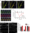
Figure 5. SNX27 mediates recycling endosome transport.
(a) Loss of SNX27 from spines following glycine treatment. Time-lapse imaging following the loss and subsequent recovery of SNX27 signal (green) with time following glycine treatment, but not in the presence of NMDA receptor antagonist APV. Glycine stimulation was applied from 0–5 min. Time is indicated in minutes and seconds. Scale bar, 4 μm. (b) Quatitative analysis of loss of SNX27 GFP signal normalized against the spine mcherry signal (n=87 and 90 spines from 5 neurons for Gly and Gly/APV treatment respectively, error bars=s.e.m.). (c) Absence of SNX27 interferes with recycling endosome transport. Wild type and SNX27-depleted neurons were saturated with Alx-Tf for 30 min, washed and further incubated in neurobasal media, glycine or glycine/APV for 25 min. Representative images showing the loss of Alx-Tf from wild type and SNX27-deficient neurons following 25 min treatment with neurobasal media, glycine or glycine/APV. Scale bar, 5 μm. (d) Quantitative analysis of intracellular fluorescence remaining following 25 min treatment with neurobasal media, glycine or glycine/APV; n=10 neurons respectively, error bars=s.e.m. ** indicates P<0.05 for WT vs SNX27-/-(glycine treated), unpaired t-test.
Figure 6
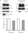
Figure 6. GluN1 expression in wild type and SNX27−/− mice.
(a) GluN1 amounts in synaptosomes and PSD fractions derived from wild type and SNX27−/− hippocampus. (b) Graph shows the GluN1 levels in synaptosomes and PSD fractions of SNX27−/− hippocampus relative to wild-type hippocampus. n=3; **indicates P<0.05, unpaired t-test; error bars=s.e.m.
Figure 7
Figure 7. Ras-binding properties of SNX27.
(a) Upper panel: selective interaction of endogenous K-ras with SNX27 in hippocampal lysate. Lower panel: immunoprecipitation of HEK293 cells expressing HA-K-ras and Myc-SNX27 full-length or SNX27 lacking RA domain (Myc-SNX27ΔRA). (b) Localization of SNX27 and K-ras with and without glycine treatment (200 μm, 5 min). The graph represents the mean of five independent experiments with 10 images each. (n=5, ** indicates P<0.05, unpaired t-test, error bars=s.e.m.). Scale bar, 2 μm. (c) Time dependence of K-ras binding to calmodulin following Gly, Gly/APV and Gly/EGTA stimulation (upper). Quantitative analysis of time course of K-ras binding to CaM following Gly (▪), Gly/APV (□) and Gly/EGTA (Δ) stimulation (lower). n=3. All error bars represent s.e.m.
Figure 8
Figure 8. AMPAR binding properties of SNX27.
(a) Endogenous coimmunoprecipitation with SNX27 antibody. SNX27 interacts with GluA1 receptor, but not GluA2 receptor. (b) Reverse coimmunoprecipitation with GluA2 antibody to verify interaction of GluA2 with GluA1. GluA2 interacts with GluA1, but not SNX27. (c) Immunoprecipitation of HEK293 cells expressing GFP-GluA1 c-terminal tail and Myc-SNX27 or Myc-SNX27 without the PDZ domain (Myc-SNX27ΔPDZ). (d) Time dependence of enhanced K-ras and GluA1 binding to SNX27 following Gly stimulation (upper). Quantitative analysis of time course of K-ras and GluA1 binding to SNX27 following Gly stimulation (lower). The density bands at each time point for GluA1 and K-ras were normalized against the respective SNX27 density band at the same time point. This result would give the normalized intensity ratio on the y axis and these were plotted against time on the x axis. n=3, ** indicates P≤0.01, unpaired t-test; error bars=s.e.m. (e) Recruitment of GluA1 by SNX27 is inhibited by CaM kinase inhibitor (KN93) and farnesyltransferase inhibitors (FTI-276 and FTI-277). The GluA1 density bands were normalized against the respective SNX27 density band. This result would give the normalized intensity ratio on the y axis. n=3, ** indicates P≤0.05, unpaired t-test; error bars=s.e.m.
Figure 9
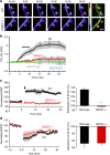
Figure 9. Disrupting SNX27 interferes with AMPA receptor surface delivery and LTP.
(a) Time-lapse SEP-GluA1 fluorescence images from wild type and SNX27-deficient neurons with 5 min glycine treatment. mCherry-PSD95 was used to identify spines (right-most image). Scale bar, 1 μm. (b) The normalized increase in SEP-GluA1 fluorescence (Ft/F0) in spines was monitored in wild type, wild type treated with FTI-276 and FTI-277 and SNX27-deficient neurons; n=10 neurons respectively. Bar indicates presence of glycine stimulus. (c) Hippocampal slices were freshly prepared from wild type and SNX27 knockout mice and LTP was induced using high‐frequency stimulation in normal (squares) and SNX27-deficient slices (triangles); n=8 slices respectively. Bar graph summarizing the effect of SNX27 deletion on the magnitude of LTP at 30 min (** indicates P<0.001, t-test, error bars=s.e.m.). (d) LTD in wild type versus knockout slices with 1 Hz stimulation for 15 min; n=7 slices respectively. Bar graph summarizing the effect of SNX27 deletion on the magnitude of LTD at 30 min (P>0.9, t-test, error bars=s.e.m.).
Figure 10
Figure 10. Schematic model for LTP induction.
PSD, postsynaptic density; ExoZ, exocytic zone. During synaptic activity, binding of Ca to CaM triggers the translocation of K-ras from the plasma membrane to SNX27 on recycling endosomes. This initial binding step promotes the recruitment of GluA1 receptors to SNX27 on recycling endosomes and subsequently drives the synaptic delivery of GluA1 AMPARs.

Similar articles

Cited by

References

    1. Valdes J. L. et al. Sorting nexin 27 protein regulates trafficking of a p21-activated kinase (PAK) interacting exchange factor (β-Pix)-G protein-coupled receptor kinase interacting protein (GIT) complex via a PDZ domain interaction. J. Biol. Chem. 286, 39403–39416 (2011). - PMC - PubMed
    1. Wang X. et al. Loss of sorting nexin 27 contributes to excitatory synaptic dysfunction by modulating glutamate receptor recycling in Down’s syndrome. Nat. Med. 19, 473–480 (2013). - PMC - PubMed
    1. Ghai R. et al. Phox homology band 4.1/ezrin/radixin/moesin-like proteins function as molecular scaffolds that interact with cargo receptors and Ras GTPases. Proc. Natl Acad. Sci. USA 108, 7763–7768 (2011). - PMC - PubMed
    1. Zhu J. J., Qin Y., Zhao M., Van Aelst L. & Malinow R. Ras and Rap control AMPA receptor trafficking during synaptic plasticity. Cell 110, 443–455 (2002). - PubMed
    1. Fivaz M. & Meyer T. Reversible intracellular translocation of KRas but not HRas in hippocampal neurons regulated by Ca2+/calmodulin. J. Cell Biol. 170, 429–441 (2005). - PMC - PubMed

Publication types

LinkOut - more resources