Oestrogen increases haematopoietic stem-cell self-renewal in females and during pregnancy
- PMID: 24451543
- PMCID: PMC4015622
- DOI: 10.1038/nature12932
Oestrogen increases haematopoietic stem-cell self-renewal in females and during pregnancy
Abstract
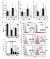
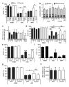
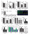
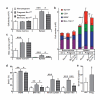
Sexually dimorphic mammalian tissues, including sexual organs and the brain, contain stem cells that are directly or indirectly regulated by sex hormones. An important question is whether stem cells also exhibit sex differences in physiological function and hormonal regulation in tissues that do not show sex-specific morphological differences. The terminal differentiation and function of some haematopoietic cells are regulated by sex hormones, but haematopoietic stem-cell function is thought to be similar in both sexes. Here we show that mouse haematopoietic stem cells exhibit sex differences in cell-cycle regulation by oestrogen. Haematopoietic stem cells in female mice divide significantly more frequently than in male mice. This difference depends on the ovaries but not the testes. Administration of oestradiol, a hormone produced mainly in the ovaries, increased haematopoietic stem-cell division in males and females. Oestrogen levels increased during pregnancy, increasing haematopoietic stem-cell division, haematopoietic stem-cell frequency, cellularity, and erythropoiesis in the spleen. Haematopoietic stem cells expressed high levels of oestrogen receptor-α (ERα). Conditional deletion of ERα from haematopoietic stem cells reduced haematopoietic stem-cell division in female, but not male, mice and attenuated the increases in haematopoietic stem-cell division, haematopoietic stem-cell frequency, and erythropoiesis during pregnancy. Oestrogen/ERα signalling promotes haematopoietic stem-cell self-renewal, expanding splenic haematopoietic stem cells and erythropoiesis during pregnancy.
Figures


Comment in
-
Stem cells: Sex specificity in the blood.Nature. 2014 Jan 23;505(7484):488-90. doi: 10.1038/505488a. Nature. 2014. PMID: 24451537 No abstract available.
-
Girl power: estrogen promotes HSC self-renewal.Cell Stem Cell. 2014 Feb 6;14(2):137-8. doi: 10.1016/j.stem.2014.01.016. Cell Stem Cell. 2014. PMID: 24506879 Free PMC article.
-
Sex hormone drives blood stem cell reproduction.EMBO J. 2014 Mar 18;33(6):534-5. doi: 10.1002/embj.201487976. Epub 2014 Feb 20. EMBO J. 2014. PMID: 24562387 Free PMC article.
Similar articles
-
Girl power: estrogen promotes HSC self-renewal.Cell Stem Cell. 2014 Feb 6;14(2):137-8. doi: 10.1016/j.stem.2014.01.016. Cell Stem Cell. 2014. PMID: 24506879 Free PMC article.
-
Oestrogen receptor alpha and beta immunoreactive cells in the suprachiasmatic nucleus of mice: distribution, sex differences and regulation by gonadal hormones.J Neuroendocrinol. 2008 Nov;20(11):1270-7. doi: 10.1111/j.1365-2826.2008.01787.x. Epub 2008 Aug 22. J Neuroendocrinol. 2008. PMID: 18752649
-
Dynamics of Bone Marrow VSELs and HSCs in Response to Treatment with Gonadotropin and Steroid Hormones, during Pregnancy and Evidence to Support Their Asymmetric/Symmetric Cell Divisions.Stem Cell Rev Rep. 2018 Feb;14(1):110-124. doi: 10.1007/s12015-017-9781-x. Stem Cell Rev Rep. 2018. PMID: 29168113
-
Designer cytokines for human haematopoietic progenitor cell expansion: impact for tissue regeneration.Handb Exp Pharmacol. 2006;(174):229-47. Handb Exp Pharmacol. 2006. PMID: 16370330 Review.
-
Intrinsic and extrinsic control of haematopoietic stem-cell self-renewal.Nature. 2008 May 15;453(7193):306-13. doi: 10.1038/nature07038. Nature. 2008. PMID: 18480811 Review.
Cited by
-
Evaluation of male reproductive hormones of Kalahari Red and Kalawad goat bucks as affected by age and season under tropical conditions.Trop Anim Health Prod. 2024 Oct 28;56(8):364. doi: 10.1007/s11250-024-04215-4. Trop Anim Health Prod. 2024. PMID: 39466348
-
27-Hydroxycholesterol Negatively Affects the Function of Bone Marrow Endothelial Cells in the Bone Marrow.Int J Mol Sci. 2024 Sep 29;25(19):10517. doi: 10.3390/ijms251910517. Int J Mol Sci. 2024. PMID: 39408846 Free PMC article.
-
Hematopoietic and leukemic stem cells homeostasis: the role of bone marrow niche.Explor Target Antitumor Ther. 2024;5(5):1027-1055. doi: 10.37349/etat.2024.00262. Epub 2024 Aug 15. Explor Target Antitumor Ther. 2024. PMID: 39351440 Free PMC article. Review.
-
A Flow Cytometry-Based Examination of the Mouse White Blood Cell Differential in the Context of Age and Sex.Cells. 2024 Sep 20;13(18):1583. doi: 10.3390/cells13181583. Cells. 2024. PMID: 39329764 Free PMC article.
-
Lipoprotein metabolism mediates hematopoietic stem cell responses under acute anemic conditions.Nat Commun. 2024 Sep 16;15(1):8131. doi: 10.1038/s41467-024-52509-w. Nat Commun. 2024. PMID: 39284836 Free PMC article.
References
-
- Williams TM, Carroll SB. Genetic and molecular insights into the development and evolution of sexual dimorphism. Nature Reviews Genetics. 2009;10:797–804. - PubMed
-
- Asselin-Labat ML, et al. Control of mammary stem cell function by steroid hormone signalling. Nature. 2010;465:798–802. - PubMed
-
- Joshi PA, et al. Progesterone induces adult mammary stem cell expansion. Nature. 2010;465:803–807. - PubMed
EXTENDED DATA REFERENCES
-
- Azzi L, El-Alfy M, Martel C, Labrie F. Gender differences in mouse skin morphology and specific effects of sex steroids and dehydroepiandrosterone. The Journal of Investigative Dermatology. 2005;124:22–27. - PubMed
-
- Kühn R, Schwenk F, Aguet M, Rajewsky K. Inducible gene targeting in mice. Science. 1995;269:1427–1429. - PubMed
-
- de Boer J, et al. Transgenic mice with hematopoietic and lymphoid specific expression of Cre. European journal of immunology. 2003;33:314–325. - PubMed
Publication types
MeSH terms
Substances
Associated data
- Actions
Grants and funding
LinkOut - more resources
Full Text Sources
Other Literature Sources
Medical
Molecular Biology Databases

