. 2014 Feb 28;114(5):806-22.
doi: 10.1161/CIRCRESAHA.114.303119. Epub 2014 Jan 15.

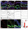
Pulmonary lymphangiectasia resulting from vascular endothelial growth factor-C overexpression during a critical period

Affiliations

Pulmonary lymphangiectasia resulting from vascular endothelial growth factor-C overexpression during a critical period

Li-Chin Yao et al. Circ Res. .

Abstract

Rationale: Lymphatic vessels in the respiratory tract normally mature into a functional network during the neonatal period, but under some pathological conditions they can grow as enlarged, dilated sacs that result in the potentially lethal condition of pulmonary lymphangiectasia.

Objective: We sought to determine whether overexpression of the lymphangiogenic growth factor (vascular endothelial growth factor-C [VEGF-C]) can promote lymphatic growth and maturation in the respiratory tract. Unexpectedly, perinatal overexpression of VEGF-C in the respiratory epithelium led to a condition resembling human pulmonary lymphangiectasia, a life-threatening disorder of the newborn characterized by respiratory distress and the presence of widely dilated lymphatics.

Methods and results: Administration of doxycycline to Clara cell secretory protein-reverse tetracycline-controlled transactivator/tetracycline operator-VEGF-C double-transgenic mice during a critical period from embryonic day 15.5 to postnatal day 14 was accompanied by respiratory distress, chylothorax, pulmonary lymphangiectasia, and high mortality. Enlarged sac-like lymphatics were abundant near major airways, pulmonary vessels, and visceral pleura. Side-by-side comparison revealed morphological features similar to pulmonary lymphangiectasia in humans. The condition was milder in mice given doxycycline after age postnatal day 14 and did not develop after postnatal day 35. Mechanistic studies revealed that VEGF recptor (VEGFR)-3 alone drove lymphatic growth in adult mice, but both VEGFR-2 and VEGFR-3 were required for the development of lymphangiectasia in neonates. VEGFR-2/VEGFR-3 heterodimers were more abundant in the dilated lymphatics, consistent with the involvement of both receptors. Despite the dependence of lymphangiectasia on VEGFR-2 and VEGFR-3, the condition was not reversed by blocking both receptors together or by withdrawing VEGF-C.

Conclusions: The findings indicate that VEGF-C overexpression can induce pulmonary lymphangiectasia during a critical period in perinatal development.

Keywords: VEGFR-2; VEGFR-3; chylothorax; lung; lymphangiogenesis; lymphangiomatosis, pulmonary; lymphatic vessels; pulmonary edema.


Figures
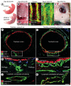
Figure 1
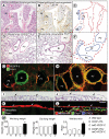
Figure 1. Lymphangiectasia in trachea of neonatal CCSP-VEGF-C mice
A, Normal skin color of control newborn mouse and cyanotic skin of newborn CCSP-VEGF-C mouse on doxycycline from E16.5 to P0. B, Pleural effusion (asterisk) in newborn CCSP-VEGF-C mouse on doxycycline from E16.5 to P0. C, Segmented pattern of lymphatics (LYVE-1, green) and blood vessels (PECAM-1, red) in normal trachea of control newborn mouse (CCSP-VEGF-C on water). D, Widespread lymphangiectasia in trachea of CCSP-VEGF-C mouse on doxycycline from E16.5 to P0. E, Chylothorax (asterisk) in CCSP-VEGF-C mouse on doxycycline from P0 to P14. F-I, Tracheal cross-sections stained for epithelial Clara cells (CCSP, red), lymphatics (LYVE-1 green), and blood vessels (PECAM-1, blue) in CCSP-VEGF-C mice on water (F, H) or doxycycline (G, I) from P0 to P14. F, Lymphatics in control mouse are scattered around the trachea. G, Lymphatics in CCSP-VEGF-C mouse on doxycycline surround most of the trachea beneath the epithelium. Boxes in F and G mark regions in H and I that show the differences in lymphatics (LYVE-1, green, arrows) and similarities of blood vessels (PECAM-1, blue, arrowheads) in the two conditions. J-K, Podocalyxin (red) is restricted to the luminal surface and LYVE-1 (green) is on both surfaces of lymphatic endothelium in CCSP-VEGF-C mice on water (J) or doxycycline (K) from P0 to P14, demonstrating that lymphatic sacs in lymphangiectasia have a lumen (K). Scale bar: 100 μm (C-D); 150 μm (F-G); 50 μm (H-I); 10 μm (J-K).
Figure 2
Figure 2. Comparison of lungs in child with pulmonary lymphangiectasia and in neonatal CCSP-VEGF-C mice
A-B, Widely dilated lymphatics (asterisks) in lung from a 1-month-old girl with pulmonary vein stenosis accompanied by lymphangiectasia (Case A). Sections from autopsy specimen stained by H&E (A) or D2-40/podoplanin (B, brown). C, Enlarged lymphatics in A and B are marked by dashed red lines. Other landmarks are shown by solid blue lines. Br, bronchiole; PA, pulmonary artery; and PV, pulmonary vein. D-E, Lung sections stained by H&E from neonatal CCSP-VEGF-C mice on water (D) or doxycycline (E) from P0 to P14. F, Regions of lymphangiectasia in E (asterisks) around bronchi and pulmonary vessels are outlined by dashed red lines. G-H, Sparse lymphatics around major bronchus and pulmonary vessels (arrows) in lung of control mouse on water (G) compared to widespread lymphatics in lung of CCSP-VEGF-C mouse on doxycycline from P0 to P14 (H). Sections stained for VEGFR-3 (red) and Prox1 (green). Green staining of bronchial smooth muscle does not reflect Prox1. I-J, Lung section with no pleural lymphatics in neonate on water (I) compared to subpleural lymphangiectasia (asterisks) in lung of neonate on doxycycline from P0 to P14 (J). Arrows mark surface of visceral pleura. Stained by H&E. K-L, Visceral pleura stained for mesothelin (red, arrows) has no lymphatics in control lung (K) but overlies subpleural lymphangiectasia (Prox1, green nuclei, asterisks) in lung of CCSP-VEGF-C mouse on doxycycline (L). M, Measurements of wet lung weight (mg), dry lung weight (mg), and ratio of wet-to-dry lung weight for wild-type mice (A), single transgenic mice (B, C), and CCSP-VEGF-C double transgenic mice on doxycycline from P0 to P35 (D). All three measurements in CCSP-VEGF-C mice are abnormal. *P < 0.05 vs. wild-type. Scale bar: 250 μm (D-E); 150 μm (G-J); 50 μm (K-L).
Figure 3
Figure 3. Abnormal lymphatic junctions and barrier function in lymphangiectasia
A-B, Adherens junctions (VE-cadherin, black) between endothelial cells of tracheal lymphatics are shown in inverted grayscale images. Discontinuous, button-like junctions (arrows) in lymphatic of normal mouse at P14 (A) are contrasted with continuous zipper-like junctions (arrows) in lymphatic of CCSP-VEGF-C mouse on doxycycline from P0 to P14 (B). C-D, Adherens junctions (VE-cadherin, red) between endothelial cells of lung lymphatics (Prox1, green), compare button-like junctions (arrows) in CCSP-VEGF-C mouse on water (C) with zipper-like junctions (arrows) in mouse on doxycycline from P0 to P14 (D). E-G, Image pairs comparing thoracic duct in normal Prox1-GFP mouse at P0 and Prox1-GFP/CCSP-VEGF-C mouse on doxycycline from E16.5 to P0. E, Thoracic duct normally filled with chyle (left) is compared to almost invisible thoracic duct in mouse on doxycycline (dashed lines, right). F, Thoracic duct with strong Prox1-GFP fluorescence in normal valves (left) is compared to thoracic duct with herniations (arrows) and abnormal valves in mouse on doxycycline (right). G, Normal thoracic duct filled with Dil-labeled chyle from milk (left) is compared to DiI extravasated from thoracic duct in mouse on doxycycline. Scale bar: 20 μm (A-D); 900 μm (E); 250 μm (F-G).
Figure 4
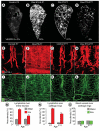
Figure 4. Age-related decrease in severity of VEGF-C driven lymphangiectasia
A-D, Decreased severity of lymphangiectasia in CCSP-VEGF-C mice on doxycycline beginning at E16.5, P0, or P70. Photographic montages of lungs showing age-related differences in abundance of lymphatics (VEGFR-3 immunoreactivity, white), and corresponding area densities of white pixels (%), in CCSP-VEGF-C mice on water at P3 (A) or on doxycycline from E16.5 to P1 (B), P0 to P7 (C), or P70 to P77 (D). E-L, Lymphatics (E-H, LYVE-1, red) and blood vessels (I-L, PECAM-1, green) in tracheal whole mounts from newborn (E, F, I, J) and adult (G, H, K, L) CCSP-VEGF-C mice on water (E, G, I, K) or doxycycline (F, H, J, L) for 7 days. E, G, Segmented pattern of lymphatics in neonatal and adult mice on water. F, Sheet-like lymphangiectasia in neonatal mouse on doxycycline from P0 to P7. H, Sprouting lymphangiogenesis in adult mouse on doxycycline from P70 to P77. I-L, No apparent differences in blood vessels of neonatal or adult mice on water or doxycycline. M-O, Area density of lymphatics over entire trachea (M), over cartilage rings (N), and corresponding blood vessels over cartilage rings (O). * P < 0.05 vs. water. Scale bar: 450 μm (A, C); 350 μm (B); 600 μm (D); 200 μm (E-L).
Figure 5
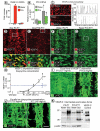
Figure 5. VEGF-C comparison in neonatal and adult CCSP-VEGF-C mice
A-B, VEGF-C (A) and rtTA (B) mRNA expression in trachea of CCSP-VEGF-C mice at P7 or P77 after doxycycline for 7 days and in corresponding controls (water). *P < 0.05 vs. neonates on water; †P < 0.05 vs. neonates on water or doxycycline. C, Intensity of VEGF-C immunofluorescence, measured along dashed line with ImageJ, in tracheal epithelium imaged by confocal microscopy with the same settings. D-G, VEGF-C immunoreactivity (red) in epithelium of tracheal whole mounts of neonatal and adult mice shown alone (D, E, upper) and with CCSP (green) immunoreactivity. White boxes in D-G demarcate regions enlarged below. H, Dose-response relationship between VEGF-C mRNA expression in trachea and doxycycline concentration in drinking water. Tracheal VEGF-C mRNA was about the same (dashed line) in neonates on doxycycline at 10−1 mg/ml and adults on doxycycline at 5 mg/ml. * P < 0.05 vs. water (0 on X-axis; 0.005 on Y-axis). I, Lymphangiectasia (LYVE-1) occurred in neonates but not in adults even when doxycycline concentration was adjusted to match VEGF-C expression at the two ages. J, Similarly, lymphangiectasia was present in neonates but not in adults when both were given doxycycline at a low concentration (10−2 mg/ml). K, Western blot showing stronger bands for intermediate forms of VEGF-C (32-35 kDa) in neonates than in adults on doxycycline for 7 days (upper panel). The mature form of VEGF-C (20 kDa, asterisk) is more abundant in adults on doxycycline (middle panel). Bands from conditioned medium harvested from cells transfected with native mouse VEGF-C serve as a reference for molecular weights . The products of full-length cDNA contain intermediate and mature forms of VEGF-C polypeptides. Intermediate forms from CCSP-VEGF-C mice are slightly larger in molecular weight than the corresponding positive control. Upper and middle panels are from different exposures of the same blot. Loading control is beta-actin (lower panel). Scale bar: 70 μm (C); 125 μm (D-G upper; I-J left); 25 μm (D-G lower); 175 μm (I-J right).
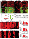
Figure 6
Figure 6. VEGFR-2 and VEGFR-3 in trachea of CCSP-VEGF-C mice
A-D, Strong VEGFR-2 immunoreactivity (red) in tracheal blood vessels (arrowheads) and weak VEGFR-2 staining in lymphatics (arrows) compared to strong VEGFR-3 staining in lymphatics of CCSP-VEGF-C mice on water or doxycycline from P0 to P7 or P70 to P77. E, Expression of VEGFR-2 and VEGFR-3 mRNA in trachea of same groups as in A-D (N = 8 per group). *P < 0.05 vs. baseline at corresponding age. F, Western blots for VEGFR-2 and VEGFR-3 in trachea of same groups as in E (N = 10 per group). Loading control is beta-actin. In neonates, VEGFR-2 protein was increased 2-fold and VEGFR-3 was increased 7-fold after doxycycline. In adults, VEGFR-2 protein was increased 2-fold and VEGFR-3 was increased 4-fold after doxycycline. G, J, Lymphatics stained for LYVE-1 (red) in tracheal whole mounts of neonatal (G) or adult (J) mice after doxycycline plus PBS, anti-VEGFR-2 antibody DC101, anti-VEGFR-3 antibody mF4-31C1, or both antibodies for 7 days. Scale bar: 80 μm (A-D); 200 μm (G, J). H, I, K, Extent of inhibition of lymphangiogenesis in CCSP-VEGF-C mice treated with doxycycline and inhibitory antibodies used in G and J. LYVE-1-positive lymphatics were assessed over entire trachea of neonatal mice at P7 (H) or by measuring sprouts over cartilage rings at P7 (I) or P77 (K). * P < 0.05 vs. PBS. † P < 0.05 vs. mF4-31C1.
Figure 7
Figure 7. VEGFR-2/VEGFR-3 heterodimers revealed in lymphatic endothelial cells by proximity ligation assay (PLA)
A-D, Confocal micrographs comparing the number of PLA red dots (VEGFR-2/VEGFR-3 heterodimers) in tracheal lymphatics (LYVE-1, green) of neonatal (A-B) and adult (C-D) CCSP-VEGF-C mice on water or doxycycline for 7 days. Nuclei are stained with DAPI. Inverted grayscale versions of the same images emphasize the larger number of PLA dots in the neonate (B, lower) than in the adult (D, lower). E, PLA dots marking lymphatic endothelial cells in trachea of neonatal CCSP-VEGF-C mouse on doxycycline for 7 days, shown alone (left panel), with LYVE-1 (middle panel), and with LYVE-1 and DAPI-stained nuclei (right panel). Scale bar: 20 μm. F, Number of PLA dots per lymphatic endothelial cell nucleus. *P < 0.05 vs. water; †P < 0.05 vs. adults on doxycycline.
Figure 8
Figure 8. Limited reversibility of lymphangiectasia
A-B, Lymphatics stained for LYVE-1 immunoreactivity in tracheal whole mounts from CCSP-VEGF-C mice at baseline (A) or after doxycycline from P0 to P7 and then no doxycycline from P7 to P14 (B). Lymphangiectasia is still widespread after one week off doxycycline. C, Expression of VEGF-C mRNA in trachea of CCSP-VEGF-C mice (N = 5 per group) at baseline (green) or after doxycycline from P0 to P7 and then no doxycycline for 0 to 4 weeks (red). D, E, Extent of lymphangiectasia in trachea shown by LYVE-1 staining after doxycycline from P0 to P7 followed by inhibition of VEGFR-2, VEGFR-3, and both receptors by DC101 or mF4-31C1 given from P7 to P14 (D). Area density of LYVE-1 immunoreactivity over the entire trachea under these conditions (E). * P < 0.05 vs. PBS. F, Persistence of lymphangiectasia in trachea of a mouse on doxycycline from P0 to P7 and then off doxycycline for 19 months. Amount at 19 months (E, yellow bar) is only 24% less than at P7. Scale bar: 450 μm (A-B, D); 700 μm (F).

Similar articles

Cited by

  • A census of the lung: CellCards from LungMAP.
    Sun X, Perl AK, Li R, Bell SM, Sajti E, Kalinichenko VV, Kalin TV, Misra RS, Deshmukh H, Clair G, Kyle J, Crotty Alexander LE, Masso-Silva JA, Kitzmiller JA, Wikenheiser-Brokamp KA, Deutsch G, Guo M, Du Y, Morley MP, Valdez MJ, Yu HV, Jin K, Bardes EE, Zepp JA, Neithamer T, Basil MC, Zacharias WJ, Verheyden J, Young R, Bandyopadhyay G, Lin S, Ansong C, Adkins J, Salomonis N, Aronow BJ, Xu Y, Pryhuber G, Whitsett J, Morrisey EE; NHLBI LungMAP Consortium. Sun X, et al. Dev Cell. 2022 Jan 10;57(1):112-145.e2. doi: 10.1016/j.devcel.2021.11.007. Epub 2021 Dec 21. Dev Cell. 2022. PMID: 34936882 Free PMC article.
  • Immune cell trafficking, lymphatics and hypertension.
    Balasubbramanian D, Lopez Gelston CA, Rutkowski JM, Mitchell BM. Balasubbramanian D, et al. Br J Pharmacol. 2019 Jun;176(12):1978-1988. doi: 10.1111/bph.14370. Epub 2018 Jun 25. Br J Pharmacol. 2019. PMID: 29797446 Free PMC article. Review.
  • Heterogeneity in the lymphatic vascular system and its origin.
    Ulvmar MH, Mäkinen T. Ulvmar MH, et al. Cardiovasc Res. 2016 Sep;111(4):310-21. doi: 10.1093/cvr/cvw175. Epub 2016 Jun 29. Cardiovasc Res. 2016. PMID: 27357637 Free PMC article. Review.
  • Hyperoxia Disrupts Lung Lymphatic Homeostasis in Neonatal Mice.
    Shankar N, Thapa S, Shrestha AK, Sarkar P, Gaber MW, Barrios R, Shivanna B. Shankar N, et al. Antioxidants (Basel). 2023 Mar 2;12(3):620. doi: 10.3390/antiox12030620. Antioxidants (Basel). 2023. PMID: 36978868 Free PMC article.
  • CD146, from a melanoma cell adhesion molecule to a signaling receptor.
    Wang Z, Xu Q, Zhang N, Du X, Xu G, Yan X. Wang Z, et al. Signal Transduct Target Ther. 2020 Aug 11;5(1):148. doi: 10.1038/s41392-020-00259-8. Signal Transduct Target Ther. 2020. PMID: 32782280 Free PMC article. Review.

References

    1. Leak LV. Lymphatic removal of fluids and particles in the mammalian lung. Environ Health Perspect. 1980;35:55–75. - PMC - PubMed
    1. Randolph GJ, Angeli V, Swartz MA. Dendritic-cell trafficking to lymph nodes through lymphatic vessels. Nat Rev Immunol. 2005;5:617–628. - PubMed
    1. El-Chemaly S, Levine SJ, Moss J. Lymphatics in lung disease. Ann N Y Acad Sci. 2008;1131:195–202. - PMC - PubMed
    1. Bredt H. lymphangiectasia pulmonum congenita. Virchows Arch. 1952;321:517–530. - PubMed
    1. Laurence KM. Congenital pulmonary cystic lymphangiectasis. J Pathol Bacteriol. 1955;70:325–333. - PubMed

Publication types

MeSH terms

Substances

Supplementary concepts