

Manipulation of cellular DNA damage repair machinery facilitates propagation of human papillomaviruses
- PMID: 24412279
- PMCID: PMC4050178
- DOI: 10.1016/j.semcancer.2013.12.003
Manipulation of cellular DNA damage repair machinery facilitates propagation of human papillomaviruses
Abstract
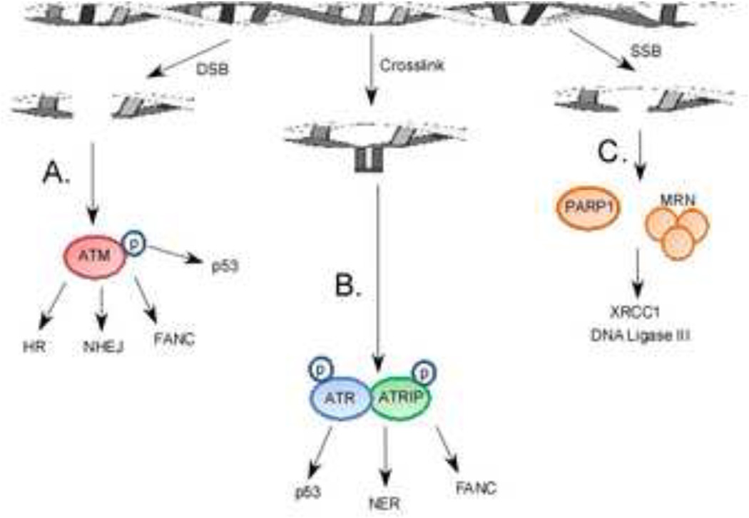
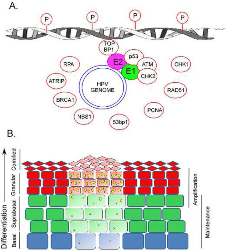
In general, the interplay among viruses and DNA damage repair (DDR) pathways can be divided based on whether the interaction promotes or inhibits the viral lifecycle. The propagation of human papillomaviruses is both promoted and inhibited by DDR proteins. As a result, HPV proteins both activate repair pathways, such as the ATM and ATR pathways, and inhibit other pathways, most notably the p53 signaling pathway. Indeed, the role of HPV proteins, with regard to the DDR pathways, can be divided into two broad categories. The first set of viral proteins, HPV E1 and E2 activate a DNA damage response and recruit repair proteins to viral replication centers, where these proteins are likely usurped to replicate the viral genome. Because the activation of the DDR response typically elicits a cell cycle arrest that would impeded the viral lifecycle, the second set of HPV proteins, HPV E6 and E7, prevents the DDR response from pausing cell cycle progression or inducing apoptosis. This review provides a detailed account of the interactions among HPV proteins and DDR proteins that facilitate HPV propagation.
Keywords: DNA damage repair; HPV; HPV replication.
Copyright © 2013 Elsevier Ltd. All rights reserved.
Conflict of interest statement
The authors declare that they have no conflicts of interest.
Figures
Similar articles
-
Why Human Papillomaviruses Activate the DNA Damage Response (DDR) and How Cellular and Viral Replication Persists in the Presence of DDR Signaling.Viruses. 2017 Sep 21;9(10):268. doi: 10.3390/v9100268. Viruses. 2017. PMID: 28934154 Free PMC article. Review.
-
Spatial and Functional Organization of Human Papillomavirus Replication Foci in the Productive Stage of Infection.mBio. 2021 Dec 21;12(6):e0268421. doi: 10.1128/mBio.02684-21. Epub 2021 Nov 9. mBio. 2021. PMID: 34749533 Free PMC article.
-
Regulation of the Human Papillomavirus Life Cycle by DNA Damage Repair Pathways and Epigenetic Factors.Viruses. 2020 Jul 10;12(7):744. doi: 10.3390/v12070744. Viruses. 2020. PMID: 32664381 Free PMC article. Review.
-
Human papillomaviruses recruit cellular DNA repair and homologous recombination factors to viral replication centers.J Virol. 2012 Sep;86(17):9520-6. doi: 10.1128/JVI.00247-12. Epub 2012 Jun 27. J Virol. 2012. PMID: 22740399 Free PMC article.
-
Human Papillomaviruses Preferentially Recruit DNA Repair Factors to Viral Genomes for Rapid Repair and Amplification.mBio. 2018 Feb 13;9(1):e00064-18. doi: 10.1128/mBio.00064-18. mBio. 2018. PMID: 29440569 Free PMC article.
Cited by
-
Genomic Landscape of Primary and Recurrent Anal Squamous Cell Carcinomas in Relation to HPV Integration, Copy-Number Variation, and DNA Damage Response Genes.Mol Cancer Res. 2021 Aug;19(8):1308-1321. doi: 10.1158/1541-7786.MCR-20-0884. Epub 2021 Apr 21. Mol Cancer Res. 2021. PMID: 33883185 Free PMC article.
-
FANCD2 Binds Human Papillomavirus Genomes and Associates with a Distinct Set of DNA Repair Proteins to Regulate Viral Replication.mBio. 2017 Feb 14;8(1):e02340-16. doi: 10.1128/mBio.02340-16. mBio. 2017. PMID: 28196964 Free PMC article.
-
The Interplay between Antiviral Signalling and Carcinogenesis in Human Papillomavirus Infections.Cancers (Basel). 2020 Mar 10;12(3):646. doi: 10.3390/cancers12030646. Cancers (Basel). 2020. PMID: 32164347 Free PMC article. Review.
-
Understanding the Role of Intrinsic Disorder of Viral Proteins in the Oncogenicity of Different Types of HPV.Int J Mol Sci. 2018 Jan 9;19(1):198. doi: 10.3390/ijms19010198. Int J Mol Sci. 2018. PMID: 29315236 Free PMC article.
-
DNA repair gene expression is increased in HPV positive head and neck squamous cell carcinomas.Virology. 2020 Sep;548:174-181. doi: 10.1016/j.virol.2020.07.004. Epub 2020 Jul 22. Virology. 2020. PMID: 32838940 Free PMC article.
References
-
- Lindahl T. Instability and decay of the primary structure of DNA. Nature. 1993;362:709–715. - PubMed
-
- Boyer J, Rohleder K, Ketner G. Adenovirus E4 34k and E4 11k inhibit double strand break repair and are physically associated with the cellular DNA-dependent protein kinase. Virology. 1999;263:307–312. - PubMed
-
- Stracker TH, Carson CT, Weitzman MD. Adenovirus oncoproteins inactivate the Mre11-Rad50-NBS1 DNA repair complex. Nature. 2002;418:348–352. - PubMed
Publication types
MeSH terms
Substances
Grants and funding
LinkOut - more resources
Full Text Sources
Other Literature Sources
Medical
Research Materials
Miscellaneous
