

Imaging of an inflammatory injury in the newborn rat brain with photoacoustic tomography
- PMID: 24386140
- PMCID: PMC3873292
- DOI: 10.1371/journal.pone.0083045
Imaging of an inflammatory injury in the newborn rat brain with photoacoustic tomography
Abstract
Background: The precise assessment of cerebral saturation changes during an inflammatory injury in the developing brain, such as seen in periventricular leukomalacia, is not well defined. This study investigated the impact of inflammation on locoregional cerebral oxygen saturation in a newborn rodent model using photoacoustic imaging.
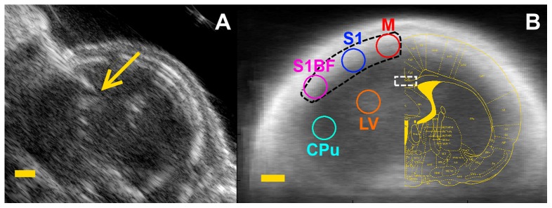
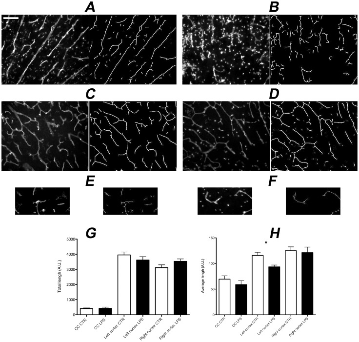
Methods: 1 mg/kg of lipopolysaccharide(LPS) diluted in saline or saline alone was injected under ultrasound guidance directly in the corpus callosum of P3 rat pups. Coronal photoacoustic images were carried out 24 h after LPS exposure. Locoregional oxygen saturation (SO2) and resting state connectivity were assessed in the cortex and the corpus callosum. Microvasculature was then evaluated on cryosection slices by lectin histochemistry.
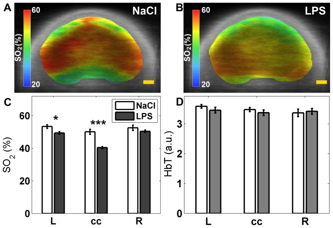
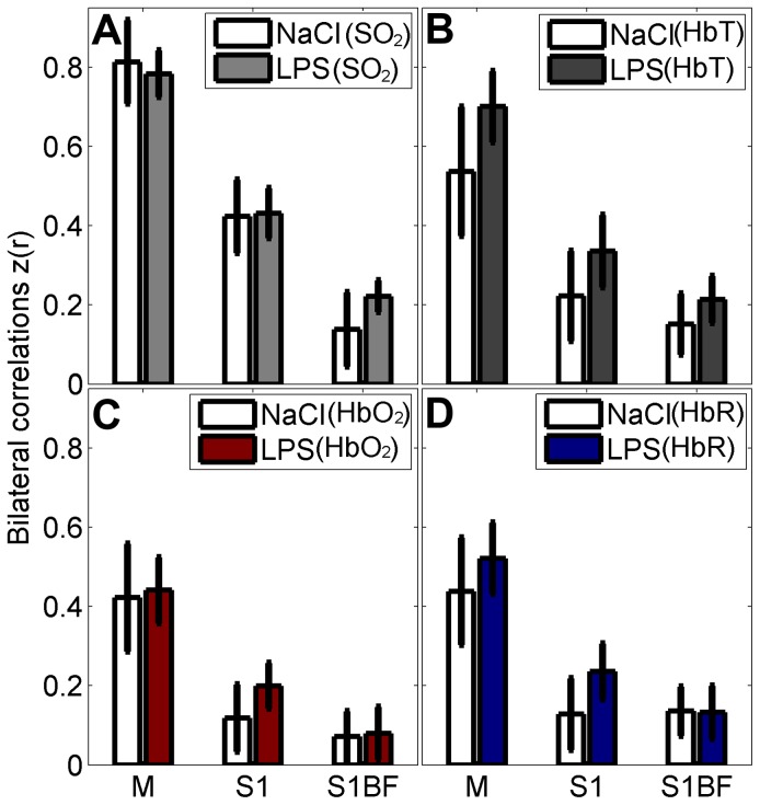
Results: Significant reduction of SO2 was found in the corpus callosum; reduced SO2 was also found in the cortex ipsilateral to the injection site. Seed-based functional connectivity analysis showed that bilateral connectivity was not affected by LPS exposure. Changes in locoregional oxygen saturation were accompanied by a significant reduction in the average length of microvessels in the left cortex but no differences were observed in the corpus callosum.
Conclusion: Inflammation in the developing brain induces marked reduction of locoregional oxygen saturation, predominantly in the white matter not explained by microvascular degeneration. The ability to examine regional saturation offers a new way to monitor injury and understand physiological disturbance non-invasively.
Conflict of interest statement
Figures
Similar articles
-
Alpha-Phenyl-n-tert-butyl-nitrone attenuates lipopolysaccharide-induced neuronal injury in the neonatal rat brain.Neuroscience. 2008 Feb 6;151(3):737-44. doi: 10.1016/j.neuroscience.2007.09.087. Epub 2007 Nov 29. Neuroscience. 2008. PMID: 18191905 Free PMC article.
-
Prophylactic maternal N-acetylcysteine in rats prevents maternal inflammation-induced offspring cerebral injury shown on magnetic resonance imaging.Am J Obstet Gynecol. 2013 Mar;208(3):213.e1-6. doi: 10.1016/j.ajog.2013.01.023. Am J Obstet Gynecol. 2013. PMID: 23433325
-
Erythropoietin attenuates lipopolysaccharide-induced white matter injury in the neonatal rat brain.Neonatology. 2007;92(4):269-78. doi: 10.1159/000105493. Epub 2007 Jul 11. Neonatology. 2007. PMID: 17627093
-
In vivo photoacoustic imaging dynamically monitors the structural and functional changes of ischemic stroke at a very early stage.Theranostics. 2020 Jan 1;10(2):816-828. doi: 10.7150/thno.38554. eCollection 2020. Theranostics. 2020. PMID: 31903152 Free PMC article.
-
In vivo MRI analysis of an inflammatory injury in the developing brain.Brain Behav Immun. 2010 Jul;24(5):759-67. doi: 10.1016/j.bbi.2009.11.005. Epub 2009 Nov 27. Brain Behav Immun. 2010. PMID: 19945527 Free PMC article.
Cited by
-
Simultaneous photoacoustic and optically mediated ultrasound microscopy: an in vivo study.Biomed Opt Express. 2015 Jan 29;6(2):631-8. doi: 10.1364/BOE.6.000631. eCollection 2015 Feb 1. Biomed Opt Express. 2015. PMID: 25780752 Free PMC article.
-
Persistent reduction in sialylation of cerebral glycoproteins following postnatal inflammatory exposure.J Neuroinflammation. 2018 Dec 5;15(1):336. doi: 10.1186/s12974-018-1367-2. J Neuroinflammation. 2018. PMID: 30518374 Free PMC article.
-
Multimodal Contrast Agents for Optoacoustic Brain Imaging in Small Animals.Front Bioeng Biotechnol. 2021 Sep 28;9:746815. doi: 10.3389/fbioe.2021.746815. eCollection 2021. Front Bioeng Biotechnol. 2021. PMID: 34650961 Free PMC article. Review.
-
Altered Functional Connectivity Following an Inflammatory White Matter Injury in the Newborn Rat: A High Spatial and Temporal Resolution Intrinsic Optical Imaging Study.Front Neurosci. 2017 Jul 4;11:358. doi: 10.3389/fnins.2017.00358. eCollection 2017. Front Neurosci. 2017. PMID: 28725174 Free PMC article.
-
Photostability of Contrast Agents for Photoacoustics: The Case of Gold Nanorods.Nanomaterials (Basel). 2021 Jan 6;11(1):116. doi: 10.3390/nano11010116. Nanomaterials (Basel). 2021. PMID: 33419130 Free PMC article. Review.
References
-
- Dalitz P, Harding R, Rees SM, Cock ML (2003) Prolonged reductions in placental blood flow and cerebral oxygen delivery in preterm fetal sheep exposed to endotoxin: possible factors in white matter injury after acute infection. J Soc Gynecol Investig 10: 283–290. - PubMed
Publication types
MeSH terms
Grants and funding
LinkOut - more resources
Full Text Sources
Other Literature Sources
