IFI16 DNA sensor is required for death of lymphoid CD4 T cells abortively infected with HIV
- PMID: 24356113
- PMCID: PMC3976200
- DOI: 10.1126/science.1243640
IFI16 DNA sensor is required for death of lymphoid CD4 T cells abortively infected with HIV
Abstract
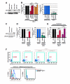
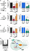
The progressive depletion of quiescent "bystander" CD4 T cells, which are nonpermissive to HIV infection, is a principal driver of the acquired immunodeficiency syndrome (AIDS). These cells undergo abortive infection characterized by the cytosolic accumulation of incomplete HIV reverse transcripts. These viral DNAs are sensed by an unidentified host sensor that triggers an innate immune response, leading to caspase-1 activation and pyroptosis. Using unbiased proteomic and targeted biochemical approaches, as well as two independent methods of lentiviral short hairpin RNA-mediated gene knockdown in primary CD4 T cells, we identify interferon-γ-inducible protein 16 (IFI16) as a host DNA sensor required for CD4 T cell death due to abortive HIV infection. These findings provide insights into a key host pathway that plays a central role in CD4 T cell depletion during disease progression to AIDS.
Figures


Comment in
-
Viral pathogenesis: HIV-1 adds fuel to the fire.Nat Rev Microbiol. 2014 Feb;12(2):74-5. doi: 10.1038/nrmicro3201. Epub 2014 Jan 2. Nat Rev Microbiol. 2014. PMID: 24384600 No abstract available.
-
Immunology. The fiery side of HIV-induced T cell death.Science. 2014 Jan 24;343(6169):383-4. doi: 10.1126/science.1250175. Science. 2014. PMID: 24458634 No abstract available.
Similar articles
-
Immunology. The fiery side of HIV-induced T cell death.Science. 2014 Jan 24;343(6169):383-4. doi: 10.1126/science.1250175. Science. 2014. PMID: 24458634 No abstract available.
-
Viral pathogenesis: HIV-1 adds fuel to the fire.Nat Rev Microbiol. 2014 Feb;12(2):74-5. doi: 10.1038/nrmicro3201. Epub 2014 Jan 2. Nat Rev Microbiol. 2014. PMID: 24384600 No abstract available.
-
Blood-Derived CD4 T Cells Naturally Resist Pyroptosis during Abortive HIV-1 Infection.Cell Host Microbe. 2015 Oct 14;18(4):463-70. doi: 10.1016/j.chom.2015.09.010. Cell Host Microbe. 2015. PMID: 26468749 Free PMC article.
-
The interferon-inducible DNA-sensor protein IFI16: a key player in the antiviral response.New Microbiol. 2015 Jan;38(1):5-20. Epub 2015 Jan 1. New Microbiol. 2015. PMID: 25742143 Review.
-
The Role of Inflammasome Activation in Early HIV Infection.J Immunol Res. 2021 Sep 20;2021:1487287. doi: 10.1155/2021/1487287. eCollection 2021. J Immunol Res. 2021. PMID: 34595244 Free PMC article. Review.
Cited by
-
Host Factors in Retroviral Integration and the Selection of Integration Target Sites.Microbiol Spectr. 2014 Dec;2(6):10.1128/microbiolspec.MDNA3-0026-2014. doi: 10.1128/microbiolspec.MDNA3-0026-2014. Microbiol Spectr. 2014. PMID: 26104434 Free PMC article. Review.
-
TREX1 Knockdown Induces an Interferon Response to HIV that Delays Viral Infection in Humanized Mice.Cell Rep. 2016 May 24;15(8):1715-27. doi: 10.1016/j.celrep.2016.04.048. Epub 2016 May 12. Cell Rep. 2016. PMID: 27184854 Free PMC article.
-
Innate immunity against HIV-1 infection.Nat Immunol. 2015 Jun;16(6):554-62. doi: 10.1038/ni.3157. Nat Immunol. 2015. PMID: 25988887 Review.
-
Interferon-Inducible Protein 16 (IFI16) Has a Broad-Spectrum Binding Ability Against ssDNA Targets: An Evolutionary Hypothesis for Antiretroviral Checkpoint.Front Microbiol. 2019 Jul 4;10:1426. doi: 10.3389/fmicb.2019.01426. eCollection 2019. Front Microbiol. 2019. PMID: 31333597 Free PMC article.
-
Caspases in Cell Death, Inflammation, and Pyroptosis.Annu Rev Immunol. 2020 Apr 26;38:567-595. doi: 10.1146/annurev-immunol-073119-095439. Epub 2020 Feb 4. Annu Rev Immunol. 2020. PMID: 32017655 Free PMC article. Review.
References
-
- Roberts TL, et al. HIN-200 proteins regulate caspase activation in response to foreign cytoplasmic DNA. Science. 2009 Feb 20;323(1057) - PubMed
Publication types
MeSH terms
Substances
Grants and funding
LinkOut - more resources
Full Text Sources
Other Literature Sources
Medical
Research Materials

