. 2013 Dec 15;27(24):2663-77.
doi: 10.1101/gad.225888.113.

Nucleosome-binding activities within JARID2 and EZH1 regulate the function of PRC2 on chromatin

Affiliations

Nucleosome-binding activities within JARID2 and EZH1 regulate the function of PRC2 on chromatin

Jinsook Son et al. Genes Dev. .

Erratum in

  • Genes Dev. 2014 Apr 15;28(8):921

Abstract

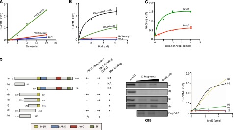
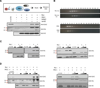
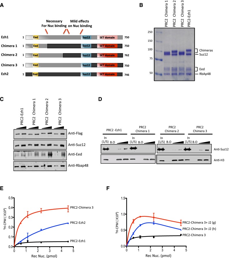
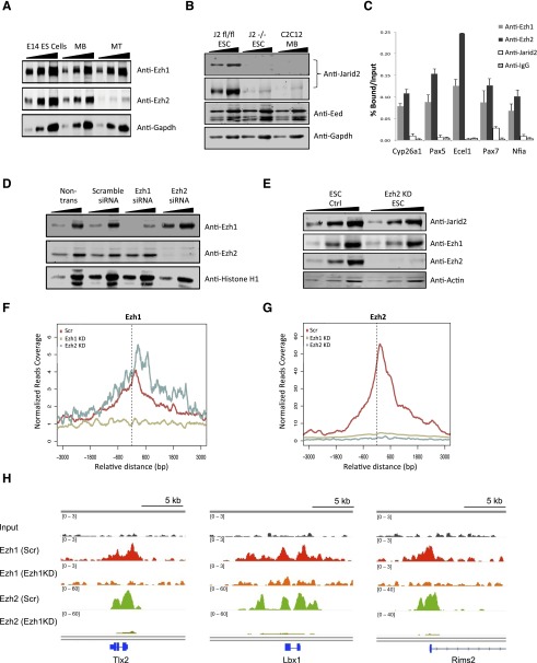
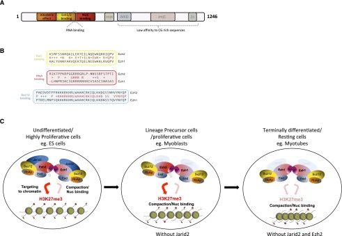
Polycomb-repressive complex 2 (PRC2) comprises specific members of the Polycomb group of epigenetic modulators. PRC2 catalyzes methylation of histone H3 at Lys 27 (H3K27me3) through its Enhancer of zeste (Ezh) constituent, of which there are two mammalian homologs: Ezh1 and Ezh2. Several ancillary factors, including Jarid2, modulate PRC2 function, with Jarid2 facilitating its recruitment to target genes. Jarid2, like Ezh2, is present in poorly differentiated and actively dividing cells, while Ezh1 associates with PRC2 in all cells, including resting cells. We found that Jarid2 exhibits nucleosome-binding activity that contributes to PRC2 stimulation. Moreover, such nucleosome-binding activity is exhibited by PRC2 comprising Ezh1 (PRC2-Ezh1), in contrast to PRC2-Ezh2. The presence of Ezh1 helps to maintain PRC2 occupancy on its target genes in myoblasts where Jarid2 is not expressed. Our findings allow us to propose a model in which PRC2-Ezh2 is important for the de novo establishment of H3K27me3 in dividing cells, whereas PRC2-Ezh1 is required for its maintenance in resting cells.

Keywords: EZH1; EZH2; JARID2; PRC2; Polycomb.


Figures

Figure 1.
Figure 1.
Minimal Jarid2 regions regulating PRC2. (A) Time course of an HMT assay using 0.67 pmol of PRC2 either alone or with 4 pmol of Aebp2 or 1 pmol of Jarid2 on 2.28 pmol of recombinant nucleosomes. The assay was incubated with 12.5 μM SAM (radiolabeled:cold = 1:45) and analyzed by scintillation counting after SDS-PAGE. (B) HMT assay performed with increasing amounts (0.39–6.25 μM) of SAM in the presence of PRC2 either alone or with Aebp2 and/or Jarid2 using recombinant nucleosomes as in A. (C) HMT assay performed with increasing amounts of Jarid2 or Aebp2 with PRC2 as in A. (D, left panel) Schematic representation of full-length Jarid2 and protein fragments analyzed for PRC2 stimulation, Ezh2 binding, and nucleosome binding. The previously characterized Jarid2 domains are also shown: Jumonji N (JmjN) (yellow), JmjC (red), ARID (blue), and zinc finger (ZF) (green). (Middle panel) Coomassie blue staining of a pull-down assay using Flag-tagged Ezh2 and Jarid2 protein fragments as shown in the left panel. Results of pull-down assays performed with Jarid2 protein fragments and Flag-tagged Ezh2 after Coomassie blue staining. (Right panel) HMT assay using 2.28 pmol of recombinant nucleosomes and 0.67 pmol of PRC2 either alone or with increasing amounts (0.68–5 pmol) of candidate Jarid2 fragments (designated with letters) as shown in the left panel.
Figure 2.
Figure 2.
Jarid2 interacts directly with nucleosomes, promoting PRC2 interaction. (A) Nucleosome-binding assay using biotin mononucleosomes and PRC2 in the presence of His-Aebp2 and/or His-Jarid2 and analyzed by Western blot using anti-Ezh2, anti-His tag, or anti-H3 antibody. (B) Sucrose gradient sedimentation assay using either nucleosomes or DNA with Jarid2 fragment g. (C) Nucleosome-binding assay using biotin mononucleosomes and His-Jarid2 fragments (as shown in Fig. 1D) followed by Western blot using anti-His tag or anti-H3 antibody. (D) Nucleosome-binding assay as in A using the Jarid2 fragments indicated.
Figure 3.
Figure 3.
PRC2 nucleosome-binding activity is enhanced when comprising Ezh1. (A) HMT assay performed with increasing amounts (1.67–20 pmol) of PRC2 containing either Ezh1 or Ezh2, as indicated. (B) Results of pull-down assays using Flag-tagged versions of Ezh1 or Ezh2 and Jarid2 fragment g after Coomassie blue staining. (C) HMT assay using 3.34 pmol of PRC2–Ezh1 in the absence or presence of 5 pmol of Jarid2 fragment g or h with increasing amounts of recombinant nucleosomes. (D) Western blot analysis of nucleosome-binding assays performed with increasing amounts (3.34–26.72 pmol) of either PRC2–Ezh1 or PRC2–Ezh2 using biotin mono- or oligo-nucleosomes and anti-Flag or anti-H3 antibody. (E) Western blot analysis of increasing amounts of PRC2–Ezh1, PRC2–Ezh2, or PRC2–Ezh1/Ezh2 using the antibodies indicated at the right. (F) Nucleosome-binding assay with increasing amounts of PRC2–Ezh1/Ezh2 using biotin mononucleosomes as in D. The signal for anti-Flag is shown in red, and the signal for anti-Ezh2 is in green.
Figure 4.
Figure 4.
Minimal Ezh1 regions regulating nucleosome binding. (A) Schematic representations of Ezh1, Ezh2, and Ezh1/Ezh2 chimera (1–3) comprising the following respective amino acids: (1) Ezh1 amino acids 1–132 and Ezh2 amino acids 129–746, (2) Ezh1 amino acids 1–274 and Ezh2 amino acids 259–746, and (3) Ezh1 amino acids 1–493 and Ezh2 amino acids 490–746. Previously characterized Eed or Suz12-binding regions are shown in yellow or blue, respectively, and the SET domain is shown in red. (B) Coomassie blue staining of the Flag tag affinity-purified PRC2 complex containing different Ezh1/Ezh2 chimera from SF9 cells, as indicated. (C) Western blot analysis of increasing amounts of PRC2–Ezh1 or PRC2 comprising the Ezh1/Ezh2 chimera indicated using the antibodies shown at the right. (D) Nucleosome-binding assays using biotin mononucleosomes with the PRC2 complexes indicated. (E) HMT assay performed with increasing amounts of recombinant nucleosomes in the presence of the PRC2 complexes indicated. (F) HMT assay performed with increasing amounts of recombinant nucleosomes using PRC2-Chimera 3 in the absence or presence of Jarid2 fragment g or h.
Figure 5.
Figure 5.
Ezh2 occupancy at PRC2 target genes in myoblasts is dependent, in part, on Ezh1. (A) Western blot analysis using 10–40 μg of cell extract prepared from embryonic day 14 (E14) ES cells, C2C12 myoblasts (MBs), and myotubes (MTs) using the antibodies indicated. (B) Western blot analysis using 20–40 μg of whole-cell extracts prepared from Jarid2 Flox/Flox ES cells, Jarid2 knockout ES cells, and C2C12 myoblasts using the antibodies indicated. (C) ChIP-qPCR analysis using C2C12 myoblasts and the antibodies indicated. Error bars represent the SD of triplicates. (D) Western blot analysis showing the efficiency of Ezh1 or Ezh2 knockdown using 10–20 μg of extracts from cells treated with the siRNA indicated. (E) Western blot analysis using 10–40 μg of cell extract prepared from wild-type or Ezh2 knockdown ES cells using the antibodies indicated. (F) Normalized density plots of selected Ezh1 target genes (±3 kb; bin, 10 bp) (see the Materials and Methods for Ezh1 proximate gene identification) in Scr, Ezh1, and Ezh2 knockdown C2C12 myoblasts. (G) Normalized density plots of Ezh2 using selected genes in F. (H) ChIP-seq binding profiles for anti-Ezh1 and anti-Ezh2 in Scr and Ezh1 knockdown C2C12 myoblasts.
Figure 6.
Figure 6.
Decreased H3K27me3 levels on PRC2 targets upon Ezh1 knockdown. (A) ChIP-qPCR analysis for anti-Ezh1 using Ezh1 or Ezh2 knockdown C2C12 myoblasts (MBs). (B) ChIP-qPCR analysis as in A using Ezh2 antibody. (C) ChIP-qPCR analysis for anti-H3 (left panel) or anti-H3K27me3 (right panel) using Scr or Ezh1 knockdown C2C12. Error bars represent the SD of triplicates.
Figure 7.
Figure 7.
(A) Schematic representation of Jarid2 indicating three functional domains located within the N terminus, as identified in this study. The RNA-binding domain is described by Kaneko et al. (in press). (B) Comparison of amino acid sequences of different domains present in Ezh1 and/or Ezh2. The conservation between the Eed-, RNA-, and Suz12-binding domains is shown. Note the low degree of conservation in the RNA-binding domain, consistent with the lack of Ezh1 RNA-binding activity. (C) Schematic model of the establishment and maintenance of H3K27me by PRC2. (Left) A dividing cell contains PRC2–Ezh2, PRC2–Ezh1, and Jarid2. PRC2–Ezh2 contains robust H3K27me3 activity, whereas PRC2–Ezh1 is less active in catalyzing H3K27me3 but can compact chromatin. Jarid2 and noncoding RNA can recruit PRC2–Ezh2 to its targets. (Middle) A lineage precursor cell lacks Jarid2; however, PRC2–Ezh2 can bind to chromatin through dimerization with PRC2–Ezh1. (Right) A differentiated cell lacks Ezh2 and Jarid2 such that the levels of H3K27me3 are maintained by PRC2–Ezh1 through its ability to bind nucleosomes in general and the allosteric stimulation of its HMT activity upon binding of its Eed subunit to pre-existing H3K27me3.

Similar articles

Cited by

References

    1. Bernstein BE, Mikkelsen TS, Xie X, Kamal M, Huebert DJ, Cuff J, Fry B, Meissner A, Wernig M, Plath K, et al. 2006. A bivalent chromatin structure marks key developmental genes in embryonic stem cells. Cell 125: 315–326 - PubMed
    1. Blais A, van Oevelen CJ, Margueron R, Acosta-Alvear D, Dynlacht BD 2007. Retinoblastoma tumor suppressor protein-dependent methylation of histone H3 lysine 27 is associated with irreversible cell cycle exit. J Cell Biol 179: 1399–1412 - PMC - PubMed
    1. Brown JL, Mucci D, Whiteley M, Dirksen ML, Kassis JA 1998. The Drosophila Polycomb group gene pleiohomeotic encodes a DNA binding protein with homology to the transcription factor YY1. Mol Cell 1: 1057–1064 - PubMed
    1. Cao R, Zhang Y 2004. SUZ12 is required for both the histone methyltransferase activity and the silencing function of the EED–EZH2 complex. Mol Cell 15: 57–67 - PubMed
    1. Cao R, Wang L, Wang H, Xia L, Erdjument-Bromage H, Tempst P, Jones RS, Zhang Y 2002. Role of histone H3 lysine 27 methylation in Polycomb-group silencing. Science 298: 1039–1043 - PubMed

Publication types

Associated data

LinkOut - more resources