. 2014 Feb 1;386(1):72-85.
doi: 10.1016/j.ydbio.2013.11.028. Epub 2013 Dec 11.

Loss of col8a1a function during zebrafish embryogenesis results in congenital vertebral malformations

Affiliations

Loss of col8a1a function during zebrafish embryogenesis results in congenital vertebral malformations

Ryan S Gray et al. Dev Biol. .

Abstract

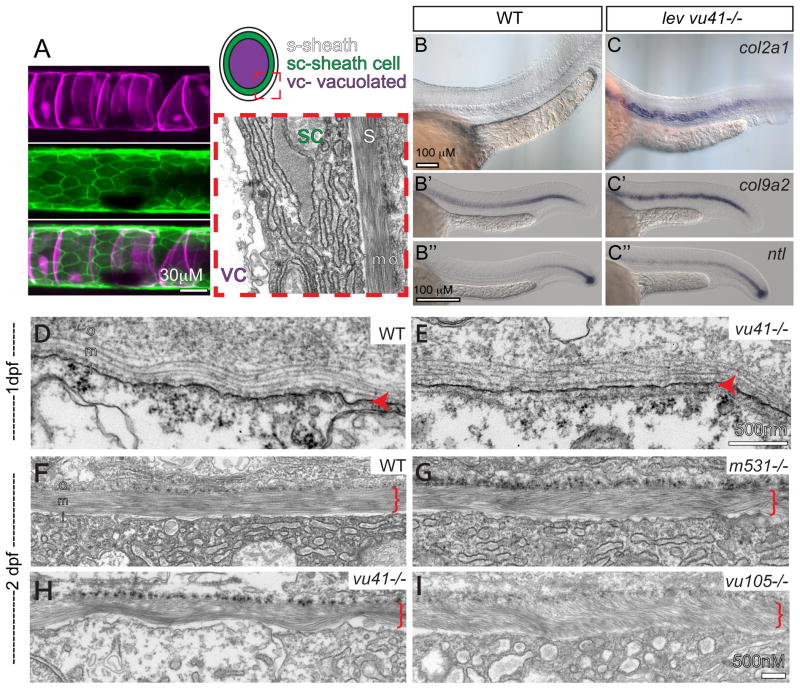
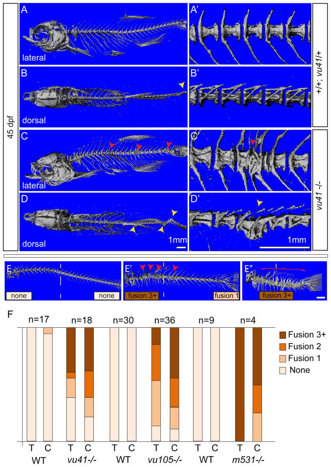
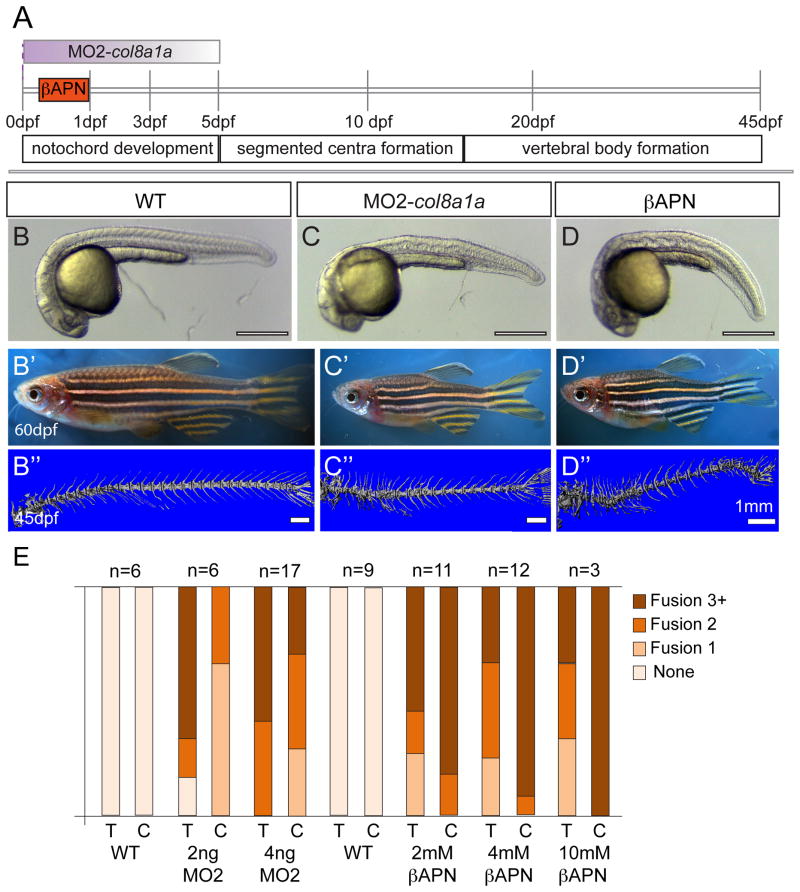
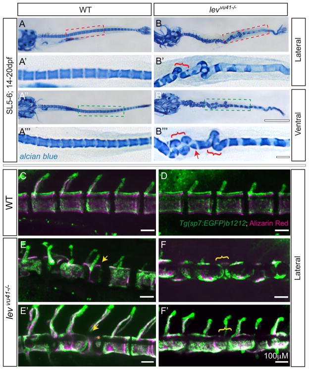
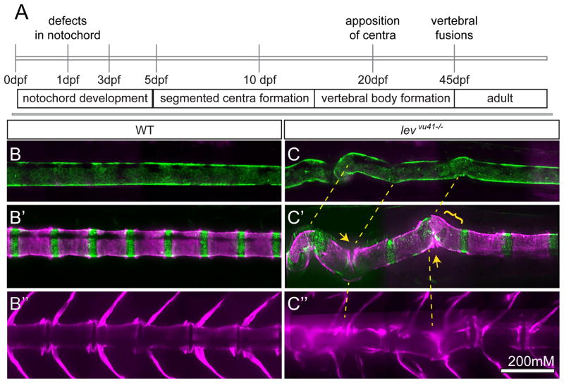
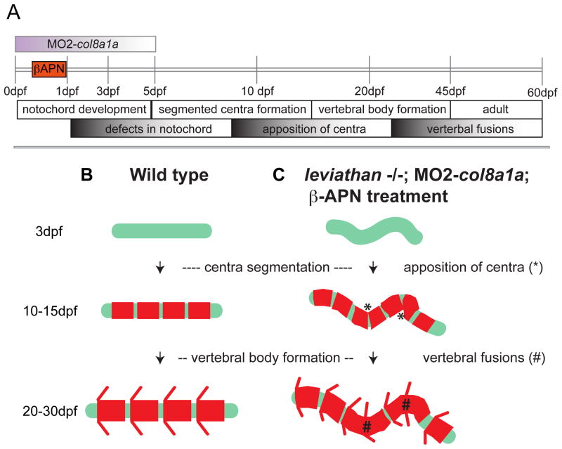
Congenital vertebral malformations (CVM) occur in 1 in 1000 live births and in many cases can cause spinal deformities, such as scoliosis, and result in disability and distress of affected individuals. Many severe forms of the disease, such as spondylocostal dystostosis, are recessive monogenic traits affecting somitogenesis, however the etiologies of the majority of CVM cases remain undetermined. Here we demonstrate that morphological defects of the notochord in zebrafish can generate congenital-type spine defects. We characterize three recessive zebrafish leviathan/col8a1a mutant alleles ((m531, vu41, vu105)) that disrupt collagen type VIII alpha1a (col8a1a), and cause folding of the embryonic notochord and consequently adult vertebral column malformations. Furthermore, we provide evidence that a transient loss of col8a1a function or inhibition of Lysyl oxidases with drugs during embryogenesis was sufficient to generate vertebral fusions and scoliosis in the adult spine. Using periodic imaging of individual zebrafish, we correlate focal notochord defects of the embryo with vertebral malformations (VM) in the adult. Finally, we show that bends and kinks in the notochord can lead to aberrant apposition of osteoblasts normally confined to well-segmented areas of the developing vertebral bodies. Our results afford a novel mechanism for the formation of VM, independent of defects of somitogenesis, resulting from aberrant bone deposition at regions of misshapen notochord tissue.

Keywords: Collagen; Notochord; Osteoblast; Vertebral malformations.


Figures

Figure 1
Figure 1. lev zebrafish mutant alleles disrupt the col8a1a gene
(A) Representative col8a1a (e.g. levm531−/−) mutant embryo displaying kinking of the notochord (red arrowhead) and shortened body axis at 1 dpf. (B) Representative homozygous col8a1a mutant adult (e.g. levvu41−/−) displays shortened body axis. (C) The lev vu41 lesion was meiotically mapped to a region of chromosome 9 between the markers z13213 and z34459 (number of recombinants are noted at each region) and was most highly associated with the SNP, rs40743937 in our bulk segregant analysis. According to the ensemble map (ZV9), 32 genes are located within this critical region flanked by the SSLP markers, including the col8a1a. (D–E′) col8a1a in situ hybridization at 1dpf (D, D′) showing robust expression in several midline structures at 1dpf including the floorplate, chordamesoderm/notochord, and hypochord. At 2dpf (E, E′), the expression is specific to the large vacuolated cells. (F) Structure of the predicted 711-amino acid zebrafish Col8a1a protein. The N-terminal NC2 domain and C-terminal NC1 domain flank the more-canonical collagenous domain. The NC2 domain is thought to be a propeptide domain lost during maturation, whereas the collagenous and NC1 domains are important for trimerization and supermolecular network formation respectively. The G309E (levm531) and G365S (levvu105) substitutions are located with in the collagenous domain (gray box); the 607 nonsense mutation (levvu41) resides just N-terminal to the NC1 domain and the previously described gulm208a mutation.
Figure 2
Figure 2. col8a1a mutations disrupt the normal morphology of the notochord sheath
(A) The primary components of the notochord tissue at embryonic maturation illustrated with a confocal (A, B, C), cartoon (D), and transmission electron micrographs (TEM) (E). The large vacuolated cell layer (VC; Tg(TK5xC twhh:memRFP) expression; magenta), surrounded by a sheath epidermis (SC; 1.75KB col2a1 GFP-CAAX; green) are shown in a lateral view with a confocal projection. The cartoon and the TEM image are transverse views highlighting the extracellular trilaminar notochord sheath (S) composed of laminin-, collagen- and elastin-rich components. (B–C″) Representative examples of in situ hybridization of chordamesodermal genes col2a1 (B, C), col9a2 (B′, C′), and ntl (B″, C″) in WT (B–B″) and levvu41 mutant (C–C″) embryos. (C) The expression of col9a2 and ntl transcripts is upregulated, especially in more anterior regions of the notochord (C′, C″) as compared to time matched sibling embryos (B′, B″). TEM of transverse sections from WT and mutant embryos at 2dpf (D–I). (D) WT notochord at 1 dpf. (E) lev vu41−/− notochord at 1dpf. (F) WT notochord sheath at 2dpf display very straight, well organized medial sheath layer. Extracellular notochord sheaths of lev m531−/− (G), lev vu41−/− (H), and lev vu105−/− (I) mutants exhibiting a wavy, disordered organization of the medial layer at 2dpf. i- inner laminin-rich layer, m-medial, and o- outer collagen-rich layers of the extracellular sheath; SC- sheath cell, S- extra-cellular sheath, and VC- vacuolated cell.
Figure 3
Figure 3. Homozyogus col8a1a mutant adults display vertebral fusions and scoliosis
MicroCT imaging of WT (A–B′) and col8a1a mutant zebrafish (C–D′) adults (45dpf) (A–E″) in both lateral views (A, C, E–E″) with insets (A′, C′) and dorsal views (B, D) with insets (B′, D′) (A, A′, B′) WT vertebrae with one neural and hemal arches per vertebral body. (B) Representative scoliosis in the tail of a WT adult (yellow arrowhead). (C, C′) Representative levvu41−/− zebrafish with VM (red arrowheads). (D, D′) Representative levvu41−/− multiple scoliosis along the axis (yellow arrowheads) and (E–E″) Representative individuals scored for the presence of individual VM (red arrowheads) or complete fusion of vertebral column (red bracket) in the thoracic or caudal regions (as number of fused regions). (F) Graphed categorical data of VM from all lev alleles and unaffected WT siblings in thoracic (T) and caudal (C) regions. All scale bars (A–D), (A′–D′), and (E–E″) = 1mM.
Figure 4
Figure 4. Embryonic inhibition of col8a1a function or lysyl oxidases is sufficient to generate vertebral defects
(A) Timeline of vertebral column development in zebrafish. In one experiment, the injection of MO2-col8a1a at the 1-cell stage, is effective in blocking the splicing of col8a1a until ~ 3–5dpf (purple gradient bar) (C–C″). In the second experiment the lysyl oxidases inhibitor, bAPN, is added at 4 hpf and washed out at 1dpf (orange bar) (D–D″). Both treatments completed well before the onset of centra formation started in the anterior regions at 5dpf. Representative brightfield images of 1dpf (B, C, D) and 60dpf (B′, C′, D′) and microCT images (45dpf) (B″, C″, D″) of WT (B–B″), MO2-col8a1a injected (C–C″), and bAPN pulsed adult zebrafish (D–D″). (D) Categorical frequency of VM for 2 and 4ng MO2-col8a1a morpholino injected adults and uninjected WT siblings and 2, 4, and 10mM bAPN treated adults and DMSO treated sibling controls for both thoracic (T) and caudal (C) regions. Scale bars for (B, C, D) = 4mm, (B′, C′, D′) = 5mm, and (B″, C″, D″) = 1mm.
Figure 5
Figure 5. Segmentation of the vertebral column and localization of osteoblasts proceeds on a dysmorphic notochord template
Alcian blue stained larvae (14dpf) (A–B‴) with lateral insets (red dashed box; A′, B′) and ventral insets (green dashed box; A‴, B‴). (A–A‴) WT larvae display well-formed and segmented centra. (B–B‴) levvu41−/− larvae display abberant apposition of centra (red brackets) and hemicentra (red arrow). Confocal projections of zebrafish vertebral column at 21dpf, illustrating boney matrix (alizarin red in magenta) and osteoblasts (Tg(sp7:EGFP)b1212 in green) (C–F′). (C, D) WT vertebrae and associated osteoblasts are well segmented along the vertebral axis. (E, E′) An individual levvu41−/− mutant displaying segmented colocalization of osteoblast and centra, including malformed hemivertebrae (yellow arrows) that continues to have mostly well segmented osteoblasts (E′), excepting some ectopic osteoblast-mineral formations in regions previously devoid of osteoblasts or mineral (E′, yellow asterix). (F, F′) An individual levvu41−/− mutant displaying regions of severe notochord bending with aberrantly apposed osteoblasts (F, yellow bracket), those progresses to a region with more complete osteoblast coverage (F′, yellow bracket). (C,D) 10dpf; ~SL3–4 (E,F) 10 dpf; ~SL3–4 (E′, F′) 14dpf; ~SL4–5. Scale bars for (A, B, A″, B″) = 1mM; (C–F′) and (A′, A‴,B′,B‴) = 100mM.
Figure 6
Figure 6. Periodic imaging of individual col8a1a mutants illustrates the progression of notochord bends towards malformed vertebral units
(A) Timeline of vertebral column development in zebrafish highlighting the timing of this particular periodic imaging method. Confocal projections of an transgenic individuals WT (B–B″) or col8a1a/levvu41−/− mutant (C–C″), and vitally stained with alizarin red (Tg[1.75KB col2a1 GFP-CAAX]; green (B,C′) and Alizarin Red; magenta (B–C″) periodically imaged over the range of vertebral column development. (B, C) mature notochord (10dpf; ~SL 3.5–4) (B′, C′) segmentation of axial centra (20dpf; ~SL 5–6) (B″, C″) mature vertebral column (39dpf; ~SL 8–9). (C) lev mutant individual at 10dpf displaying subtle bends in the notochord which transition to larger bends (yellow dashed lines (B′)). (C′) At 20dpf, these bends can display dysmorphic centra morphology (yellow bracket) or close apposition of centra edges in regions of severe bending (yellow arrows). (C″) At 39 dpf, the formation of VM and scoliosis is predicted from the previous disrupted notochord regions (yellow dashed line). Scale bar is 200mM.
Figure 7
Figure 7. The formation of vertebral fusions and scoliosis are consequences of notochord defects
(A) Timeline of vertebral column development in zebrafish. All manipulations of the notochord in this study are first observed at 1dpf but can progress for some time afterwards in a stochastic fashion (graded bar defects in notochord). The segmentation of the axial centra begin well after the effectiveness of inhibition of lysyl oxidases or col8a1a splicing (orange bar and gradient purple bar), and proceeds prior to and after the observation of centra apposition. Finally, VM are presumed to form continuously from 30dpf onward at regions of centra apposition. (B) Wild type zebrafish form a straight, stiff notochord (green line). This notochord provides a template for the segmentation of the axial centra (red rectangles). These axial centra then provide a template for the elaboration of osteoblast dependent formation of the vertebral bodies. (C) However, when the notochord becomes dysmorphic by early loss of col8a1a function or through the loss of activity of lysyl oxidases, the stereotyped segmentation of the axial centra is disrupted allowing for apposition of centra (*) in regions of severe bending. Finally, as the deposition of vertebral bodies proceeds closely apposed osteoblasts might simply bridge small gaps and generate VM and stabilize regions of bend notochord forming a region of scoliosis (#).

Similar articles

Cited by

References

    1. Adams DS, Keller R, Koehl MA. The mechanics of notochord elongation, straightening and stiffening in the embryo of Xenopus laevis. Development. 1990;110:115–30. - PubMed
    1. Bensimon-Brito A, Cardeira J, Cancela ML, Huysseune A, Witten PE. Distinct patterns of notochord mineralization in zebrafish coincide with the localization of Osteocalcin isoform 1 during early vertebral centra formation. BMC Dev Biol. 2012;12:28. - PMC - PubMed
    1. Bradley KM, Breyer JP, Melville DB, Broman KW, Knapik EW, Smith JR. An SNP-Based Linkage Map for Zebrafish Reveals Sex Determination Loci. G3 (Bethesda) 2011;1:3–9. - PMC - PubMed
    1. Bradley KM, Elmore JB, Breyer JP, Yaspan BL, Jessen JR, Knapik EW, Smith JR. A major zebrafish polymorphism resource for genetic mapping. Genome Biol. 2007;8:R55. - PMC - PubMed
    1. Bruggeman BJ, Maier JA, Mohiuddin YS, Powers R, Lo Y, Guimaraes-Camboa N, Evans SM, Harfe BD. Avian intervertebral disc arises from rostral sclerotome and lacks a nucleus pulposus: implications for evolution of the vertebrate disc. Dev Dyn. 2012;241:675–83. - PMC - PubMed

Publication types

LinkOut - more resources