. 2013 Dec;140(23):4683-90.
doi: 10.1242/dev.102798.

Hippo signaling impedes adult heart regeneration

Affiliations

Hippo signaling impedes adult heart regeneration

Todd Heallen et al. Development. 2013 Dec.

Abstract

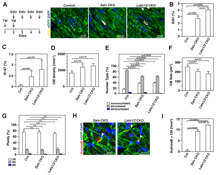
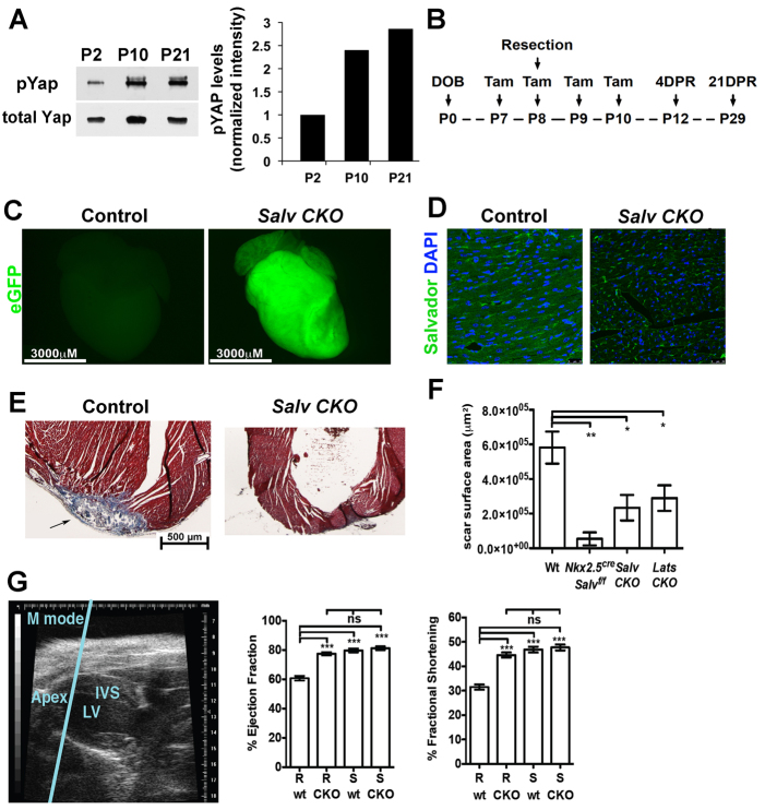
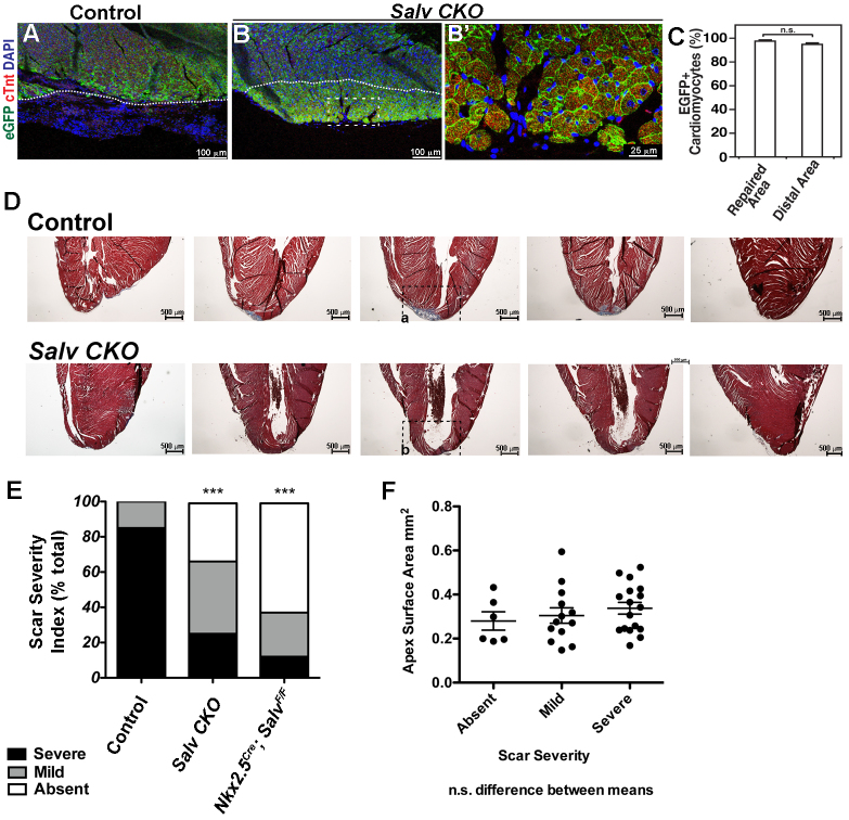
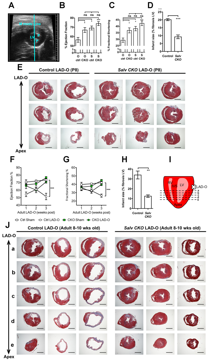
Heart failure due to cardiomyocyte loss after ischemic heart disease is the leading cause of death in the United States in large part because heart muscle regenerates poorly. The endogenous mechanisms preventing mammalian cardiomyocyte regeneration are poorly understood. Hippo signaling, an ancient organ size control pathway, is a kinase cascade that inhibits developing cardiomyocyte proliferation but it has not been studied postnatally or in fully mature adult cardiomyocytes. Here, we investigated Hippo signaling in adult cardiomyocyte renewal and regeneration. We found that unstressed Hippo-deficient adult mouse cardiomyocytes re-enter the cell cycle and undergo cytokinesis. Moreover, Hippo deficiency enhances cardiomyocyte regeneration with functional recovery after adult myocardial infarction as well as after postnatal day eight (P8) cardiac apex resection and P8 myocardial infarction. In damaged hearts, Hippo mutant cardiomyocytes also have elevated proliferation. Our findings reveal that Hippo signaling is an endogenous repressor of adult cardiomyocyte renewal and regeneration. Targeting the Hippo pathway in human disease might be beneficial for the treatment of heart disease.

Keywords: Cell cycle; Mouse; Proliferation; Regeneration.


Figures

Fig. 1.
Fig. 1.
Adult cardiomyocyte renewal via deletion of Hippo pathway genes. Lats1/2 and Salv were conditionally deleted in cardiomyocytes using the inducible Myh6-Cre/Esr line via tamoxifen injection. All analyses were performed with 3-4 month stage hearts. (A) Diagram shows tamoxifen (Tam) and EdU injection scheme. de novo DNA synthesis was monitored by EdU incorporation (yellow) in cardiomyocytes (green) using the mTmG reporter line. (B) Percentage of EdU-positive cells for each genotype (n=3). EdU-positive cells were manually counted. (C) Percentage of cardiomyocyte nuclei in cell cycle (n=3). Isolated nuclei were stained with the cardiac marker cTnI and the cell cycle marker Ki-67. (D) Cardiomyocyte density. Numbers of cardiomyocytes were counted in each section (n=3). (E) Cardiomyocyte nucleation. Nuclei per cardiomyocyte was calculated for each genotype (n=3). (F) Cardiomyocyte cross-sectional area was measured and averaged for multiple sections per specimen (n=3). (G) Ploidy analysis (n=3). (H) Immunohistochemical analysis: Aurkb (red) and cardiomyocytes (eGFP, green). Arrowheads indicate Aurkb staining. (I) Quantification of Aurkb-positive cardiomyocytes (n=3). Error bars represent s.e.m.
Fig. 2.
Fig. 2.
Salvador deletion promotes regeneration of ventricular myocardium. (A) Western blot (left) of postnatal wild-type heart extracts with designated antibodies. For quantification of western blot (right) pYAP band intensity was normalized against total Yap. (B) Strategy for cardiomyocyte-specific knockout of salvador and apical resection of P8-stage hearts. (C) Cre recombinase reporter activity (eGFP) in 4 dpr control and Salv CKO hearts. (D) 4 dpr control and Salv CKO coronal sections: salvador (green) and DAPI (blue). (E) High magnification inset images of 21 dpr control and Salv CKO trichrome-stained sections (Fig. 3D). Arrow indicates persistent fibrotic scar. (F) Scar surface area measurements were obtained from multiple specimens and averages calculated. Statistical analysis was by unpaired Student’s t-test. *P<0.05, **P<0.01. Salvf/f (n=18), Nkx2.5cre; Salvf/f (n=6), Salv CKO (n=12), Lats1-2 CKO (n=10). (G) Left: M-mode echocardiographic image from a representative 21 dpr heart. LV, left ventricle; IVS, interventricular septum. Right: ejection fraction and fractional shortening percentages of resected (R) and sham-operated (S) control (wt) and Salv CKO (CKO) hearts at 21-32 dpr. wt/R (n=9), CKO/R (n=6), wt/S (n=3), CKO/S (n=9). Statistical analysis was by one-way ANOVA. *P<0.05, **P<0.01, ***P<0.001. Error bars represent s.e.m.
Fig. 3.
Fig. 3.
Lineage tracing of 21 dpr hearts. Immunohistochemical analysis was performed on resected hearts of control and Salv CKO mice at 21 dpr. (A-B′) Representative images of eGFP (cardiomyocyte-lineage cells, green) and cTnt (cardiomyocytes, red) staining of control and Salv CKO hearts at the resection site. Dotted lines indicate plane of resection. Salv CKO hearts display repair with cTnt/eGFP-positive cells. B’ shows higher magnification of the boxed region in B. (C) Quantification of eGFP+ cardiomyocytes comparing border-zone and distal regions of the heart. No significant difference (n.s.) between repaired area and distal area were found (n=3). (D) Trichrome-stained 21 dpr control and Salv CKO serial sections from anterior (left) to posterior (right). (E) Bar graph representing qualitative differences between genotypes in scar severity. Control (n=18), Salv CKO (n=12), Nkx2.5cre; Salvf/f (n=6). Statistical analysis was by χ2 distribution. (F) Distribution of resected apices measured by surface area between groups. No significant difference was found indicating equivalent resected area between groups. Error bars represent s.e.m.
Fig. 4.
Fig. 4.
Cardiac regeneration and functional recovery following myocardial infarction. (A) B-mode echocardiographic image from representative 21 day post-occlusion heart. LV, left ventricle; P, papillary muscle. (B,C) Ejection fraction (B) and fractional shortening (C) percentages of P8 LAD-O hearts. Occluded (O) and sham-operated (S) control (ctrl) and Salv CKO (CKO) hearts at 21-32 days post myocardial infarction (dpmi). ctrl/O (n=7), CKO/O (n=9), ctrl/S (n=6), CKO/S (n=9). Statistical analysis was by one-way ANOVA with Bonferroni’s multiple comparison test. (D) Infarct surface area measurements were obtained from multiple specimens and averages calculated. Statistical analysis was by unpaired Student’s t-test. Control (n=6), Salv CKO (n=8). (E) Serial transverse sections of 4-week post-occlusion hearts. (F,G) Ejection fraction (F) and fractional shortening (G) percentages of adult LAD-O hearts. 1 week: Ctrl Sham (n=4), Ctrl LAD-O (n=6), CKO Sham (n=3), CKO LAD-O (n=3); 2 weeks: Ctrl Sham (n=3), Ctrl LAD-O (n=5), CKO Sham (n=3), CKO LAD-O (n=2); 3 weeks: Ctrl Sham (n=7), Ctrl LAD-O (n=7), CKO Sham (n=3), CKO LAD-O (n=5). (H) Adult LAD-O infarct size 3 weeks post-occlusion measured as percentage fibrosis of LV myocardium (total fibrotic area/total myocardial area×100). Ctrl LAD-O (n=6), CKO LAD-O (n=3). Statistical analysis was by two-way ANOVA with Bonferroni’s multiple comparison test. (I) Illustration of transverse section planes presented in J. Cross indicates plane of occlusion. IVS, interventricular septum; LV, left ventricle; RV, right ventricle. (J) Serial transverse sections of representative 3-week post-occlusion adult hearts. All echocardiographic measurements were collected blind from genotype. Scale bars: 1 mm. *P<0.05, **P<0.01, ***P<0.001.
Fig. 5.
Fig. 5.
Increased proliferation of cardiomyocytes in resected salvador mutant hearts. (A-F) Control and Salv CKO 4 dpr resected hearts. Cardiomyocyte lineage (eGFP positive). Border zone (A,B,E) and distal area (C,D,F) showing EdU-positive nuclei (arrowheads, yellow) after apex resection. E and F show high-magnification representative images of control and Salv CKO, respectively. (G) Proliferation rate was quantified by percentage of EdU incorporation in cardiomyocytes (n=3). Salv CKO hearts show significant increase of proliferation in both border zone and distal area. (H) Quantification of Aurora B kinase-positive cardiomyocytes (n=3). *P<0.001. (I) Quantification of EdU+ cardiomyocytes following siRNA knockdown of indicated genes in wild-type neonatal cardiomyocytes (n=3). (J,K) Cardiomyocyte proliferation, density and nucleation from unstressed control and Salv CKO mice at multiple stages were calculated. Cnt (n=3), Salv CKO (n=3). Error bars represent s.e.m.

Similar articles

Cited by

References

    1. Bergmann O., Bhardwaj R. D., Bernard S., Zdunek S., Barnabé-Heider F., Walsh S., Zupicich J., Alkass K., Buchholz B. A., Druid H., et al. (2009). Evidence for cardiomyocyte renewal in humans. Science 324, 98–102 - PMC - PubMed
    1. Boon R. A., Iekushi K., Lechner S., Seeger T., Fischer A., Heydt S., Kaluza D., Tréguer K., Carmona G., Bonauer A., et al. (2013). MicroRNA-34a regulates cardiac ageing and function. Nature 495, 107–110 - PubMed
    1. Bruneau B. G. (2012). Direct reprogramming for cardiac regeneration: from dream to reality. Circ. Res. 110, 1392–1394 - PMC - PubMed
    1. Chugh A. R., Beache G. M., Loughran J. H., Mewton N., Elmore J. B., Kajstura J., Pappas P., Tatooles A., Stoddard M. F., Lima J. A., et al. (2012). Administration of cardiac stem cells in patients with ischemic cardiomyopathy: the SCIPIO trial: surgical aspects and interim analysis of myocardial function and viability by magnetic resonance. Circulation 126 Suppl. 1, S54–S64 - PMC - PubMed
    1. Del Re D. P., Yang Y., Nakano N., Cho J., Zhai P., Yamamoto T., Zhang N., Yabuta N., Nojima H., Pan D., et al. (2013). Yes-associated protein isoform 1 (Yap1) promotes cardiomyocyte survival and growth to protect against myocardial ischemic injury. J. Biol. Chem. 288, 3977–3988 - PMC - PubMed

Publication types

Substances