. 2013 Nov;9(11):e1003940.
doi: 10.1371/journal.pgen.1003940. Epub 2013 Nov 14.

Crosstalk between NSL histone acetyltransferase and MLL/SET complexes: NSL complex functions in promoting histone H3K4 di-methylation activity by MLL/SET complexes

Affiliations

Crosstalk between NSL histone acetyltransferase and MLL/SET complexes: NSL complex functions in promoting histone H3K4 di-methylation activity by MLL/SET complexes

Xiaoming Zhao et al. PLoS Genet. 2013 Nov.

Abstract

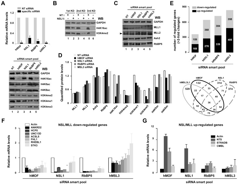
hMOF (MYST1), a histone acetyltransferase (HAT), forms at least two distinct multiprotein complexes in human cells. The male specific lethal (MSL) HAT complex plays a key role in dosage compensation in Drosophila and is responsible for histone H4K16ac in vivo. We and others previously described a second hMOF-containing HAT complex, the non-specific lethal (NSL) HAT complex. The NSL complex has a broader substrate specificity, can acetylate H4 on K16, K5, and K8. The WD (tryptophan-aspartate) repeat domain 5 (WDR5) and host cell factor 1 (HCF1) are shared among members of the MLL/SET (mixed-lineage leukemia/set-domain containing) family of histone H3K4 methyltransferase complexes. The presence of these shared subunits raises the possibility that there are functional links between these complexes and the histone modifications they catalyze; however, the degree to which NSL and MLL/SET influence one another's activities remains unclear. Here, we present evidence from biochemical assays and knockdown/overexpression approaches arguing that the NSL HAT promotes histone H3K4me2 by MLL/SET complexes by an acetylation-dependent mechanism. In genomic experiments, we identified a set of genes including ANKRD2, that are affected by knockdown of both NSL and MLL/SET subunits, suggested they are co-regulated by NSL and MLL/SET complexes. In ChIP assays, we observe that depletion of the NSL subunits hMOF or NSL1 resulted in a significant reduction of both H4K16ac and H3K4me2 in the vicinity of the ANKRD2 transcriptional start site proximal region. However, depletion of RbBP5 (a core component of MLL/SET complexes) only reduced H3K4me2 marks, but not H4K16ac in the same region of ANKRD2, consistent with the idea that NSL acts upstream of MLL/SET to regulate H3K4me2 at certain promoters, suggesting coordination between NSL and MLL/SET complexes is involved in transcriptional regulation of certain genes. Taken together, our results suggest a crosstalk between the NSL and MLL/SET complexes in cells.


Conflict of interest statement

The authors have declared that no competing interests exist.

Figures

Figure 1
Figure 1. HAT activity facilitates histone H3K4 methylation by WDR5-containing complex in vitro.
A. WDR5 is a shared subunit between HAT NSL and HMT MLL/SET complexes. Anti-Flag agarose eluate from a Flag-WDR5 stable cell line was analyzed by SDS-PAGE, and proteins were visualized by silver staining (left). Equal amounts of anti-Flag agarose eluates from Flag-PHF20 (a subunit of NSL complex), Flag-MSL3L1 (a subunit of MSL complex) and Flag-WDR5 (a shared subunit between NSL and MLL/SET complexes) were fractionated by SDS-PAGE, and proteins were identified with Western blot using the indicated antibodies (right). B. Immunoaffinity purified WDR5-containing complex possesses a dual enzymatic activity. hMOF and RbBP5 protein from Flag-WDR5 and Flag-Ash2- (a subunit of MLL/SET complexes) expressing cells were detected with Western blotting using the indicated antibodies (top). Relatively equal amounts of RbBP5 proteins in WDR5- and Ash2-containing complexes were used in HMT assays. Modified residues at H3K4 were detected with methylation specific antibodies (middle). HAT assays were performed with 0.5 µCi of [3H] AcCoA and 3 µg HeLa nucleosomes. Histone proteins were visualized by Coomassie R-250 blue stain and radioactively labeled proteins were visualized by autoradiography (bottom). C. HAT activity of the WDR5-containing complex is not changed in the presence or absence of SAM in vitro. D. HMT activity of the WDR5-containing complex is increased in an AcCoA-dependent manner. HMT assays were performed with recombinant core histones in the presence or absence of AcCoA in vitro.
Figure 2
Figure 2. The activity of histone H3K4 methylation is facilitated by hMOF-containing complexes both in vitro and in vivo.
A. Purification of hMOFwt- and hMOFmt-containing complexes. HA-purified hMOFwt- or G327E-cantaining complexes were analyzed by silver stain (left) and Western blot (right). B. hMOFmt-containing complex lost HAT activity. Relatively equal amounts of hMOF protein in the HA-purified hMOFwt (lane 2–4) or G327E (lane 5–7) complexes were subjected to a HAT assay. Histone proteins were visualized by Coomassie R-250 blue stain (bottom) and radioactively labeled proteins were visualized by autoradiography (top). C. Schematic flowchart of the HMT assay in vitro. D. The activity of H3K4me2 is facilitated by the hMOFwt-containing complex on recombinant polynucleosomes (lane 6–8). The HMT assay was carried out by mixing in the combinations indicated in the figures. Modified residues at H3K4 were detected with methylation-specific antibodies. E. Global H3K4me2 is reduced in hMOF siRNA knockdown HeLa cells. HeLa cells were transfected with 20 nM hMOF siRNA and non-targeting siRNA (as control) three times every 48 hours. Then, 48 hours after each transfection, cells were harvested for Western blot analysis and qRT-PCR check (F). hMOF protein and modified residues at histone H4 and H3 were detected with indicated antibodies. G. Global histone modifications in hMOF overexpressed 293T cells. 293T cells were transiently transfected with 2 µg hMOF cDNAs. Then, 48 hours after transfection, cells were harvested and lysed by adding 4×SDS sample buffer. Proteins were identified with Western blot.
Figure 3
Figure 3. The HAT NSL complex, but not the MSL complex, promotes H3K4me2 by Flag-Ash2 in vitro.
A. Substrate specificity of two hMOF-containing complexes on recombinant polynucleosomes. HAT assays were performed by mixing [3H]-labeled Acetyl CoA or cold AcCoA, HeLa nucleosomes or recombinant polynucleosomes and 1–4 µl of the indicated anti-Flag agarose eluates. Histones were visualized with Coomassie R250 blue stain and acetylated histones were visualized by autoradiography (top). Modified residues at histone H4 were detected with acetylation-specific antibodies (bottom). B–C. The activity of H3K4me2 by Flag-Ash2 complex is facilitated by HAT NSL, but not the MSL complex in vitro. D. The NSL complex is required for promoting H3K4me2 activity via the Flag-Ash2 complex on recombinant polynucleosomes. The experimental procedures are as shown at the top. In this experiment, reconstituted nucleosomes were prepared by mixing ∼1500 bp biotinylated oligonucleotides and recombinant human histones through serial dilution. Reconstituted nucleosomes were then immobilized on avidin-coupled Dyna-beads. HAT assays were first performed on immobilized nucleosomes, and the HMT assays were carried out after two washes with HAT buffer. Modified residues at histone H3K4 were detected with H3K4me2 antibody.
Figure 4
Figure 4. Select genes are co-regulated by NSL and RbBP5-MLL complexes.
A. Alteration of global histone H4 or H3K4 modifications in hMOF, hNSL1, hRbBP5 or hMSL3L1 siRNA knockdown HeLa cells. HeLa cells were transfected with indicated siRNAs (non-targeting siRNA as control). Next, 48 hours after transfection, cells were lysed and total RNA was isolated with Trizol. mRNA of specific genes were measured with qRT-PCR (top). Whole-cell extracts were prepared 48 hours after transfection with specific-siRNAs and were subjected to SDS-PAGE (18% gel). Modified residues at histone H3K4 or H4K5/8/K16 were detected with specific-modified antibodies (bottom). B. Reduction of global H3K4me2 was observed in NSL1 siRNA knockdown HeLa cells. HeLa cells were transfected with 20 nM NSL1 siRNA three times every 48 hours, after which cells were harvested for Western blot analysis. C. Protein of subunits of MLL/SET complexes did not change in hMOF or NSL1 siRNA knockdown HeLa cells. HeLa cells were transfected with 20 nM indicated siRNAs. Then, 48 hours after transfection, whole-cell extracts were prepared and proteins were detected using the indicated antibodies. D. Quantified protein. Western blot images were scanned and signals were quantified with densitometry using Quantity One Basic software (Bio-Rad). E. Gene expression profiles in siRNA knockdown HeLa cells. Stacked column charts represents the total number of regulated genes (>2-fold decrease or increase) in the indicated specific siRNA knockdown HeLa cells. Each bar in the stacked column chart is composed of down- and up-regulated genes (top figure). The Venn diagram depicts shared and distinct gene expression in each population (bottom figure). F–G. mRNA of select genes is regulated by NSL and RbBP5-MLL complexes. Genes indicated in figures were measured by qRT-PCR in specific-siRNA knockdown HeLa cells. Bar graphs show ratios of qRT-PCR signals (normalized to non-targeting siRNA) to actin (also normalized to non-targeting siRNA). Error bars represent the standard error of the mean of 3 independent experiments.
Figure 5
Figure 5. NSL complex is essential for targeting of RbBP5-MLL complex to ANKRD2.
A. Five primer sets in the ANKRD2 locus used for amplifying ChIP'd DNA. B. Distribution of hMOF on the ANKRD2 locus. ChIP assays were performed using hMOF antibody. ChIP'd DNA was analyzed by qPCR. Bar graph shows the ratios of ChIP'd DNA signals (normalized to input) to IgG (also normalized to input; right panel). Error bars represent the standard error of the mean of 3 independent experiments. Significant. C. Enrichment of histone H3K4me and H4K16ac on the ANKRD2 promoter region (−0.25 kb). ChIP assays were performed using H3K4me1/me2/me3 or H4K16ac antibodies. ChIP'd DNA was amplified by PCR (top). Quantified PCR signals (Quantity One software) were analyzed by t-test (bottom). Error bars indicate SE and the significant difference is expressed as **p<0.01. D. Functional cooperation of histone H4K16ac and H3K4me2 on ANKRD2 (−0.25 kb), but not on FHL1 (0.4 kb) gene promoter. ChIP assays using hMOF siRNA knockdown HeLa cells were analyzed by qPCR. E. The NSL complex is essential for binding of the RbBP5-MLL complex to the ANKRD2 gene promoter region (−0.25 kb). ChIP assays using hNSL1 and hRbBP5 siRNA knockdown HeLa cells were analyzed by qPCR. Bar graphs show ratios of ChIP signals (also normalized to input DNA and IgG) to non-targeting siRNA controls (normalized to input DNA and IgG). Error bars represent the standard error of the mean of 3 independent experiments.

Similar articles

Cited by

References

    1. Bannister A, Kouzarides T (2011) Regulation of chromatin by histone modifications. Cell Res 21: 381–395. - PMC - PubMed
    1. Jin J, Cai Y, Li B, Conaway RC, Workman JL, et al. (2005) In and out: histone variant exchange in chromatin. Trends Biochem Sci 30: 680–687. - PubMed
    1. Berger SL (2007) The complex language of chromatin regulation during transcription. Nature 447: 407–412. - PubMed
    1. Lee JS, Smith E, Shilatifard A (2010) The language of histone crosstalk. Cell 142: 682–685. - PMC - PubMed
    1. Cedar H, Bergman Y (2009) Linking DNA methylation and histone modification: patterns and paradigms. Nat Rev Genet 10: 295–304. - PubMed

Publication types

MeSH terms

LinkOut - more resources