. 2013 Oct;9(10):e1003913.
doi: 10.1371/journal.pgen.1003913. Epub 2013 Oct 24.

Loss of miR-10a activates lpo and collaborates with activated Wnt signaling in inducing intestinal neoplasia in female mice

Affiliations

Loss of miR-10a activates lpo and collaborates with activated Wnt signaling in inducing intestinal neoplasia in female mice

Gustavo Stadthagen et al. PLoS Genet. 2013 Oct.

Abstract

miRNAs are small regulatory RNAs that, due to their considerable potential to target a wide range of mRNAs, are implicated in essentially all biological process, including cancer. miR-10a is particularly interesting considering its conserved location in the Hox cluster of developmental regulators. A role for this microRNA has been described in developmental regulation as well as for various cancers. However, previous miR-10a studies are exclusively based on transient knockdowns of this miRNA and to extensively study miR-10a loss we have generated a miR-10a knock out mouse. Here we show that, in the Apc(min) mouse model of intestinal neoplasia, female miR-10a deficient mice develop significantly more adenomas than miR-10(+/+) and male controls. We further found that Lpo is extensively upregulated in the intestinal epithelium of mice deprived of miR-10a. Using in vitro assays, we demonstrate that the primary miR-10a target KLF4 can upregulate transcription of Lpo, whereas siRNA knockdown of KLF4 reduces LPO levels in HCT-116 cells. Furthermore, Klf4 is upregulated in the intestines of miR-10a knockout mice. Lpo has previously been shown to have the capacity to oxidize estrogens into potent depurinating mutagens, creating an instable genomic environment that can cause initiation of cancer. Therefore, we postulate that Lpo upregulation in the intestinal epithelium of miR-10a deficient mice together with the predominant abundance of estrogens in female animals mainly accounts for the sex-related cancer phenotype we observed. This suggests that miR-10a could be used as a potent diagnostic marker for discovering groups of women that are at high risk of developing colorectal carcinoma, which today is one of the leading causes of cancer-related deaths.


Conflict of interest statement

The authors have declared that no competing interests exist.

Figures

Figure 1
Figure 1. Generation of miR-10a KO mice.
(A) Schematic representation of the miR-10a WT locus, the targeting construct used for inactivation and the final miR-10a null allele. The targeting construct (TC) harbored a miR-10a inactivated allele, where 70 nucleotides from the pre-miRNA sequence were replaced with a neomycin resistance cassette (neo) flanked by loxP sites and long homologous regions for recombination. To obtain the final miR-10a null allele (KO), the neomycin cassette was removed in the mouse germ line by breeding heterozygous mice to transgenic mice harboring the Cre transgene. Arrowheads depict the sites recognized by different primers used in genotyping of mice. (B) Genotyping PCR of mice with all different miR-10a genotypes generated. Primers L_chkinsrtmiR10a.5d and 10a.internal amplified a 273 bp fragment corresponding to the miR-10 WT allele and 361 bp for the floxed miR-10a KO allele, L_chkinsrtmiR10a.5d and R_chkinsrtmiR10a.5 amplified 291 bp from the miR-10aneo allele. The location of all these primers is depicted in (A).
Figure 2
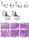
Figure 2. Disruption of miR-10a leads to enhanced intestinal tumorigenesis in ApcMin mice.
Tumor multiplicity in the small (A) and large intestines (B) of female and male miR-10a +/+;ApcMin (WT; n = 22 and n = 19 for each sex) and miR-10a−/−;ApcMin (KO; n = 15 and n = 16 for each sex) mice; each dot represents data for one mouse. Mean adenoma multiplicities per mouse for each group were: WT = 41.95 and KO = 79.33 for female mice and WT = 50.37 and KO = 55.19 for male mice in the small intestine and WT = 0.82 and KO = 2.40 for female mice and WT = 1.47 and KO = 2.44 for male mice in the large intestine. * p = 0.014, ** p = 0.0042 (two-tailed t-test) (C) Size distribution of polyps in the small intestine of female and male miR-10a +/+;ApcMin (WT; filled bars) and miR-10a−/−;ApcMin (KO; empty bars) mice. Mean tumor diameters were 1.01 and 1.04 mm for males WT and KO respectively and 1.03 and 0.94 for females WT and KO respectively (p = 0.782 and p = 0.4113, Wilcoxon rank sum test). (D) Left panel shows normal appearing small intestine with characteristic villi and well-ordered distribution of goblet cells together with basal location of epithelial nuclei. Middle panel is a typical example of a low-grade dysplasia in miR-10a +/+;ApcMin (WT) mice with accumulation of irregular goblet cells pattern (arrowheads) and some loss of nuclear polarity (indicated by “+”). Right panel shows a typical miR-10a−/−;ApcMin (KO) high-grade dysplasia with a large area of loss of goblet cells, widespread loss of nuclear polarity, nuclear pleomorphism, and almost complete loss of villus organization (indicated by “*”). Note the transition from lower-grade dysplasia area with highly irregular goblet cell distribution (arrowheads). Scalebar = 100 µM. Whole intestines were paraffin-embedded as “Swiss rolls”, sectioned and stained with hematoxylin and eosin. All animals were in a B6 background and between 110 and 160 days of age.
Figure 3
Figure 3. Lpo is transcriptionally upregulated in the intestines of miR-10a deficient female mice.
(A) Lpo mRNA is ∼29-fold upregulated in intestines of miR-10a KO compared to WT mice as shown by qRT-PCR. Lpo mRNA levels are normalized to Actb and values ± SD are shown relative to the first WT sample. (B) Western-blot from same tissue samples as in (A) confirming upregulation on protein level. Vinculin was used as loading control. As evident from Lpo and Vinculin control as well as ponceau staining (now shown), sample 4 did not contain any protein for unknown reason. (C) Representative immunohistochemistry staining of Lpo in miR-10a WT and KO intestine. Scale bar 100 µm. (D) Scoring of Lpo expression level estimated by distribution and staining intensity in Lpo stained intestines of WT and miR-10a−/− mice. Scoring is divided into low, medium or high expression. Consistent with qRT-PCR and Western blotting analysis a significant difference (p≤0.006, Pearson chi-square test with exact probability) in Lpo expression is observed between the different genotypes.
Figure 4
Figure 4. Transcription factor KLF4 is regulated by miR-10a and can regulate the LPO promoter in vitro.
HCT-116 cells were transfected with a miR-10a duplex or control for 72 h. (A) Relative mRNA levels of KLF4 were measured by qRT-PCR and ACTB was used for normalization. Data are shown as mean ± S.D. of three replicates relative to the control and are representative of three independent experiments. * p<0.05 using a two-tailed t-test. (B) Protein levels in miR-10a or control transfected cells were assessed by Western-blot using antibodies against KLF4. GAPDH was used as loading control. (C) Western Blot showing the over expression from the pcDNA3.1-KLF4 vector. GAPDH was used as loading control. (D) Luciferase reporter assay in HCT-116 cells (24 h) with pGL4-luc2 holding part of the LPO promoter (1 kb upstream TSS) or the pGL4-luc2 empty vector co-transfected with a vector over-expressing KLF4 or a control vector (pcDNA3.1+). Data are shown as mean ± S.D. of three replicates relative to the pcDNA3.1 transfected control and are representative of eleven independent experiments. **** p<0.0001 using a two-tailed t-test. (E) HCT-116 cells were transfected with KLF4 siRNA for 48 h or 72 h. Relative mRNA levels of LPO were measured by qRT-PCR and ACTB was used for normalization. Data are shown as mean ± S.D. of three replicates relative to the control and are representative of five independent experiments.
Figure 5
Figure 5. Klf4 is upregulated in miR-10a KO intestines.
(A) mRNA levels of Klf4 were measured by qRT-PCR, Actb, Ubc, Hprt and 36b4 were used for normalization. Data are shown as mean ± S.D. of miR-10a KO (n = 16) and WT (n = 13) samples relative to an average of the controls. * p<0.05 using a Mann-Whitney test. (B) Representative immunohistochemistry staining of Klf4 in miR-10a WT (n = 5) and KO (n = 8) intestine. Scale bar 100 µm (C) VisiomorphDP software scoring of Klf4 expression level estimated by distribution and staining intensity in Klf4 stained intestines of WT and miR-10a KO mice. * p = 0.019, students t-test.

Similar articles

Cited by

References

    1. Mitchell PS, Parkin RK, Kroh EM, Fritz BR, Wyman SK, et al. (2008) Circulating microRNAs as stable blood-based markers for cancer detection. Proc Natl Acad Sci U S A 105: 10513–10518. - PMC - PubMed
    1. Paranjape T, Slack FJ, Weidhaas JB (2009) MicroRNAs: tools for cancer diagnostics. Gut 58: 1546–1554. - PMC - PubMed
    1. Deng S, Calin GA, Croce CM, Coukos G, Zhang L (2008) Mechanisms of microRNA deregulation in human cancer. Cell Cycle 7: 2643–2646. - PubMed
    1. Tehler D, Hoyland-Kroghsbo NM, Lund AH (2011) The miR-10 microRNA precursor family. RNA Biol 8: 728–734. - PMC - PubMed
    1. Lund AH (2010) miR-10 in development and cancer. Cell Death Differ 17: 209–214. - PubMed

Publication types

Grants and funding

Work in the authors' laboratory is supported by the Danish National Research Foundation, the Danish National Advanced Technology Foundation, the Danish Council for Strategic Research, the Novo Nordisk Foundation, the EC FP7 programs (ONCOMIRS, grant agreement number 201102. This publication reflects only authors' views. The commission is not liable for any use that may be made of the information herein.), the Lundbeck Foundation, and the Danish Cancer Society. The funders had no role in study design, data collection and analysis, decision to publish, or preparation of the manuscript.