. 2013 Dec 20;342(6165):1484-90.
doi: 10.1126/science.1245627. Epub 2013 Oct 31.

Cryo-EM structure of a fully glycosylated soluble cleaved HIV-1 envelope trimer

Affiliations

Cryo-EM structure of a fully glycosylated soluble cleaved HIV-1 envelope trimer

Dmitry Lyumkis et al. Science. .

Abstract

The HIV-1 envelope glycoprotein (Env) trimer contains the receptor binding sites and membrane fusion machinery that introduce the viral genome into the host cell. As the only target for broadly neutralizing antibodies (bnAbs), Env is a focus for rational vaccine design. We present a cryo-electron microscopy reconstruction and structural model of a cleaved, soluble Env trimer (termed BG505 SOSIP.664 gp140) in complex with a CD4 binding site (CD4bs) bnAb, PGV04, at 5.8 angstrom resolution. The structure reveals the spatial arrangement of Env components, including the V1/V2, V3, HR1, and HR2 domains, as well as shielding glycans. The structure also provides insights into trimer assembly, gp120-gp41 interactions, and the CD4bs epitope cluster for bnAbs, which covers a more extensive area and defines a more complex site of vulnerability than previously described.


Figures

Fig. 1
Fig. 1. 5.8 Å EM reconstruction and model of Env trimer in complex with PGV04
(A) Side and (B) top views of BG505 SOSIP trimer EM reconstruction (left) and corresponding model (right). The viral membrane would be at the bottom of the figure. Segmentation and color coding is as follows: PGV04 (gray), gp120 (blue), gp41 (orange), V1/V2 (purple), and V3 (green). (C) The middle panel shows a side view of the EM map alone with the Fab density removed. The outer panels show the modeled portion corresponding to the boxed region in the middle panel.
Fig. 2
Fig. 2. Structures of gp120 V1/V2/V3 variable loops
(A) Comparison of the V1/V2 domain from the EM model (purple) and the previously published X-ray structure (3U2S, white) with key residues labeled. The structures are nearly identical except for the base of V1/V2 (strands A and D) and the V1 loop. (B) Superposition of gp120 and the V3 loop from the EM model (blue/green) onto a previous X-ray structure containing a V3 loop (2B4C, gray/white). While gp120 is structurally conserved, the V3 β-hairpin loop exhibits a different configuration. Specifically, V3 bends around a hinge formed near residues 302 and 326. The N-terminal residues of V3 up to residue 301 and the C-terminal residues after residue 327 are structurally similar. Residues 1–90, and 126–196 are omitted for clarity. (C) Quaternary arrangement of V1, V2, and V3 regions of gp120 at the top of the trimer. The positions of glycans at N156, N160, and N197 have been highlighted as red spheres. Lines denote approximate protomer boundaries. (D) Surface view of the modeled portion in C, with the basic residues (Arg and Lys) colored blue. A large number of basic amino acids are localized to the trimer apex.
Fig. 3
Fig. 3. Structure of gp41
(A) Segmented EM density map of the gp41 trimer with gp120 removed. The C-terminal half of HR1 (rust) forms a three-helix bundle at the center of the trimer, while the C-terminal half of HR2 (yellow) forms a helical structure that wraps around the trimer base. Additional density that is not assigned in the model (beige) likely corresponds to the intervening region between HR1 and HR2, including the disulfide loop, as well as C1 and C5 from gp120. Density parallel to HR1 (brown) likely corresponds to the N-terminal half of HR1, the fusion peptide proximal region (FPPR) and the fusion peptide (FP). (B) Modeled portion corresponding to the same views of the EM density maps in A. (C) EM density of the three-helix bundle formed by HR1 in the PGV04-bound trimer structure. (D) Overlay of the EM density of the three-helix bundle formed by HR1 in the PGV04-bound structure that is filtered to 9.5 Å (orange) with the 9 Å reconstruction of a 17b-bound SOSIP gp140 trimer (gray, EMDB-5462) (35). (E) An 8.2 Å reconstruction of a SOSIP trimer from which the last 14 amino acids were deleted (SOSIP.650:PGV04). The difference between the SOSIP.650 and SOSIP.664 maps corresponds to a short helical segment (red) at the end of HR2, that projects toward the adjacent protomer (see also fig. S8).
Fig. 4
Fig. 4. PGV04 interactions with Env trimer
(A) Superposition of the gp120 portion of the X-ray structure of the gp120:PGV04 complex (3SE9, white/gray) and the EM structure of the SOSIP.664 trimer PGV04 complex (blue/green) illustrates subtle differences in the PGV04:gp120 interaction. Residues 121–202, 395–410 are omitted for clarity. (B) Elaborated nature of the CD4 binding site with densities of interest highlighted (N197 is colored red; N276 is colored light blue; N363/N386 are colored blue; V2 and/or portions of neighboring protruding glycans are colored purple). Densities corresponding to glycans and V2 are positioned to influence how the CD4bs is recognized. A glycan at N276 makes extensive interactions with the light chain of PGV04. The full PGV04 epitope on the trimer within 5 Å is denoted by green cross-hatching. (C) Top and side views of EM reconstruction of one BG505 SOSIP.664 trimer (white) bound to two PGV04 (green) Fabs at 7.9 Å resolution. (D) Difference density representing the filtered subtraction of an unliganded protomer from a PGV04-liganded protomer. Raw data is shown in fig. S16. Difference density (blue) in the vicinity of N276, N363/N383 and V2 is apparent when PGV04 is bound at the CD4bs (green). (E) Effect of glycosylation on PGV04 binding to the SOSIP.664 gp140 trimer, as evaluated by ITC. PGV04 binds a deglycosylated version of the same trimer with slightly higher affinity and stoichiometry, as well as reduced entropy. Bar graphs represent the mean with standard deviation for the binding affinity, stoichiometry and entropy derived from at least two independent titrations. Non-reducing SDS-PAGE shows the difference in molecular weight when the trimer is produced in 293T cells (WT) or in 293S GntI−/− cells before and after deglycosylation with EndoH. (F) Top panels show raw data and bottom panels are the binding isotherms for representative ITC binding experiments. Mean values for stoichiometry (N), binding affinity (KD) and entropy (−TΔS), and standard deviations, calculated from at least two independent measurements.
Fig. 5
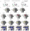
Fig. 5. Quaternary nature of the CD4 binding site
(A) The bridging sheet has a different topology in the trimer compared to gp120 monomer structures (e.g. PDB 1GC1, 3TGT), as illustrated by the cartoon models. Trimer formation alters how the base of V2 (β2–3) and β20–21 is arranged relative to the CD4bs (see also fig. S18). (B) In the EM reconstruction, the adjacent protomer appears to form additional contacts with CD4 and CD4bs bnAbs. The gp120/CD4bs Fab X-ray structures were docked into the EM map using the gp120 portion of the structure. All regions of the EM map within 5 Å of PGV04, VRC01 and CD4 have been colored yellow (fig. S20 shows all areas within 2 Å). (C) Major clashes with some CD4bs antibodies differ dramatically in the trimeric context. Areas of the EM map within 2 Å of the Fabs have been colored red (fig. S20 shows all areas within 5 Å). The minimal clashes with PGV04 serve as a comparator. b12 has only a few clashes with V1/V2, glycans, and V3 from the adjacent protomer, while b13 and F105 have extensive clashes. We note that, although b12 is typically a bnAb it does not neutralize the BG505 virus or bind the BG505 SOSIP.664 trimer (19). Thus, the clashes that we visualize here are consistent with each antibody being unable to neutralize the corresponding virus (19). (D) Areas in the EM map within 5 Å of docked Fabs, all of which contain acidic HFR3 insertions, are colored yellow (fig. S20 shows all areas within 2 Å). In addition to their previously known CD4bs epitopes, these bnAbs also interact with the V3 tip and proximal region near the protomer interface (red box). (E) bnAbs from D contain acidic HFR3 residues/insertions that interact with the basic residues in the highlighted area. Close up views of the antibody HFR3 interactions with basic residues in the trimer highlighted in the red boxed regions in D. Acidic residues in the HFR3 are displayed as sticks while regions in the EM map within 3 Å of basic residues have been colored blue and labeled accordingly.

Similar articles

Cited by

References

    1. Kwong PD, Mascola JR, Nabel GJ. Broadly neutralizing antibodies and the search for an HIV-1 vaccine: the end of the beginning. Nat Rev Immunol. 2013;13:693. - PubMed
    1. Kwong PD, et al. Structure of an HIV gp120 envelope glycoprotein in complex with the CD4 receptor and a neutralizing human antibody. Nature. 1998;393:648. - PMC - PubMed
    1. Joyce MG, et al. Outer domain of HIV-1 gp120: antigenic optimization, structural malleability, and crystal structure with antibody VRC-PG04. J Virol. 2013;87:2294. - PMC - PubMed
    1. Kwon YD, et al. Unliganded HIV-1 gp120 core structures assume the CD4-bound conformation with regulation by quaternary interactions and variable loops. Proc Natl Acad Sci USA. 2012;109:5663. - PMC - PubMed
    1. Klein F, et al. Somatic mutations of the immunoglobulin framework are generally required for broad and potent HIV-1 neutralization. Cell. 2013;153:126. - PMC - PubMed

Publication types

MeSH terms

Substances

Associated data