Intracellular Shigella remodels its LPS to dampen the innate immune recognition and evade inflammasome activation
- PMID: 24167293
- PMCID: PMC3832022
- DOI: 10.1073/pnas.1303641110
Intracellular Shigella remodels its LPS to dampen the innate immune recognition and evade inflammasome activation
Erratum in
- Proc Natl Acad Sci U S A. 2013 Dec 17;110(51):20843
Abstract
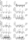
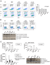
LPS is a potent bacterial effector triggering the activation of the innate immune system following binding with the complex CD14, myeloid differentiation protein 2, and Toll-like receptor 4. The LPS of the enteropathogen Shigella flexneri is a hexa-acylated isoform possessing an optimal inflammatory activity. Symptoms of shigellosis are produced by severe inflammation caused by the invasion process of Shigella in colonic and rectal mucosa. Here we addressed the question of the role played by the Shigella LPS in eliciting a dysregulated inflammatory response of the host. We unveil that (i) Shigella is able to modify the LPS composition, e.g., the lipid A and core domains, during proliferation within epithelial cells; (ii) the LPS of intracellular bacteria (iLPS) and that of bacteria grown in laboratory medium differ in the number of acyl chains in lipid A, with iLPS being the hypoacylated; (iii) the immunopotential of iLPS is dramatically lower than that of bacteria grown in laboratory medium; (iv) both LPS forms mainly signal through the Toll-like receptor 4/myeloid differentiation primary response gene 88 pathway; (v) iLPS down-regulates the inflammasome-mediated release of IL-1β in Shigella-infected macrophages; and (vi) iLPS exhibits a reduced capacity to prime polymorfonuclear cells for an oxidative burst. We propose a working model whereby the two forms of LPS might govern different steps of the invasive process of Shigella. In the first phases, the bacteria, decorated with hypoacylated LPS, are able to lower the immune system surveillance, whereas, in the late phases, shigellae harboring immunopotent LPS are fully recognized by the immune system, which can then successfully resolve the infection.
Keywords: PAMPs/PRRs; enteric pathogen; immune evasion; innate immunity.
Conflict of interest statement
The authors declare no conflict of interest.
Figures


Similar articles
-
Secretory IgA-mediated neutralization of Shigella flexneri prevents intestinal tissue destruction by down-regulating inflammatory circuits.J Immunol. 2009 Nov 1;183(9):5879-85. doi: 10.4049/jimmunol.0901838. Epub 2009 Oct 14. J Immunol. 2009. PMID: 19828639
-
Role of a fluid-phase PRR in fighting an intracellular pathogen: PTX3 in Shigella infection.PLoS Pathog. 2018 Dec 7;14(12):e1007469. doi: 10.1371/journal.ppat.1007469. eCollection 2018 Dec. PLoS Pathog. 2018. PMID: 30532257 Free PMC article.
-
Two msbB genes encoding maximal acylation of lipid A are required for invasive Shigella flexneri to mediate inflammatory rupture and destruction of the intestinal epithelium.J Immunol. 2002 May 15;168(10):5240-51. doi: 10.4049/jimmunol.168.10.5240. J Immunol. 2002. PMID: 11994481
-
The Orchestra and Its Maestro: Shigella's Fine-Tuning of the Inflammasome Platforms.Curr Top Microbiol Immunol. 2016;397:91-115. doi: 10.1007/978-3-319-41171-2_5. Curr Top Microbiol Immunol. 2016. PMID: 27460806 Review.
-
Shigella interaction with intestinal epithelial cells determines the innate immune response in shigellosis.Int J Med Microbiol. 2003 Apr;293(1):55-67. doi: 10.1078/1438-4221-00244. Int J Med Microbiol. 2003. PMID: 12755366 Review.
Cited by
-
Liujunanwei decoction attenuates cisplatin-induced nausea and vomiting in a Rat-Pica model partially mediated by modulating the gut micsrobiome.Front Cell Infect Microbiol. 2022 Aug 19;12:876781. doi: 10.3389/fcimb.2022.876781. eCollection 2022. Front Cell Infect Microbiol. 2022. PMID: 36061858 Free PMC article.
-
Synthetic bottom-up approach reveals the complex interplay of Shigella effectors in regulation of epithelial cell death.Proc Natl Acad Sci U S A. 2018 Jun 19;115(25):6452-6457. doi: 10.1073/pnas.1801310115. Epub 2018 Jun 4. Proc Natl Acad Sci U S A. 2018. PMID: 29866849 Free PMC article.
-
The Immunostimulatory Capacity of Nontypeable Haemophilus influenzae Lipooligosaccharide.Pathog Immun. 2017 Feb 16;2(1):34-49. doi: 10.20411/pai.v2i1.162. eCollection 2017. Pathog Immun. 2017. PMID: 30993246 Free PMC article.
-
The role of macrophage polarization in infectious and inflammatory diseases.Mol Cells. 2014 Apr;37(4):275-85. doi: 10.14348/molcells.2014.2374. Epub 2014 Mar 12. Mol Cells. 2014. PMID: 24625576 Free PMC article. Review.
-
Bacterial secretion systems and regulation of inflammasome activation.J Leukoc Biol. 2017 Jan;101(1):165-181. doi: 10.1189/jlb.4MR0716-330R. Epub 2016 Nov 3. J Leukoc Biol. 2017. PMID: 27810946 Free PMC article. Review.
References
-
- Hoshino K, et al. Cutting edge: Toll-like receptor 4 (TLR4)-deficient mice are hyporesponsive to lipopolysaccharide: evidence for TLR4 as the Lps gene product. J Immunol. 1999;162(7):3749–3752. - PubMed
-
- Takeda K, Akira S. Microbial recognition by Toll-like receptors. J Dermatol Sci. 2004;34(2):73–82. - PubMed
-
- Schletter J, Heine H, Ulmer AJ, Rietschel ET. Molecular mechanisms of endotoxin activity. Arch Microbiol. 1995;164(6):383–389. - PubMed
Publication types
MeSH terms
Substances
LinkOut - more resources
Full Text Sources
Other Literature Sources
Molecular Biology Databases
Research Materials

