. 2013 Oct 24:10:119.
doi: 10.1186/1742-4690-10-119.

Reactivation of latent HIV-1 in central memory CD4⁺ T cells through TLR-1/2 stimulation

Affiliations

Reactivation of latent HIV-1 in central memory CD4⁺ T cells through TLR-1/2 stimulation

Camille L Novis et al. Retrovirology. .

Abstract

Background: Toll-like receptors (TLRs) are crucial for recognition of pathogen-associated molecular patterns by cells of the innate immune system. TLRs are present and functional in CD4⁺ T cells. Memory CD4⁺ T cells, predominantly central memory cells (TCM), constitute the main reservoir of latent HIV-1. However, how TLR ligands affect the quiescence of latent HIV within central memory CD4⁺ T cells has not been studied.

Results: We evaluated the ability of a broad panel of TLR agonists to reactivate latent HIV-1. The TLR-1/2 agonist Pam3CSK4 leads to viral reactivation of quiescent HIV in a model of latency based on cultured TCM and in resting CD4⁺ T cells isolated from aviremic patients. In addition, we investigated the signaling pathway associated with Pam3CSK4 involved in HIV-1 reactivation. We show that the transcription factors NFκB, NFAT and AP-1 cooperate to induce viral reactivation downstream of TLR-1/2 stimulation. Furthermore, increasing levels of cyclin T1 is not required for TLR-mediated viral reactivation, but induction of viral expression requires activated pTEFb. Finally, Pam3CSK4 reactivates latent HIV-1 in the absence of T cell activation or proliferation, in contrast to antigen stimulation.

Conclusions: Our findings suggest that the signaling through TLR-1/2 pathway via Pam3CSK4 or other reagents should be explored as an anti-latency strategy either alone or in combination with other anti-latency drugs.


Figures
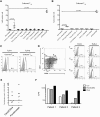
Figure 1
Figure 1
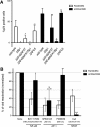
Reactivation of latent HIV-1 through toll-like receptors agonists. (A) Cultured TCM were treated with the toll-like receptors agonists indicated between parentheses for different TLRs or costimulated with αCD3/αCD28 and assessed for intracellular p24Gag expression by flow cytometry. Experiments were done in 5 different donors. Each dot represents a different donor and mean and SD are indicated with horizontal lines. Significance was calculated by 2-tailed paired samples t test analysis (P vales provided). (B) Cultured TCM were treated with different TLR-2 agonists indicated between parentheses or costimulated with αCD3/αCD28 and assessed for intracellular p24Gag expression by flow cytometry. Experiments were done in 3 donors from A. Significance was calculated by 2-tailed paired samples t test analysis (P vales provided). (C) Cultured TCM were stained with specific antibodies against TLR-1 and TLR-2 (open black histogram) and analyzed by flow cytometry. Isotype controls were used as control (closed grey histogram). The percentage of TLR-1 and TLR-2 positive cells is indicated in each panel. (D) Ex vivo isolated memory CD4+ T cells were stained with specific antibodies against CCR7, CD27, TLR-1 and TLR-2 and analyzed by flow cytometry. Expression of TLR-1 and TLR-2 was analyzed in each subset of memory CD4+ T cells (open black histogram). Isotype controls were used as control (closed grey histogram). The percentage of TLR-1 and TLR-2 positive cells in each subset is indicated in each panel. (E) Cells isolated from seven aviremic patients were treated with Pam3CSK4 or Panobinostat and the levels of HIV-1 US RNA were measured three days later. Each symbol corresponds to a different patient. Data is represented as fold induction over the IL-2 treatment control (F) Cells isolated from three aviremic patients were treated with IL-2, Pam3CSK4 or PHA for 24 hours and supernatants were subject to the Q-VOA assay.
Figure 2
Figure 2
Pam3CSK4 requires NFAT, AP-1 and NF-κB to induce viral reactivation in cultured TCM. (A) Cultured TCM cells were infected with wild-type DHIV or with different LTR mutants. 9 days after infection, cells were treated with Pam3CSK4 or costimulated with antibodies to CD3 and CD28 (αCD3/αCD28) for 3 days and assessed for intracellular p24Gag expression by flow cytometry. Percentage of reactivation was normalized to that of wild-type DHIV for each treatment. Bar graph corresponds to mean and SD of experiments performed with three donors. (B) Cultured TCM cells were infected with wild-type DHIV and treated with Pam3CSK4 or stimulated with αCD3/αCD28 in the presence of the indicated inhibitor for the protein or transcription factor indicated and assessed for intracellular p24 Gag expression by flow cytometry. Bar graph corresponds to mean and SD of experiments performed with four different donors. Significance was calculated by 2-tailed paired samples t test analysis (* < 0.05, ** < 0.01, *** < 0.001).
Figure 3
Figure 3
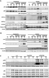
Signaling triggered by Pam3CSK4 in cultured TCM. Culture TCM were either left unstimulated (0 h) or stimulated with IL-2, Pam3CSK4 and αCD3/αCD28 after 30 minutes or 3 hours. Nuclear and cytoplasmic fractions were isolated as indicated in the experimental procedure section. Proteins were loaded on a SDS-polyacrylamide gel, transferred to a membrane and western-blotted against p50, p65, NFATc1, NFATc2, α-tubulin, histone H3 and β-actin (A) or against cJun, cFos, histone H3 and β-actin (B). Data is representative of two donors. (C) Culture TCM were left unstimulated (0 h) or stimulated with IL-2, Pam3CSK4 and αCD3/αCD28 after 30 minutes or 3 hours. Whole cell extracts were isolated as indicated in the experimental procedure section. Proteins were loaded on a SDS-polyacrylamide gel, transferred to a membrane and western-blotted against JNK, phospho-JNK, ERK, phospho-ERK and β-actin. Data is representative of two donors.
Figure 4
Figure 4
Pam3CSK4 triggers intracellular Ca2+ influx. Cultured TCM were loaded with the Ca2+ dye Fluo3-AM and cells were tested for their ability to increase intracellular calcium levels after treatment with ionomycin, or with increasing concentrations of Pam3CSK4, Pam2CSK4 or LAM-MS by flow cytometry. The arrows indicate when the stimuli were added. Data is representative of two donors.
Figure 5
Figure 5
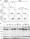
Pam3CSK4 induces viral reactivation in a Tat-dependent manner. (A) Cultured TCM cells were infected with wild-type DHIV and treated with Pam3CSK4 or stimulated with αCD3/αCD28 in the presence of 50 nM and 100 nM of Flavopiridol and assessed for intracellular p24 Gag expression by flow cytometry. Data is representative of two donors. (B) Culture TCM were either left unstimulated (0 h) or stimulated with IL-2, Pam3CSK4 and αCD3/αCD28 for 12 h, 24 h or 48 h. Whole cell extracts were isolated as indicated in the experimental procedure section. Proteins were loaded on a SDS-polyacrylamide gel, transferred to a membrane and western-blotted against CyclinT1, phospho-CDK9, CDK9, and β-actin. Data is representative of two donors.
Figure 6
Figure 6
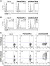
Pam3CSK4 induces viral reactivation in the absence of T cell activation or T cell proliferation. Cultured TCM cells were treated with IL-2, Pam3CSK4 or costimulated with antibodies to CD3 and CD28 (αCD3/αCD28) for 3 days and assessed for the induction of CD69 (A) and CD25 (B) by flow cytometry (open black histograms). Isotype control was used as control (closed gray histogram). The percentage of CD69 and CD25 positive cells is indicated in each panel and the mean fluorescence intensity (MFI) is indicated between parentheses for CD25. Data is representative of three donors. (C) Cultured TCM cells were infected with wild-type DHIV or left uninfected. At 9 days after infection, cells were stained with the cell-proliferation dye CPD eFluor670. Stained cells were treated with IL-2, Pam3CSK4 or costimulated with antibodies to CD3 and CD28 (αCD3/αCD28) for 3 days and assessed for intracellular p24Gag expression and CPD eFluor670 staining by flow cytometry. Numbers indicate percentage of cells. Data is representative of three donors.

Similar articles

Cited by

References

    1. Finzi D, Hermankova M, Pierson T, Carruth LM, Buck C, Chaisson RE, Quinn TC, Chadwick K, Margolick J, Brookmeyer R. et al.Identification of a reservoir for HIV-1 in patients on highly active antiretroviral therapy. Science. 1997;278:1295–1300. doi: 10.1126/science.278.5341.1295. - DOI - PubMed
    1. Chun TW, Stuyver L, Mizell SB, Ehler LA, Mican JA, Baseler M, Lloyd AL, Nowak MA, Fauci AS. Presence of an inducible HIV-1 latent reservoir during highly active antiretroviral therapy. Proc Natl Acad Sci U S A. 1997;94:13193–13197. doi: 10.1073/pnas.94.24.13193. - DOI - PMC - PubMed
    1. Wong JK, Hezareh M, Gunthard HF, Havlir DV, Ignacio CC, Spina CA, Richman DD. Recovery of replication-competent HIV despite prolonged suppression of plasma viremia. Science. 1997;278:1291–1295. doi: 10.1126/science.278.5341.1291. - DOI - PubMed
    1. Brenchley JM, Hill BJ, Ambrozak DR, Price DA, Guenaga FJ, Casazza JP, Kuruppu J, Yazdani J, Migueles SA, Connors M. et al.T-cell subsets that harbor human immunodeficiency virus (HIV) in vivo: implications for HIV pathogenesis. J Virol. 2004;78:1160–1168. doi: 10.1128/JVI.78.3.1160-1168.2004. - DOI - PMC - PubMed
    1. Chomont N, El-Far M, Ancuta P, Trautmann L, Procopio FA, Yassine-Diab B, Boucher G, Boulassel MR, Ghattas G, Brenchley JM. et al.HIV reservoir size and persistence are driven by T cell survival and homeostatic proliferation. Nat Med. 2009;15:893–900. doi: 10.1038/nm.1972. - DOI - PMC - PubMed

Publication types