. 2014 Jan;88(1):456-68.
doi: 10.1128/JVI.02572-13. Epub 2013 Oct 23.

Measles virus C protein impairs production of defective copyback double-stranded viral RNA and activation of protein kinase R

Affiliations

Measles virus C protein impairs production of defective copyback double-stranded viral RNA and activation of protein kinase R

Christian K Pfaller et al. J Virol. 2014 Jan.

Abstract

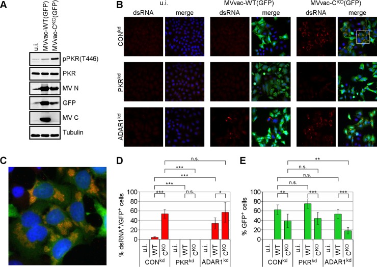
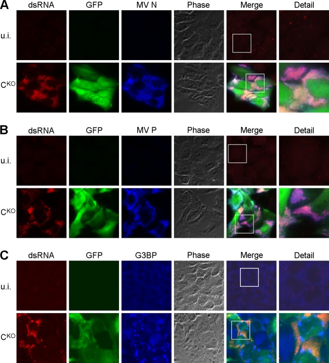
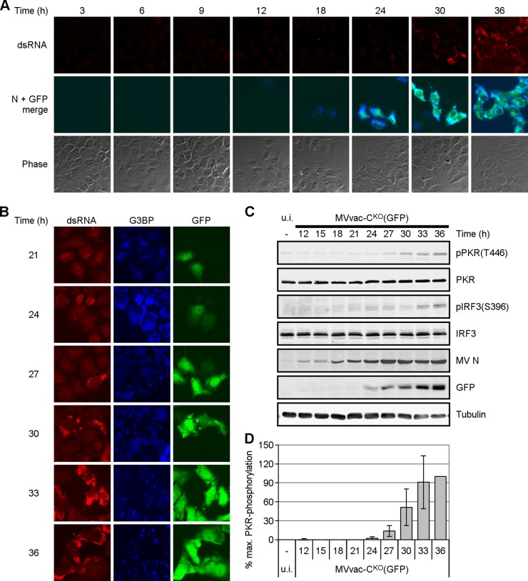
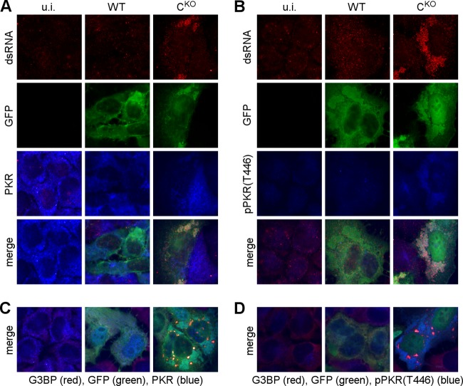
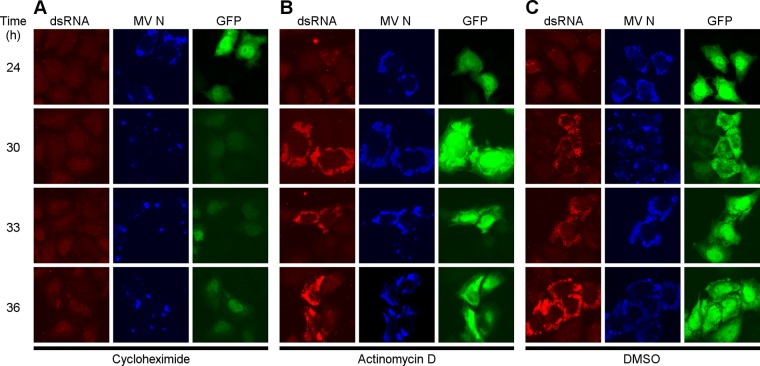
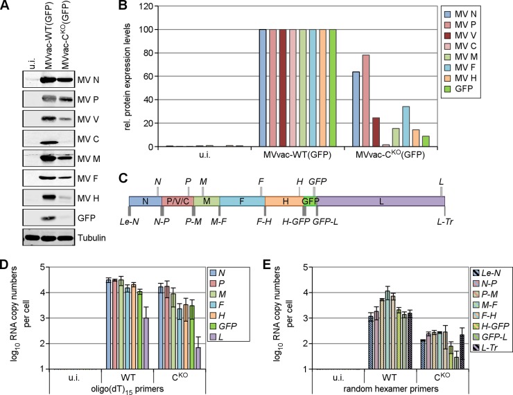
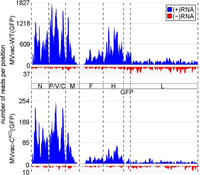
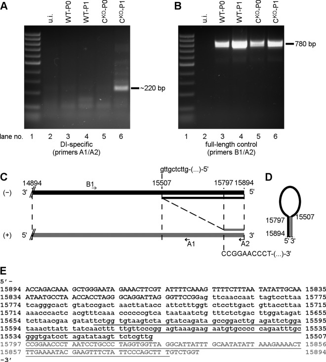
Measles virus (MV) lacking expression of C protein (C(KO)) is a potent activator of the double-stranded RNA (dsRNA)-dependent protein kinase (PKR), whereas the isogenic parental virus expressing C protein is not. Here, we demonstrate that significant amounts of dsRNA accumulate during C(KO) mutant infection but not following parental virus infection. dsRNA accumulated during late stages of infection and localized with virus replication sites containing N and P proteins. PKR autophosphorylation and stress granule formation correlated with the timing of dsRNA appearance. Phospho-PKR localized to dsRNA-containing structures as revealed by immunofluorescence. Production of dsRNA was sensitive to cycloheximide but resistant to actinomycin D, suggesting that dsRNA is a viral product. Quantitative PCR (qPCR) analyses revealed reduced viral RNA synthesis and a steepened transcription gradient in C(KO) virus-infected cells compared to those in parental virus-infected cells. The observed alterations were further reflected in lower viral protein expression levels and reduced C(KO) virus infectious yield. RNA deep sequencing confirmed the viral RNA expression profile differences seen by qPCR between C(KO) mutant and parental viruses. After one subsequent passage of the C(KO) virus, defective interfering RNA (DI-RNA) with a duplex structure was obtained that was not seen with the parental virus. We conclude that in the absence of C protein, the amount of PKR activator RNA, including DI-RNA, is increased, thereby triggering innate immune responses leading to impaired MV growth.


Figures

FIG 1
FIG 1
MV deficient in C protein expression causes increased phosphorylation of PKR and production of dsRNA. (A) Immunoblot of whole-cell lysates against indicated proteins expressed in HeLa cells infected with MVvac-WT(GFP) or MVvac-CKO(GFP) or left uninfected (u.i.) for 48 h. (B) Immunofluorescence staining against dsRNA (red), GFP (green), and nuclei (DAPI; blue) in HeLa cells stably expressing short hairpin RNA (shRNA) directed against PKR (PKRkd), ADAR1 (ADAR1kd), or an unspecific control (CONkd) and infected for 48 h or left uninfected (u.i.). The merge shows combination of all three channels. Images were taken at ×40 magnification. (C) Magnification of the framed section in panel B. (D) Quantification of dsRNA-expressing cells as a percentage of WT or CKO mutant virus-infected (GFP-positive) cells. (E) Quantification of infection as the percentage of GFP-positive cells. Averages and standard deviations were determined from six fields from two independent experiments with ∼100 cells/field for each condition. Statistical significance was determined by Student's t test (two-tailed, two-sample test with equal variance) and P values are marked: *, P < 0.05; **, P < 0.01; ***, P < 0.005. n.s., not significant.
FIG 2
FIG 2
dsRNA localizes to sites of viral replication but not to stress granules. HeLa CONkd cells were infected with CKO mutant virus or left uninfected (u.i.) and fixed 48 h after infection. Immunofluorescence was performed against dsRNA (red), GFP (green), and viral N protein (A; blue), viral P protein (B; blue), or the cellular stress granule marker G3BP (C; blue). The merge column shows the combination of all three colors. The detail column shows the expanded framed regions in the merge column. Images were taken at ×64 magnification and are representative of three independent experiments.
FIG 3
FIG 3
dsRNA accumulates with kinetics similar to PKR activation and stress granule formation. HeLa CONkd cells were infected with MVvac-CKO(GFP) mutant virus and fixed at the indicated times (h) after infection. (A) Cells were simultaneously stained with antibodies against dsRNA (red), GFP (green), and viral N protein (blue). The first row shows dsRNA, the second row shows the merged GFP and N signals, and the third row shows phase contrast. Images were taken at ×64 magnification. (B) Immunofluorescence was performed using antibodies directed against dsRNA (red; left column), G3BP (blue; middle column), and GFP (green; right column). Images were taken at ×64 magnification. (C) Immunoblot analysis of the indicated proteins. Blots are representative of three independent experiments. (D) Quantification of pPKR levels. Levels are averages and standard deviations from two independent experiments. They were normalized to the intensity of the corresponding tubulin band in each lane and are expressed as the percentage of the value at 36 h.
FIG 4
FIG 4
Phospho-PKR localizes to sites of dsRNA accumulation. HeLa CONkd cells were infected with parental MVvac-WT(GFP) or mutant MVvac-CKO(GFP) virus and fixed 48 h after infection. (A) Immunofluorescence against dsRNA (red), GFP (green), and total PKR (blue). (B) Immunofluorescence against dsRNA (red), GFP (green), and pPKR(T446) (blue). (C) Immunofluorescence against G3BP (red), GFP (green), and total PKR (blue). (D) Immunofluorescence against G3BP (red), GFP (green), and pPKR(T446) (blue). The merge row shows the combination of all three colors.
FIG 5
FIG 5
dsRNA is a viral product. HeLa CONkd cells infected with MVvac-CKO(GFP) mutant virus and treated with 10 μg/ml cycloheximide (A), 10 μg/ml actinomycin D (B), or DMSO (C) at 24 h after infection. Cells were fixed at the indicated time points, and immunofluorescence was performed in parallel with antibodies directed against dsRNA (red; left column), viral N protein (blue; middle column), or GFP (green; right column). Images were taken at ×64 magnification and are representative of three independent experiments.
FIG 6
FIG 6
Knockout of C protein slows viral macromolecular synthesis and results in a steeper transcription gradient. HeLa CONkd cells were infected with parental MVvac-WT(GFP) or mutant MVvac-CKO(GFP) virus or left uninfected (u.i.). Cells were harvested at 48 h after infection. (A) Immunoblot analysis with antibodies directed against the indicated viral and cellular proteins. Blots are representative of three independent experiments. (B) Quantification of the proteins from panel A. Protein levels were normalized to the corresponding tubulin levels in each lane and are relative to the levels of MVvac-WT(GFP)-infected cells, which were set as 100% for each protein. (C) Schematic representation of the genome organization of MVvac-WT(GFP). The relative size and location of the viral gene-specific (light-gray boxes) and intercistronic (dark-gray boxes) qPCR products are shown. (D) qPCR analysis of viral mRNA from infected cells using cDNA prepared with oligo(dT)15 primers. (E) qPCR analysis of intercistronic viral sequences from infected cells using cDNA prepared with random hexamer primers. The resulting cDNAs were analyzed for product copy number as described in Materials and Methods. Data shown are average numbers ± standard deviations from three independent experiments.
FIG 7
FIG 7
RNA-Seq analyses of RNA from CKO mutant and parental virus-infected cells. cDNA libraries prepared using 10 μg total RNA from virus-infected cells depleted of ribosomal RNAs were analyzed by Ion Torrent sequencing as described in Materials and Methods. Diagrams show the alignment of reads to the viral genome sequence for parental MVvac-WT(GFP)-infected cells (top) and mutant MVvac-CKO(GFP)-infected cells (bottom). Reads are separated corresponding to their polarity with +RNA shown in blue (mRNA, antigenome) and −RNA (genome) shown in red. Dashed lines correspond to gene borders.
FIG 8
FIG 8
CKO mutant virus generates copyback DI-RNA. cDNA generated from HeLa CONkd cells infected with original virus stocks of WT or CKO mutant viruses (P0) or with viruses that have been passaged once on Vero cells (P1) were analyzed for copyback DI-RNAs by PCR. (A) DI-specific PCR using primers A1 and A2, both of which are negative polarity with respect to virus genome sequence. (B) Control PCR using primers B1 (positive polarity) and A2 (negative polarity). (C) Schematic diagram of the sequence obtained for the 220-bp fragment observed with CKO-P1. Breakpoint (genome position −15507) and reinitiation point (genome position +15797) of copyback DI are indicated, as well as relative binding sites of primers A1, A2, and B1. (D) Schematic diagram of the DI-RNA stem-loop model with the predicted 98-bp stem. (E) Sequence of the detected copyback DI-RNA. Complementary ends are shown in uppercase font, and the loop sequence of the DI-RNA is shown in lowercase font. The PCR fragment that was sequenced is shown in underlined font. Antigenomic +RNA is shown in gray; −RNA (genomic RNA) is shown in black in panels C, D, and E. Nucleotide positions correspond to the Moraten vaccine strain of MV (GenBank accession number AF266287).

Similar articles

Cited by

References

    1. Griffin DE. 2007. Measles virus, p 1551–1585 In Knipe DM, Howley PM. (ed), Fields virology, 5 ed, vol 2 Lippincott Williams & Wilkins, Philadelphia, PA
    1. Cattaneo R, McChesney M. 2008. Measles virus, p 285–291 In Mahy BWJ, van Regenmortel MHV. (ed), Encyclopedia of virology, 3rd ed. Academic Press, Oxford, United Kingdom
    1. Cattaneo R, Kaelin K, Baczko K, Billeter MA. 1989. Measles virus editing provides an additional cysteine-rich protein. Cell 56:759–764. 10.1016/0092-8674(89)90679-X - DOI - PubMed
    1. Bellini WJ, Englund G, Rozenblatt S, Arnheiter H, Richardson CD. 1985. Measles virus P gene codes for two proteins. J. Virol. 53:908–919 - PMC - PubMed
    1. Escoffier C, Manie S, Vincent S, Muller CP, Billeter M, Gerlier D. 1999. Nonstructural C protein is required for efficient measles virus replication in human peripheral blood cells. J. Virol. 73:1695–1698 - PMC - PubMed

Publication types