. 2014 Feb:67:235-47.
doi: 10.1016/j.freeradbiomed.2013.10.014. Epub 2013 Oct 17.

Treatment with the reactive oxygen species scavenger EUK-207 reduces lung damage and increases survival during 1918 influenza virus infection in mice

Affiliations

Treatment with the reactive oxygen species scavenger EUK-207 reduces lung damage and increases survival during 1918 influenza virus infection in mice

John C Kash et al. Free Radic Biol Med. 2014 Feb.

Abstract

The 1918 influenza pandemic caused over 40 million deaths worldwide, with 675,000 deaths in the United States alone. Studies in several experimental animal models showed that 1918 influenza virus infection resulted in severe lung pathology associated with dysregulated immune and cell death responses. To determine if reactive oxygen species produced by host inflammatory responses play a central role in promoting severity of lung pathology, we treated 1918 influenza virus-infected mice with the catalytic catalase/superoxide dismutase mimetic, salen-manganese complex EUK-207 beginning 3 days postinfection. Postexposure treatment of mice infected with a lethal dose of the 1918 influenza virus with EUK-207 resulted in significantly increased survival and reduced lung pathology without a reduction in viral titers. In vitro studies also showed that EUK-207 treatment did not affect 1918 influenza viral replication. Immunohistochemical analysis showed a reduction in the detection of the apoptosis marker cleaved caspase-3 and the oxidative stress marker 8-oxo-2'-deoxyguanosine in lungs of EUK-207-treated animals compared to vehicle controls. High-throughput sequencing and RNA expression microarray analysis revealed that treatment resulted in decreased expression of inflammatory response genes and increased lung metabolic and repair responses. These results directly demonstrate that 1918 influenza virus infection leads to an immunopathogenic immune response with excessive inflammatory and cell death responses that can be limited by treatment with the catalytic antioxidant EUK-207.

Keywords: Antioxidants; Free radicals; Immune response; Influenza; Pathogenesis; Reactive oxygen species; Virus.


Figures

Fig. 1
Fig. 1. EUK-207 treatment increases survival in 1918 influenza virus infected mice
(A) Survival curves for EUK-207- and vehicle treatment during 1918 influenza virus infection. Administration of vehicle and drug by IP injection occurred on 3 to 10 dpi. (B) Weight loss during 1918 influenza virus infection in vehicle and EUK-207-treated mice. Data presented are from single experiment with 15 animals per group infected with 500 PFU (4x LD50) of the 1918 influenza virus, and are representative of 3 independent experiments with total N=69 for EUK-207-treated groups and N=55 for vehicle-treated groups. (C) Measurement of influenza M gene mRNA by qRT-PCR relative to GAPDH mRNA at 3 (onset of treatment), 6 and 8 dpi. Data are presented as average expression values ± standard deviation. (D) Viral replicatior kinetics in MDCK cells in the presence and absence of EUK-207 treatment. MDCK cells were infected with an MOI=0.01 and were cultured continuously in the presence or absence of 1.5 μg/ml EUK-207, as described in Materials and Methods. At 12h, 24h, 48h and 72h post-infection virus present in the supernatant was quantified by plaque assay. Kinetics were performed in triplicate and data are presented as mean ± standard deviation. (E) Chemical structure of EUK-207. The axial ligand, “OAc”, is an acetoxy group.
Fig. 2
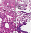
Fig. 2. EUK-207 treatment reduces severity of lung pathology compared to vehicle-treated mice
Hematoxylin and eosin stains demonstrating representative histopathology differences between drug treated and untreated mice at 8 dpi. Original magnifications for each image are noted in their bottom right hand corners. A) A lung from an untreated mouse shows severe histopathologic changes. B) Severe lymphohistocytic and neutrophilic alveolitis, pulmonary edema and necrotizing bronchitis in an untreated lung. C) Close up of bronchial epithelium in an untreated lung showing transmural inflammation, white asterisk, and multifocal necrosis. The lumen is clogged with necrotic debris. D) A lung from a EUK-207-treated mouse showing less severe histopathologic changes than an untreated lung. E) Mild to moderate alveolitis with saimflmmxed cellularity infiltrate, pulmonary edema and mild bronchitis in an EUK-207-treated lung. F) Close up of bronchial epithelium in a treated lung showing marked epithelial cell jumbling in the normally pseudostratified columnar epithelium indicating epithelial reproliferation, black arrow. The bronchial lumen is denoted by the “b.” The broken line boxes in images A and D indicate the zoom regions shown in B and E, aligned vertically. The solid line boxes in same images indicate the zoom regions shown in C and F.
Fig. 3
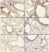
Fig. 3. EUK-207 treatment does not affect distribution of 1918 influenza virus infection in lungs of infected animals
An immunohistochemical viral antigen positive is indicated by brown labeling of a cell's cytoplasm and/or nucleus. Cellular debris is also frequently positive. All figures were taken at an original magnification of 200×. A) Untreated mouse, 6 dpi; B) untreated mouse, 8 dpi; C) no primary antibody, negative reagent control; D) treated mouse, 6 dpi; E) treated mouse, 8 dpi; and F) uninfected mouse, biological negative control.
Fig. 4
Fig. 4. EUK-207 treatment decreases cell death in lung of 1918 influenza virus infected animals
Immunohistochemistry for apoptosis marker cleaved caspase 3 (cCASP3). (A) Bronchial epithelium as well as alveoli in the lung from an untreated mouse shows stronger, more frequent cytoplasmic labeling. (B) Close up of the bronchial epithelium outlined by the broken line box in A. (C) Bronchial epithelium and alveoli in a lung from a treated mouse show less labeling. (D) Close up of the bronchial epithelium outlined by the broken line box in C. The black asterisk denotes a mitotic figure. Original magnifications for each image are noted in their bottom right hand corners.
Fig. 5
Fig. 5. EUK-207 treatment reduces oxidative damage during 1918 influenza virus infection
Immunohistochemistry for oxidation marker 8-Oxo-2'-deoxyguanosine (8-OHdG). (A) Staining of mock infected, untreated mouse lung. (B) Close up of the alveolar epithelium from mock infected, untreated mouse lung from panel A. (C) Staining of vehicle-treated 1918 influenza virus-infected mouse lung showing prominent 8-OHdG staining in bronchial epithelium as well as alveolar epithelium. (D) Close up view of alveolar epithelium from panel C showing 8-OHdG staining. Prominently staining alveolar epithelial cells showing type II hyperplasia are shown also with staining in inflammatory cells in interstitial infiltrates. (E) Staining of EUK-207-treated 1918 influenza virus-infected mouse lung showing marked reduction in 8-OHdG staining in bronchial epithelium as well as alveolar epithelium as compared to the lung from the vehicle-treated animal in panel C. (F) Close up view of alveolar epithelium from panel E showing staining in alveolar macrophages and a marked reduction in 8OH-dG staining in alveolar epithelium. Original magnifications for each image are noted in their bottom right hand corners.
Fig. 6
Fig. 6. EUK-207 treatment results in increased expression of lung repair related genes and decreased expression of inflammatory response and cell death related genes
Gene ontology comparison of genes identified using next-generation sequencing (NGS) that were induced in pooled mRNA isolated from lungs of EUK-207-treated mice or in pooled mRNA isolated from lungs of vehicle-treated animals at 8 dpi. * Fishers exact test, ** Fishers exact test with FDR method ofBenjamini and Hochberg.
Fig. 7
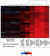
Fig. 7. Identification of RNA biomarkers in vehicle-treated, fatal 1918 influenza virus infected animals
Two-dimensional hierarchical clustering diagram of genes preferentially induced in individual vehicle-treated animals at 8 dpi, but not in lungs of individual mice infected with 100 PFU of 1918 influenza virus at 8 dpi. Also shown is the expression of this population of genes in individual EUK-207-treated animals. These animals show a range of expression that was either similar to lethal or non-lethal 1918 influenza virus infections. Genes shown in red were up-regulated and genes shown in blue were down-regulated and genes shown in black showed no change in expression compared to a pool RNA isolated from mock infected mice.

Similar articles

Cited by

References

    1. Johnson NP, Mueller J. Updating the accounts: global mortality of the 1918-1920 “Spanish” influenza pandemic. Bull Hist Med. 2002;76:1–05-115. - PubMed
    1. Taubenberger JK, Morens DM. 1918 Influenza: the mother of all pandemics. Emerg Infect Dis. 2006;12:1–5-22. - PMC - PubMed
    1. Morens DM, Taubenberger JK, Fauci AS. Predominant role of bacterial pneumonia as a cause of death in pandemic influenza: implications for pandemic influenza preparedness. J Infect Dis. 2008;198:9–62-970. - PMC - PubMed
    1. Tumpey TM, Basler CF, Aguilar PV, Zeng H, Solorzano A, Swayne DE, Cox NJ, Katz JM, Taubenberger JK, Palese P, Garcia-Sastre A. Characterization of the reconstructed 1918 Spanish influenza pandemic virus. Science. 2005;310:7–7-80. - PubMed
    1. Taubenberger JK, Reid AH, Lourens RM, Wang R, Jin G, Fanning TG. Characterization of the 1918 influenza virus polymerase genes. Nature. 2005;437:8–89-893. - PubMed

Publication types

MeSH terms