. 2013 Oct 31;5(2):331-9.
doi: 10.1016/j.celrep.2013.09.020. Epub 2013 Oct 17.

CDIP1-BAP31 complex transduces apoptotic signals from endoplasmic reticulum to mitochondria under endoplasmic reticulum stress

Affiliations

CDIP1-BAP31 complex transduces apoptotic signals from endoplasmic reticulum to mitochondria under endoplasmic reticulum stress

Takushi Namba et al. Cell Rep. .

Abstract

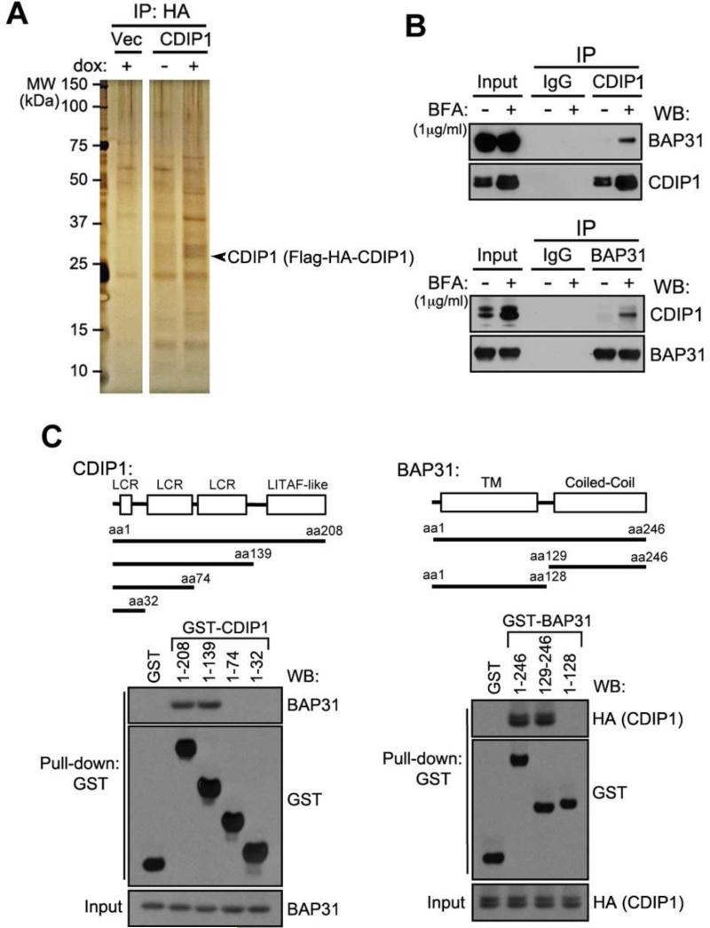
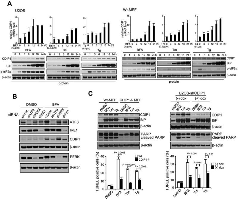
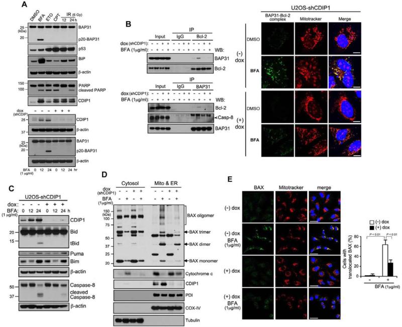
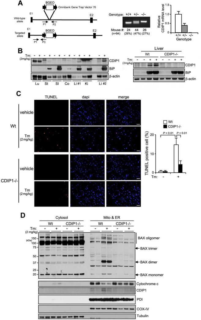
Resolved endoplasmic reticulum (ER) stress response is essential for intracellular homeostatic balance, but unsettled ER stress can lead to apoptosis. Here, we show that a proapoptotic p53 target, CDIP1, acts as a key signal transducer of ER-stress-mediated apoptosis. We identify B-cell-receptor-associated protein 31 (BAP31) as an interacting partner of CDIP1. Upon ER stress, CDIP1 is induced and enhances an association with BAP31 at the ER membrane. We also show that CDIP1 binding to BAP31 is required for BAP31 cleavage upon ER stress and for BAP31-Bcl-2 association. The recruitment of Bcl-2 to the BAP31-CDIP1 complex, as well as CDIP1-dependent truncated Bid (tBid) and caspase-8 activation, contributes to BAX oligomerization. Genetic knockout of CDIP1 in mice leads to impaired response to ER-stress-mediated apoptosis. Altogether, our data demonstrate that the CDIP1/BAP31-mediated regulation of mitochondrial apoptosis pathway represents a mechanism for establishing an ER-mitochondrial crosstalk for ER-stress-mediated apoptosis signaling.


Figures

Figure 1
Figure 1. CDIP1 interacts with BAP31 and ER stress increases this interaction
(A) Affinity purification of CDIP1 interacting proteins. The CDIP1 interacting proteins were visualized by silver staining. (B) Co-immunoprecipitation of BAP31 by CDIP1. U2OS cells were treated with or without Brefeldin A (BFA) (1 μg ml−1) for 24 h. Proteins were cross-linked with DSP prior to protein extraction. (C)Mapping of CDIP1-BAP31 binding sites by GST pull-down with indicated GST-CDIP1 deletion proteins (aa 1-32, 1-74, 1-139 and 1–208) and GST-BAP31 deletion proteins (aa 1-129, 128-246 and 1–246) were generated for GST pull-down experiments. LCR: low complexity region; TM: transmembrane; and Coiled-Coil: coiled-coil domain. See also Figure S1 and Table S1 & S2.
Figure 2
Figure 2. The effect of ER stress-mediated CDIP1 induction on ER stress-induced apoptosis
(A) ER stress induction of CDIP1 in U2OS cells (left panel) and MEFs (right panel). U2OS cells or Wt-MEFs were incubated with BFA (1 μg ml−1), Tm (0.5 μg ml−1) or Tg (0.1 μM) for indicated periods. Total RNAs were extracted and subjected to real-time quantitative PCR analysis using a specific primer set for CDIP1 and normalized to β-actin (MEFs) or GAPDH (U2OS). Data shown are mean ± s.d. (in triplicates and measured at the same time). Cell lysates were analyzed by western blotting with indicated antibodies. The blot was cut based on the size of proteins or stripped. (B) ER stress-mediated CDIP1 induction is regulated by ATF6 and IRE1 pathways. U2OS cells with knockdown using siCont., siATF6, siPERK and siIRE1 were treated with BFA (1 μg ml−1) or DMSO for 18 hours. Whole cell lysates were analyzed by western blotting with indicated antibodies. (C) CDIP1 deficiency precludes ER stress-induced apoptosis. Cells were treated with ER stress agents (BFA, Tm, or Tg) as described above for 18 h. Cell lysates were analyzed for the expression of indicated proteins. PARP cleavage was used as an apoptosis marker. Apoptosis was determined by TUNEL assay, followed by enumeration of dUTP-TMR red-positive cells by flow cytometry. Values shown are mean ± s.d. of three different experiments measured at the same time. P-value was calculated using Student's t-test. Left panel: Wt (CDIP1+/+) and CDIP1−/− MEFs. Right panel: U2OS cells with doxycycline-regulatable shCDIP1 were exposed with or without dox for 48 h, prior to ER stress treatment. See also Figure S1 & S2.
Figure 3
Figure 3. CDIP1 promotes Bax oligomerization and mitochondrial translocation during ER stress
(A) BAP31 cleavage is predominantly ER stress- and CDIP1-dependent. Top panel: U2OS cells were treated with BFA (1 μg ml−1), ETO (25 μM), CPT (500 nM) for 24 hours or γ-irradiation (6 Gy) for 12 and 24 hours. Bottom panel: U2OS cells with dox-regulatable shCDIP1 were treated with or without dox for 48 h prior to BFA treatment. Cell lysates were analyzed for the expression of indicated proteins. Same amounts of proteins were analyzed in two different gels (BAP31, p20BAP31, BiP, p53, β-actin; CDIP1 and PARP). The blot was cut based on the size of proteins. (B) CDIP1 depletion suppresses the BAP31 binding to Bcl-2 and procaspase-8 and BAP31-Bcl-2 interaction on the mitochondria upon ER stress. Left panel: U2OS cells with dox-regulatable shCDIP1 were treated with or without dox (1 μg ml−1) prior to BFA treatment (16 h), followed by Bcl-2 and BAP31 co-immunoprecipitation. Right panel: Representative images of DuoLink using BAP31 and Bcl-2 antibodies in U2OS-shCDIP1 cells upon BFA treatment. The detected endogenous BAP31 and Bcl-2 interaction by proximity ligation assay is shown as green dots. MitoTracker was used as mitochondria-specific marker. Merged images are also shown, and yellow color represents co-localization between BAP31 and Bcl-2 interaction (green) and MitoTracker (red) (scale bar, 10 μm). (C) The effect of CDIP1 depletion on ER stress-induced expression of BH3-only domain proteins. Using the same procedure described in (A), whole cell lysates were analyzed for the expression of indicated proteins. Same amounts of proteins were analyzed in three different gels (CDIP1 and Bid; Puma, Bim and β-actin; caspase-8 and β-actin). (D) ER stress-dependent Bax oligomerization and cytochrome c release requires CDIP1. U2OS cells with dox-regulatable shCDIP1 were grown with or without dox for 24 h and treated with BFA for additional 24 h. Cell lysates were then fractionated into two organellar fractions (cytosol and mitochondria/ER) and blotted with antibodies against BAX, cytochrome c, CDIP1, PDI (ER-specific marker), COX-IV (mitochondria-specific marker) and α-tubulin (cytosol marker). (E) The effect of CDIP1 depletion on Bax translocation to mitochondria upon ER stress. Using the same procedure described in (D), endogenous active form of Bax was visualized by immunostaining with anti-Bax antibody (N-20) by confocal microscopy. The strength of the laser (561nm) used to visualize the MitoTracker staining was determined at a level insufficient to fluoresce the endogenous RFP (data not shown). Merged images are also shown, and yellow color represents co-localization between Bax (green) and MitoTraker (red) (scale bar, 50 μm). The percentage of BAX translocation was determined by counting three different fields (30-50 cells/field). Values shown are mean ± s.d. of three different experiments. P-value was calculated using two-way ANOVA. See also Figures S3, S4 and S5.
Figure 4
Figure 4. CDIP1 deficiency leads to impaired ER stress-induced cell death in mouse
(A) A gene trap vector was inserted into the first intron of CDIP1 (left panel). PCR assay was shown using tail genomic DNA from three different genotypes: Wt (+/+), CDIP1 heterozygous (+/−) and CDIP1-null (−/−) littermates. Relative CDIP1 mRNA levels were measured by real-time quantitative PCR analysis (right panel). (B) CDIP1 is predominantly induced in liver tissue upon ER stress. Wt or CDIP1-null mice were intraperitoneally administrated with Tm (2 mg kg−1) or vehicle (150 mM dextrose). After 24 hours, tissues were removed and subjected to western blot analysis: Lu: lung, St: stomach, SI: small intestine, Co: colon, Li #1 & Li #2: liver, and Ki: kidney. (C) CDIP1-deficient mouse is resistant to ER stress-induced apoptosis. Wt- or CDIP1-null mice were injected with Tm as described above. Frozen sections of liver from vehicle- or Tm- treated mice were subjected to TUNEL staining to detect apoptotic cells (scale bar, 50 μm). The percentage of TUNEL-positive cells was determined by counting four different sections (approximate 100 cells/section) per liver of four different mice. Values shown are mean ± s.d. (n=4). P-value was calculated using two-way ANOVA. (D) ER stress-dependent Bax oligomerization and cytochrome c release requires CDIP1 in vivo. Liver tissues from vehicle- or Tm-administered Wt and CDIP1-null mice were fractionated and analyzed for the expression of the indicated proteins. See also Figure S5.

Similar articles

Cited by

References

    1. Breckenridge DG, Nguyen M, Kuppig S, Reth M, Shore GC. The procaspase-8 isoform, procaspase-8L, recruited to the BAP31 complex at the endoplasmic reticulum. Proc Natl Acad Sci U S A. 2002;99:4331–4336. - PMC - PubMed
    1. Breckenridge DG, Stojanovic M, Marcellus RC, Shore GC. Caspase cleavage product of BAP31 induces mitochondrial fission through endoplasmic reticulum calcium signals, enhancing cytochrome c release to the cytosol. J Cell Biol. 2003;160:1115–1127. - PMC - PubMed
    1. Brown L, Ongusaha PP, Kim HG, Nuti S, Mandinova A, Lee JW, Khosravi-Far R, Aaronson SA, Lee SW. CDIP, a novel proapoptotic gene, regulates TNFalpha-mediated apoptosis in a p53-dependent manner. EMBO J. 2007;26:3410–3422. - PMC - PubMed
    1. Chen L, Willis SN, Wei A, Smith BJ, Fletcher JI, Hinds MG, Colman PM, Day CL, Adams JM, Huang DC. Differential targeting of prosurvival Bcl-2 proteins by their BH3-only ligands allows complementary apoptotic function. Mol Cell. 2005;17:393–403. - PubMed
    1. Friedman JR, Lackner LL, West M, DiBenedetto JR, Nunnari J, Voeltz GK. ER tubules mark sites of mitochondrial division. Science. 2011;334:358–362. - PMC - PubMed

Publication types

MeSH terms

Substances