Review
. 2013 Oct;93(4):1543-62.
doi: 10.1152/physrev.00011.2013.

Physiological roles of aquaporin-4 in brain

Review

Physiological roles of aquaporin-4 in brain

Erlend A Nagelhus et al. Physiol Rev. 2013 Oct.

Abstract

Aquaporin-4 (AQP4) is one of the most abundant molecules in the brain and is particularly prevalent in astrocytic membranes at the blood-brain and brain-liquor interfaces. While AQP4 has been implicated in a number of pathophysiological processes, its role in brain physiology has remained elusive. Only recently has evidence accumulated to suggest that AQP4 is involved in such diverse functions as regulation of extracellular space volume, potassium buffering, cerebrospinal fluid circulation, interstitial fluid resorption, waste clearance, neuroinflammation, osmosensation, cell migration, and Ca(2+) signaling. AQP4 is also required for normal function of the retina, inner ear, and olfactory system. A review will be provided of the physiological roles of AQP4 in brain and of the growing list of data that emphasize the polarized nature of astrocytes.


Figures

Figure 1.
Figure 1.
Distribution of AQP4 in brain. A: electron micrograph showing distribution of AQP4 immunogold reactivity in cerebellar cortex. Gold particles signaling AQP4 are seen over glial membranes, with highest density over subpial endfoot membranes (double arrowheads). Glial membranes ensheathing synapses (arrowhead) also contain AQP4 but in more modest amounts. Inset displays the principle of immunogold cytochemistry. B: high-magnification micrograph of the blood-brain interface confirms that AQP4 is restricted to glia and has a polarized distribution. The highest density of AQP4 is found along the perivascular glial endfoot membrane (arrow). Labeling obtained by preembedding immunogold cytochemistry. Asterisk denotes vessel lumen. [A and B from Nielsen et al. (129).] C: AQP4 is anchored to the perivascular basal lamina by the dystrophin-associated protein complex (DAPC), consisting of α-syntrophin (α-Syn), α-dystrobrevin (α-DB), the 71-kDa isoform of dystrophin (DP71), β-dystroglycan (β-DG), and α-dystroglycan (αDG). The COOH-terminal SXV-sequence of AQP4 interacts directly or indirectly with the PDZ domain of α-syntrophin. α-Dystroglycan is attached to the basal lamina by direct binding to laminin as well as through agrin. O-glycosylation of α-dystroglycan is necessary for extracellular matrix ligand binding. D: schematic illustration of AQP4 expression in brain. AQP4 (blue symbols) is expressed in astrocytes and ependymocytes.
Figure 2.
Figure 2.
AQP4 structure and plasma membrane organization. A: proposed membrane topology based on the hourglass model for aquaporins. AQP4 has six bilayer-spanning domains and five connecting loops (A–E). Loops B and E contain highly conserved Asn-Pro-Ala (NPA) motifs that overlap midway between the leaflets of the bilayer, creating a narrow aqueous pathway (74). B: molecular surface of monomeric AQP4, with carbon in gray, oxygen in red, nitrogen in blue, and sulfate in yellow. The water molecules are shown as red spheres. C: diagram showing AQP4 with eight water molecules (W1–W8). The narrowest constriction of the pore is located close to the extracellular entrance and is ∼2.8 Å in diameter, i.e., identical to the diameter of a water molecule (196). Arg216 is part of this constriction site. The HE and HB hemipores, shown as rods with their electrostatic dipoles in blue and red, form an electrostatic field that causes water molecules to take opposite orientations in the two half channels. The water molecules form hydrogen bonds with neighboring water molecules or with main chain carbonyl oxygens. The channel does not conduct protons, since the central water molecule forms transient hydrogen bonds with the two amide groups of the Asn residues and thus prevents the water molecules from serving as a proton wire. D: tetrameric organization of AQP4 shown in ribbon diagram with lipids in ball-and-stick representation and the water molecules as red spheres. [B–D from Tani et al. (184). Copyright 2009, with permission from Elsevier.] E: AQP4 tetramers cluster in the plasma membrane as square arrays, also termed orthogonal arrays of particles (OAPs). F: freeze-fracture electron micrograph of OAPs in an astrocyte membrane. [From Wolburg et al. (200). Copyright 2011, with permission from Elsevier.]
Figure 3.
Figure 3.
The perivascular astroglial sheath provides a complete covering of brain microvessels. A: electron microscopic image of capillary with four endfoot profiles (pve I–IV). The hatched area (enlarged in B) shows endfoot-endfoot overlap. The double arrow in B represents the projection of the overlap, while the black arrow indicates the angle of view in C. C: 3D reconstruction. Endfoot IV is removed to view the remaining endfeet from the inside of the vessel. In the hatched frame, enlarged in D, endfoot I (pve I) is made semitransparent to visualize the underlying endfoot III (pve III). The space between the two rows of arrowheads represents the extent of overlap. E: definition of the intercellular cleft length (IC) and the length of its projection (P). F: a semitransparent 3D reconstruction of a perivascular endfoot (pve I) seen from an abluminal perspective. The footplate is complete and covers about half the circumference of the vessel wall. Elongated mitochondria (green, red, and yellow) enter the endfoot tangentially through the left process (asp) and leave through four processes on the right hand side. Scale bar = 1 μm unless otherwise indicated. [Modified from Mathiisen et al. (101), copyright 2010. This material is reproduced with permission of John Wiley & Sons, Inc.]
Figure 4.
Figure 4.
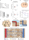
AQP4 regulates interstitial fluid resorption and solute clearance. A, top panel: baseline brain water content of glial-conditional Aqp4 knockout (cAqp4−/−) mice and litter controls (f/f) at various stages of postnatal development suggests a role for AQP4 in resorption of brain water. At postnatal days P15, P28, and P70, the brain water content was significantly higher in cAqp4−/− mice than in litter controls. Bottom panel: brain water content is increased also in adult cAqp4−/− mice. [From Haj-Yasein et al. (55), with permission.] B: Aqp4 deletion increases extracellular space (ECS) volume without affecting tortuosity, as measured by real-time iontophoresis with tetramethylammonium. [From Yao et al. (204), with permission.] C: clearance of intraparenchymally injected mannitol is reduced in mice lacking AQP4. D: hypothetical pathway for brain water flow and solute clearance, based on two-photon imaging of intracisternally injected fluorescent tracers and accumulation of intraparenchymally injected radiotracer (see text for explanation). [C and D from Iliff et al. (69). Reprinted with permission from AAAS.]
Figure 5.
Figure 5.
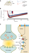
AQP4 regulates extracellular space (ECS) volume during neuronal activity. A: schematic drawing of a hippocampal slice showing the localization of the stimulating and extracellular voltage (Vo, green) and ion-sensitive recording (TMA+, tetramethylammonium) electrodes. Representative traces from synaptic responses (fEPSP) of wild-type (blue) and Aqp4−/− mice are shown. B: deletion of Aqp4 accentuated ECS shrinkage in mouse hippocampal CA1 region during activation of Schaffer collateral/commissural fibers. [A and B are from Haj-Yasein et al. (53), copyright 2012. This material is reproduced with permission of John Wiley & Sons, Inc.] C: astrocytic channels and transporters relevant for water transport during synaptic activity. Clearance of K+ and glutamate from the synaptic cleft imposes a water load on astrocytes and shrinkage of ECS. The data in A indicate that AQP4 helps maintain ECS volume during synaptic activity by facilitating water efflux from astrocytes. Kir4.1, inwardly rectifying potassium channel 4.1; GLT1, glutamate transporter 1 (EAAT2); GluR, ionotropic glutamate receptor; NKCC1, Na+/K+/2Cl cotransporter 1.

Similar articles

Cited by

References

    1. Adams ME, Mueller HA, Froehner SC. In vivo requirement of the alpha-syntrophin PDZ domain for the sarcolemmal localization of nNOS and aquaporin-4. J Cell Biol 155: 113–122, 2001 - PMC - PubMed
    1. Agre P, King LS, Yasui M, Guggino WB, Ottersen OP, Fujiyoshi Y, Engel A, Nielsen S. Aquaporin water channels: from atomic structure to clinical medicine. J Physiol 542: 3–16, 2002 - PMC - PubMed
    1. Alvestad S, Hammer J, Hoddevik EH, Skare O, Sonnewald U, Amiry-Moghaddam M, Ottersen OP. Mislocalization of AQP4 precedes chronic seizures in the kainate model of temporal lobe epilepsy. Epilepsy Res 105: 30–41, 2013 - PubMed
    1. Amiry-Moghaddam M, Otsuka T, Hurn PD, Traystman RJ, Haug FM, Froehner SC, Adams ME, Neely JD, Agre P, Ottersen OP, Bhardwaj A. An alpha-syntrophin-dependent pool of AQP4 in astroglial end-feet confers bidirectional water flow between blood and brain. Proc Natl Acad Sci USA 100: 2106–2111, 2003 - PMC - PubMed
    1. Amiry-Moghaddam M, Ottersen OP. The molecular basis of water transport in the brain. Nat Rev Neurosci 4: 991–1001, 2003 - PubMed

Publication types